Review
. 2020 Feb 21;25(4):978.
doi: 10.3390/molecules25040978.

Glycosaminoglycan-Inspired Biomaterials for the Development of Bioactive Hydrogel Networks

Affiliations
Review

Glycosaminoglycan-Inspired Biomaterials for the Development of Bioactive Hydrogel Networks

Mariana I Neves et al. Molecules. .

Abstract

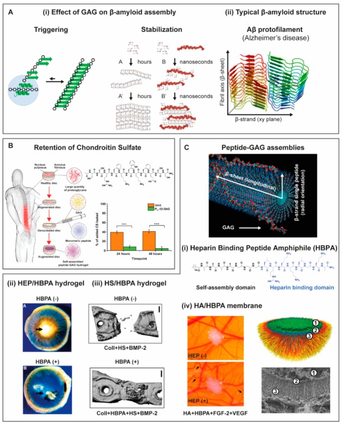
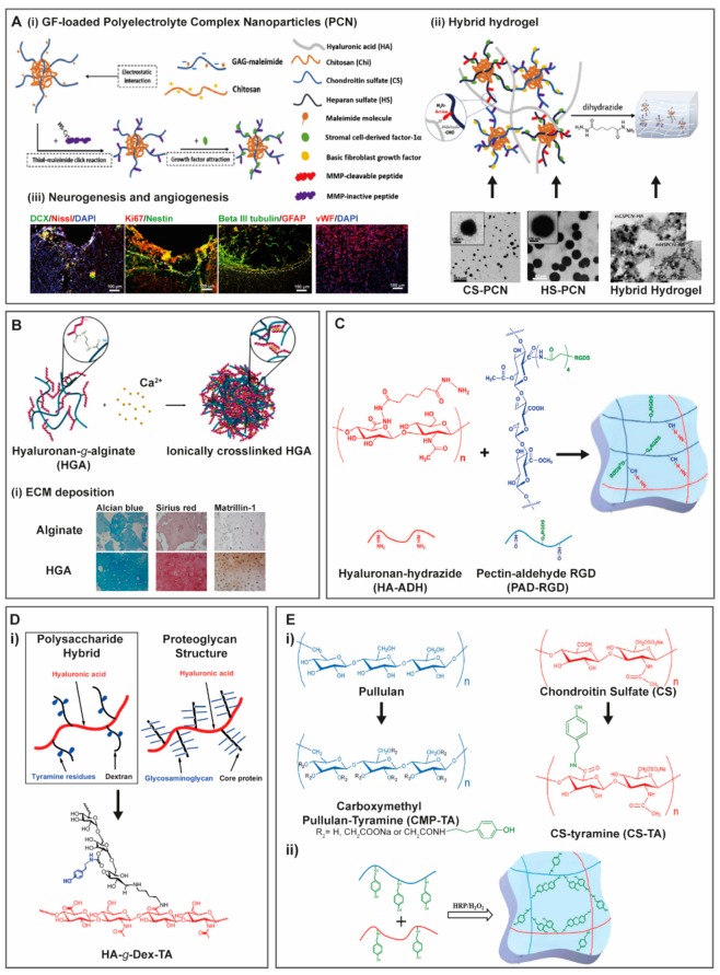
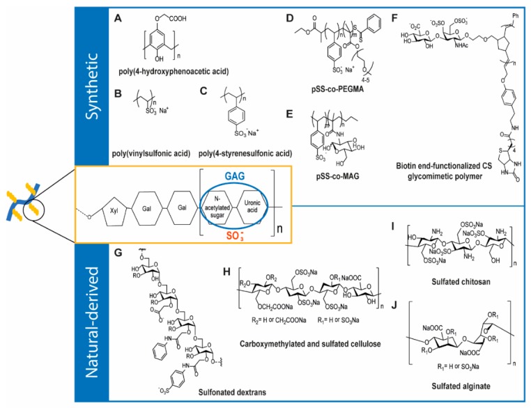
Glycosaminoglycans (GAG) are long, linear polysaccharides that display a wide range of relevant biological roles. Particularly, in the extracellular matrix (ECM) GAG specifically interact with other biological molecules, such as growth factors, protecting them from proteolysis or inhibiting factors. Additionally, ECM GAG are partially responsible for the mechanical stability of tissues due to their capacity to retain high amounts of water, enabling hydration of the ECM and rendering it resistant to compressive forces. In this review, the use of GAG for developing hydrogel networks with improved biological activity and/or mechanical properties is discussed. Greater focus is given to strategies involving the production of hydrogels that are composed of GAG alone or in combination with other materials. Additionally, approaches used to introduce GAG-inspired features in biomaterials of different sources will also be presented.

Keywords: GAG; GAG-mimetics; biomaterials; hybrid systems; hydrogels; polysaccharides; proteins; self-assembly peptides.


Conflict of interest statement

The authors declare no conflict of interest.

Figures

Figure 1
Figure 1
Structure of Glycosaminoglycans (GAG) molecules according to main disaccharide composition. GAG are composed of combined uronic acids [β-d-glucuronic acid (GlcA) or α-l-iduronic acid (IdoA)] and amino sugars [α-d- or β-d-glucosamine (GlcN), N-acetylglucosamine (GlcNac) or N-acetyl-galactosamine GalNac)]. From the combination of the different uronic acid and amino sugars, four main groups of GAG can be distinguished. (1) Heparin and Heparan Sulfate, R1=COCH3 or SO3 and R2=H or SO3; Heparan Sulfate, R1=COCH3 or SO3 and R2=H or SO3. Heparin and Heparan Sulfate share a common amino sugar (α-D-GlcN) but differ in their major uronic acid unit (α-l-IdoA in heparin and β-d-GlcA in heparan sulfate), linked by 1-4 glycosidic bonds. (2) Chondroitin Sulfate and Dermatan Sulfate also share the same amino sugar unit (GalNac) but their uronic unit is β-d-GlcA and α-l-IdoA, respectively. Chondroitin sulfates can be classified according to the position of the sulfates: R1=R2=R3=H (nonsulfated chondroitin), R1=SO3 and R2=R3=H (chondroitin-4-sulfate, CSA), R1=R3=SO3 and R2=H (chondroitin-2,4-disulfate, CSB), R2=SO3 and R1=R3=H (chondroitin-6-sulfate, CSC), R2=R3=SO3 and R1=H (chondroitin-2,6-disulfate, CSD), R1=R2=SO3 and R3=H (chondroitin-4,6-disulfate, CSE), R1=R2=R3=SO3 (trisulfated chondroitin). CS and DS can be found as co-polymeric structures and despite the difference in the uronic unit, DS is also known as chondroitin sulfate B (CSB). (3) Hyaluronan, or hyaluronic acid, is composed of β-1,4-d-GlcA and β-1,3-N-acetyl-d-GlcNac repeating units and is the only non-sulfated GAG. (4) Keratan sulfate has the particularity of not containing uronic acids, being composed of alternating units of galactose (β-1,3-d-Gal) and β-1,4-N-acetyl-d-GlcNac.
Figure 2
Figure 2
Localization of GAG and PG in native tissues. GAG associate covalently to core proteins forming proteoglycans (PG), with exception of HA. Instead, HA physically interacts with other GAG and PG through specific binding domains, as well as particular cell receptors (e.g., CD44). PG can be classified according to their location: They can be found inside cells in secretory compartments where they support storage of positively charged biomolecules (intracellular PG); at the cell membrane as membrane-associated PG where they are involved in cell–cell and cell–ECM interactions (membrane-associated PG) or be secreted to the ECM, where they play both biological and structural roles (extracellular PG).
Figure 3
Figure 3
Schematic representation of main strategies to modulate network formation of GAG-based hydrogels. GAG-hydrogels can be produced from chemical or physical crosslinking between GAG chains, requiring the production of GAG-derivatives with new functionalities. Such modifications can be performed to include new functional groups [e.g., (meth)acrylates, thiols, tyramines, coumarins, peptide sequences] able to crosslink when exposed to light/temperature (A) or enzymes (B). Incorporation of protein-binding domains (C) with high selectivity towards one another can also be used as strategy to covalently crosslink GAG. Combination of GAG with smaller synthetic polymer crosslinkers (D) can be achieved for non-modified GAG or their derivatives. Additionally, GAG-based hydrogels can be designed to have reversible crosslinking, obtained by covalent dynamic crosslinking (e.g., hydrazone bonds or coumarin photo-induced reversible cyclodimerization) (E), or by producing physical hydrogels with GAG derivatives able to crosslink via inclusion complexes (e.g., host-guest cyclodextrin complexes) (F).
Figure 4
Figure 4
(A) Representation of coumarin derivatized HA undergoing crosslinking when exposed to near-UV light stimulus (λ = 365 nm) via photocycloaddition reaction between coumarin moieties and without any catalysts or radical initiators [45]. (B) Synthesis of HA derivatives incorporating transglutaminase (TG) peptides; thiol groups were incorporated in HA backbone via carbodiimide reaction (HA-SH); thiol groups were then substituted with vinyl sulfones (HA-VS) and subsequently with TG/glutamine (HA-TG/Gln) and TG/lysine (HA-TG-Lys) peptides via Michael addition; MMP cleavage site marked by an arrow, cysteines (C) that provide thiols for conjugation onto HA-VS are in bold and the Lys (K) and Gln (Q) covalently coupled to each other on their side chains by FXIIIa are underlined [39]. (C) Crosslinking steps of a HA-tyramine derivative bioink throughout the bioprinting process: step 1, the bioink is partially enzymatically crosslinked in the presence of HRP and H2O2 to improve extrusion; step 2, during extrusion, the bioink is exposed to green light to trigger photocrosslinking and stabilize the construct; step3, post-bioprinting, enzymatically driven functionalization of the scaffold to incorporate cell adhesive RGD motives [53]. (D) (i) SpyTag/SpyCatcher and SnoopTagJr/SnoopCatcher spontaneous amide bond formation; (ii) hydrogel network formation and functionalization with ligand of interest. Central sequence in TriCatchers may incorporate RGDSP-containing or MMP-cleavable linkers [56]. (E) (i) Representation of the HA/CS/PEGDA hydrogel and gelation process that leads to a polymeric network covalently crosslinked via thiol-ene Michael addition click reactions; (ii) chemical crosslinking of GAG thiol derivative mixtures, HA-SH (blue) and CS-SH (red), with PEGDA (green) by conjugate addition [58] (Reproduced with permission from [39,45,53,56,58]).
Figure 5
Figure 5
(A) (i) Modulation of dynamic properties of hydrazone crosslinks by catalyst in injectable hydrogel: upon injection, the catalyst promotes rapid exchange of hydrazone crosslinks and rearrangement of network that facilitate flow; after injection, the catalyst rapidly diffuses away to slow down hydrazone exchange, improving structural stability; (ii) catalyst-accelerated hydrazone equilibrium (catalyst in blue) [63]. (B) Example of GAG hydrogel based on cyclodextrin-based inclusion complexes with norbornene. (i) HA was derivatized to HA-TBA (tetrabutyl ammonium) salt and modified with norbornene groups (NorHA) or β-cyclodextrin groups (CD-HA). (ii) Elastic hydrogels produced with covalent crosslinks between norbornene groups were introduced using di-thiol crosslinkers via light-mediated thiol-ene addition; viscoelastic hydrogels, thiol-ene photochemistry was used to introduce supramolecular interactions between CD-HA and adamantane (guest) groups on thiolated peptides, besides the dithiol-mediated covalent cross-links between the norbornenes [72]. (Reproduced with permission from [63,72]).
Figure 6
Figure 6
Schematic representation of main strategies to modulate network degradation of GAG-base hydrogels. To modulate network degradation, strategies can include enzyme sensitive sequences within crosslink bonds (e.g., MMP-sensitive peptide sequences) (A), or, oppositely, degradation can be inhibited at the level of GAG backbone by incorporation of functional groups that reduce GAG susceptibility to enzymatic cleavage (B). Crosslinking strategies can also be designed to be sensitive to redox agents (C).
Figure 7
Figure 7
(A) HA/collagen IPN developed to mimic fibrillarity and viscoelasticity of the ECM. (i) Scheme of formation viscoelastic HA single network hydrogel via dynamic covalent hydrazone crosslinking and chemical structures of modified HA; (ii) representation of composition and network structure of ECM, dynamic HA-collagen IPN and conventional statically crosslinked hydrogel. (iii) MSC spreading is more pronounced in these dynamic IPN hydrogels with higher stress relaxation in comparison with more conventional hydrogels that have very low stress relaxation. [91]. (B) (i) Chemical structure of gelatin/tyramine (G/T) derivative and gelatin/tyramine/HEP (G/T/H) hydrogels; (ii) crosslinking mechanism by HRP and H2O2 addition to form injectable G/T/H hydrogels and (iii) comparison of new blood vessel formation of G/T and G/T/H hydrogels using an in vivo chicken chorioallantoic membrane (CAM) assay and after 5 days of incubation [103]. (Reproduced with permission from [91,103]).
Figure 8
Figure 8
(A) (i) Schematic representation of GAGs interference on peptide self-assembly, either triggering formation or stabilizing preformed β-amyloid fibrils. (ii) Typical crystalline structure of Alzheimer’s disease Aβ plaques. Peptide β-strand segments orient parallel to the fibril cross-section (xy plane) and interact laterally to form long β-sheets aligned with the fibril long axis. Reproduced/adapted from [129,130] (i) and [128] (ii), with permission. (B) Positively charged, serine-rich peptide sequence bearing a pattern of alternating charged/polar and aromatic amino acids. CS induces self-assembling into β-sheet tapes improving GAG retention and restored compressive stiffness in an ex vivo model of denucleated intervertebral discs. Reproduced/adapted from [138], with permission. (C) Schematic representation of a peptide amphiphile (PA) self-assembling into nanofibers, which surface binds GAG molecules. Nanofiber cross-section shows PAs β-strands distributed radially and β-strands interacting laterally to form β-sheets aligned with the nanofiber long axis. (i) Chemical structure of a PA bearing a heparin binding sequence (HBPA). (ii) Hydrogels formed by HBPA and HEP showed FGF-2/VEGF retention, improving angiogenesis in a rat cornea model. (iii) Hydrogels formed by HBPA and HS showed BMP-2 retention and improved bone healing. (iv) Membranes formed by self-assembly of HBPA with HA and HEP showed FGF-2 and VEGF retention and enhanced angiogenesis in a CAM model. Membranes showed a hierarchical structure comprising a HBPA hydrogel region (1), a permeable membrane (2) and perpendicular nanofibers formed by GAG/peptide electrostatic interactions (3). (Reproduced with permission from [146] (i and ii), [153] (iii), [156,157] (vi)).
Figure 9
Figure 9
(A) (i) Formation of growth factor-loaded polyelectrolyte complex nanoparticles (PCN) through electrostatic interactions with chitosan and PCN coupling with MMP-sensitive/inactive peptide sequences via thiol-maleimide coupling reaction. bFGF and SDF-1a were sequestered on CS-PCN and HS-PCN, respectively; (ii) PCN covalent incorporation within HA-aldehyde hydrogel network through the amine terminated peptides on PCN and the aldehyde groups on HA and hydrogel was covalent crosslinking via hydrazone bonding; TEM images of CS-PCN, HS-PCN and hybrid hydrogels with CS-PCN and HSP-PCN (inset) within HA matrix; (iii) immunohistochemistry results of coronal sections of infarcted brain stained with doublecortin (DCX, migrating neural precursor cells), Niss1 (neuronal cells), DAPI (nuclei), Ki67 (cell proliferation) and Nestin (neural stem/progenitor cells) beta III tubulin (immature neurons), glial fibrillary acidic protein (GFAP, astrogliosis) indicating neurogenesis and with von Willebrand factor (vWF, endothelial cells) indicating angiogenesis (21 days after treatment) [161]. (B) Schematic representation of hyaluronate-g-alginate (HGA) hydrogel formation and (i) histological images of tissue after six weeks of hydrogel transplantation with primary chondrocytes showing increased ECM deposition in HGA samples than in alginate, indicated by higher intensity of Alcian blue, Sirius red and Matrillin-1 protein staining [184]. (C) Chemical structure of HA-hydrazide (red) and RGD-functionalized pectin-aldehyde (blue) and hydrogel crosslinking strategy [200]. (D) (i) representation of the chemical structure of the polysaccharide hybrid based on hyaluronic acid and dextran-tyramine (HA-g-Dex-TA) and comparison with structure of proteoglycan [202]. (E) Chemical structure of pullulan and CS and their corresponding derivatives, carboxymethyl pullulan-tyramine (CMP-TA) and CS-tyramine (CS-TA) and covalent hydrogel formation using these derivatives [217]. (Reproduced with permission from [161,184,200,202,217]).
Figure 10
Figure 10
Synthetic (AF) and natural-derived (GJ) GAG mimetic polymers. Approaches based on fully synthetic polymers comprise non-sulfated (A) or sulfated side chains (B,C,D). Alternatively, glycosidic units are displayed as side chains on synthetic polymer backbones (E,F). Approaches based on natural-derived polymers rely on the modification of polysaccharides backbone with sulfate groups (GJ).
Scheme 1
Scheme 1
(A) GAGs can influence peptide self-assembly through two different general mechanisms, i.e., either nucleation or stabilization of self-assembled structures. (B) Interaction with self-assembling peptides has both biological and technological implications, since de novo designed peptide self-assembly systems retain structural hallmarks of beta-amyloids.

Similar articles

Cited by

References

    1. Pomin V.H., Mulloy B. Glycosaminoglycans and Proteoglycans. Pharmaceuticals (Basel) 2018;11:27. doi: 10.3390/ph11010027. - DOI - PMC - PubMed
    1. Soares da Costa D., Reis R.L., Pashkuleva I. Sulfation of Glycosaminoglycans and Its Implications in Human Health and Disorders. Annu. Rev. Biomed. Eng. 2017;19:1–26. doi: 10.1146/annurev-bioeng-071516-044610. - DOI - PubMed
    1. Hogwood J., Naggi A., Torri G., Page C., Rigsby P., Mulloy B., Gray E. The effect of increasing the sulfation level of chondroitin sulfate on anticoagulant specific activity and activation of the kinin system. PLoS ONE. 2018;13:e0193482. doi: 10.1371/journal.pone.0193482. - DOI - PMC - PubMed
    1. Corsuto L., Rother S., Koehler L., Bedini E., Moeller S., Schnabelrauch M., Hintze V., Schiraldi C., Scharnweber D. Sulfation degree not origin of chondroitin sulfate derivatives modulates keratinocyte response. Carbohydr. Polym. 2018;191:53–64. doi: 10.1016/j.carbpol.2018.02.072. - DOI - PubMed
    1. Pan J., Qian Y., Zhou X., Lu H., Ramacciotti E., Zhang L. Chemically oversulfated glycosaminoglycans are potent modulators of contact system activation and different cell signaling pathways. J. Biol. Chem. 2010;285:22966–22975. doi: 10.1074/jbc.M109.063735. - DOI - PMC - PubMed

MeSH terms

LinkOut - more resources