. 2020 Feb 22;21(4):1486.
doi: 10.3390/ijms21041486.

Galectin 3 Deficiency Alters Chondrocyte Primary Cilium Formation and Exacerbates Cartilage Destruction via Mitochondrial Apoptosis

Affiliations

Galectin 3 Deficiency Alters Chondrocyte Primary Cilium Formation and Exacerbates Cartilage Destruction via Mitochondrial Apoptosis

Narjès Hafsia et al. Int J Mol Sci. .

Abstract

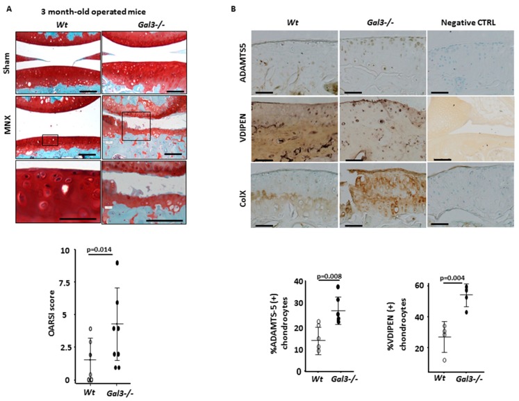
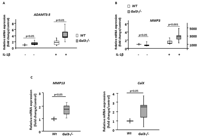
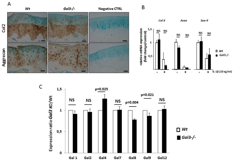
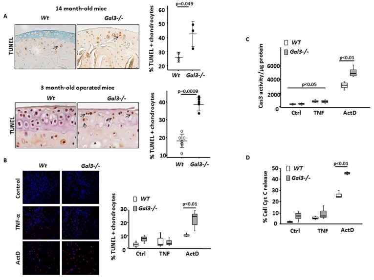
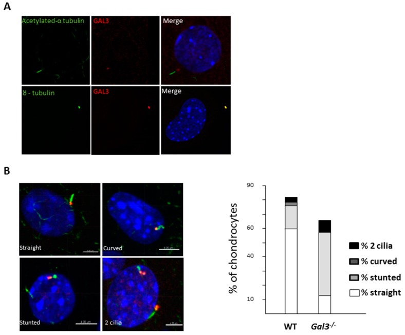
Mechanical overload and aging are the main risk factors of osteoarthritis (OA). Galectin 3 (GAL3) is important in the formation of primary cilia, organelles that are able to sense mechanical stress. The objectives were to evaluate the role of GAL3 in chondrocyte primary cilium formation and in OA in mice. Chondrocyte primary cilium was detected in vitro by confocal microscopy. OA was induced by aging and partial meniscectomy of wild-type (WT) and Gal3-null 129SvEV mice (Gal3-/-). Primary chondrocytes were isolated from joints of new-born mice. Chondrocyte apoptosis was assessed by Terminal deoxynucleotidyl transferase dUTP nick end labeling (TUNEL), caspase 3 activity and cytochrome c release. Gene expression was assessed by qRT-PCR. GAL3 was localized at the basal body of the chondrocyte primary cilium. Primary cilia of Gal3-/- chondrocytes were frequently abnormal and misshapen. Deletion of Gal3 triggered premature OA during aging and exacerbated joint instability-induced OA. In both aging and surgery-induced OA cartilage, levels of chondrocyte catabolism and hypertrophy markers and apoptosis were more severe in Gal3-/- than WT samples. In vitro, Gal3 knockout favored chondrocyte apoptosis via the mitochondrial pathway. GAL3 is a key regulator of cartilage homeostasis and chondrocyte primary cilium formation in mice. Gal3 deletion promotes OA development.

Keywords: Galectin 3; apoptosis; chondrocyte; mechanical stress; osteoarthritis; primary cilium.


Conflict of interest statement

The authors declare no conflict of interest.

Figures

Figure 1
Figure 1
Galectin 3 (GAL3) deficiency induces osteoarthritis (OA) during aging. (A) Knee cartilage sections stained with safranin-O from non-operated Gal3−/− (n = 5 mice) and wild-type (WT) (n = 7 mice) 14-month-old mice. Low (top panels) and high (low panels) magnification are presented. OA lesions were assessed by OARSI score (right panel). (B) Immunohistochemistry of expression of ADAMTS-5 and type X collagen (n = 4 WT and n = 7 Gal3−/− mice). The percentage of ADAMTS-5-positive chondrocytes in Gal3−/− and WT cartilage was counted ((B), right panel). Negative controls were nonspecific IgG antibodies. Scale bars: 100 µm. Two-tailed Mann–Whitney U test between WT and Gal3−/−.
Figure 2
Figure 2
GAL3 deficiency induces OA during mechanical stress. (A) Knee cartilage sections stained with safranin-O after joint meniscectomy (MNX) or sham operation in 3-month-old mice (n = 8 mice for WT and Gal3−/−). Low (top panels) and high (low panels) magnification are presented. OA lesions were assessed by OARSI score (low panel). (B) Immunohistochemistry of expression of ADAMTS-5 (n = 5 WT and Gal3−/− mice), VDIPEN (n = 4 WT and Gal3−/− mice) and type X collagen (n = 8 WT and Gal3−/− mice). The percentage of ADAMTS-5– and VDIPEN-positive chondrocytes in Gal3−/− and WT cartilage was counted ((B), low panel). Negative controls were nonspecific IgG antibodies. Scale bars: 100 µm. Two-tailed Mann Whitney U test between WT and Gal3−/−.
Figure 3
Figure 3
GAL3 deletion induces chondrocyte catabolism. qRT-PCR analysis of Adamts-5 (A), Mmp3 (B), Mmp13 and type X collagen (C) mRNA expression in cultured chondrocytes from articular cartilage of Gal3−/− and WT newborn mice with and without IL-1β stimulation (10 ng/mL) (201-LB/CF, R&D Systems, Lille, France) (n = 5 independent experiments). Horizontal bars were medians, box edges are Q1–Q3 and whiskers are range. Two-tailed Mann–Whitney U test between WT and Gal3−/−.
Figure 4
Figure 4
GAL3 deletion does not modulate chondrocyte anabolism. (A) Immunohistochemistry of expression of type II collagen and aggrecan in knee cartilage of WT and Gal3−/− cartilages after MNX (n = 5 of WT and Gal3−/− mice). Scale bars: 100 µm (B) qRT-PCR analysis of Coll II, Acan and Sox9 mRNA expression in cultured chondrocytes from articular cartilages of Gal3−/− and WT newborn mice with and without IL-1β stimulation (10 ng/mL) (201-LB/CF, R&D Systems, Lille, France) (n = 5 independent experiments). (C) qRT-PCR analysis of Lgals1, Lgals2, Lgals4, Lgals7, Lgals8, Lgals9 and Lgals12 expression in cultured chondrocytes from articular cartilage of Gal3−/− newborn mice (n = 5 independent experiments). Two-tailed Mann–Whitney U test between WT and Gal3−/−.
Figure 5
Figure 5
GAL3 deletion induces chondrocyte apoptosis via mitochondrial pathway. (A) Ex vivo, chondrocyte apoptosis was assessed by TUNEL labeling in 14-month-old mouse cartilages (n = 3 WT and Gal3−/− mice) (left and top panel) and 3-month-old mouse cartilages after MNX (n = 8 WT and Gal3−/− mice). (B) In vitro, articular cartilage chondrocytes from Gal3−/− and WT newborn mice were stimulated with TNF-α (20 ng/mL) or actinomycin (ActD, 50 nM) or left untreated (Ctrl) (n = 3 independent experiments). Percentage of TUNEL positive chondrocytes (A and B, right panels). (C) Caspase 3 (Cas3) activity (expressed in arbitrary units (AUs)) assessed 48 h after stimulation with TNF-α or ActD and normalized for protein contain. (D) Percentage of cytochrome c (Cyt c) release in chondrocyte cultures assessed by immunostaining and fluorescence microscopy. Scale bars: 100 µm. Horizontal bars were medians, box edges are Q1–Q3 and whiskers are range. Two-tailed Mann–Whitney U test between WT and Gal3−/−.
Figure 6
Figure 6
Deletion of galectin 3 (Gal3) induces alteration of articular chondrocyte primary cilium. (A) Confocal microscopy of acetylated α-tubulin (green) and GAL3 (red) (top pictures) or γ-tubulin (green) and GAL3 (red) (bottom pictures) in primary cilium of cultured wild-type (WT) chondrocytes (n = 5 independent experiments). (B) Various shapes of primary cilia were observed: straight, stunted, curved and double cilia. The relative proportions of each shape in WT versus Gal3−/− cultures are indicated. n = 642 WT and n = 769 Gal3−/− chondrocytes. Scale bars: 4 µm.

References

    1. Wei Y., Bai L. Recent advances in the understanding of molecular mechanisms of cartilage degeneration, synovitis and subchondral bone changes in osteoarthritis. Connect. Tissue Res. 2016;57:45–61. doi: 10.1080/03008207.2016.1177036. - DOI - PubMed
    1. Loeser R.F., Collins J.A. Aging and the pathogenesis of osteoarthritis. Nat. Rev. Rheumatol. 2016;12:412–420. doi: 10.1038/nrrheum.2016.65. - DOI - PMC - PubMed
    1. Silverwood V., Blagojevic-Bucknall M. Current evidence on risk factors for knee osteoarthritis in older adults: A systematic review and meta-analysis. Osteoarthr. Cartil. 2015;23:507–515. doi: 10.1016/j.joca.2014.11.019. - DOI - PubMed
    1. Kraan P.M., van der Berenbaum F. Translation of clinical problems in osteoarthritis into pathophysiological research goals. RMD Open. 2016;2:e000224. doi: 10.1136/rmdopen-2015-000224. - DOI - PMC - PubMed
    1. Thompson C.L., Chapple J.P. Primary cilia disassembly down-regulates mechanosensitive hedgehog signalling: A feedback mechanism controlling ADAMTS-5 expression in chondrocytes. Osteoarthr. Cartil. 2014;22:490–498. doi: 10.1016/j.joca.2013.12.016. - DOI - PMC - PubMed

LinkOut - more resources