

Functional Bioglass-Biopolymer Double Nanostructure for Natural Antimicrobial Drug Extracts Delivery
- PMID: 32098412
- PMCID: PMC7075305
- DOI: 10.3390/nano10020385
Functional Bioglass-Biopolymer Double Nanostructure for Natural Antimicrobial Drug Extracts Delivery
Abstract
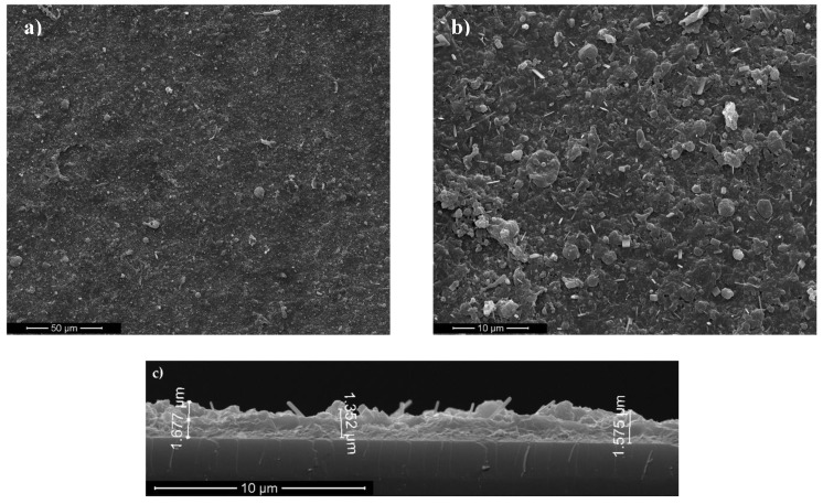
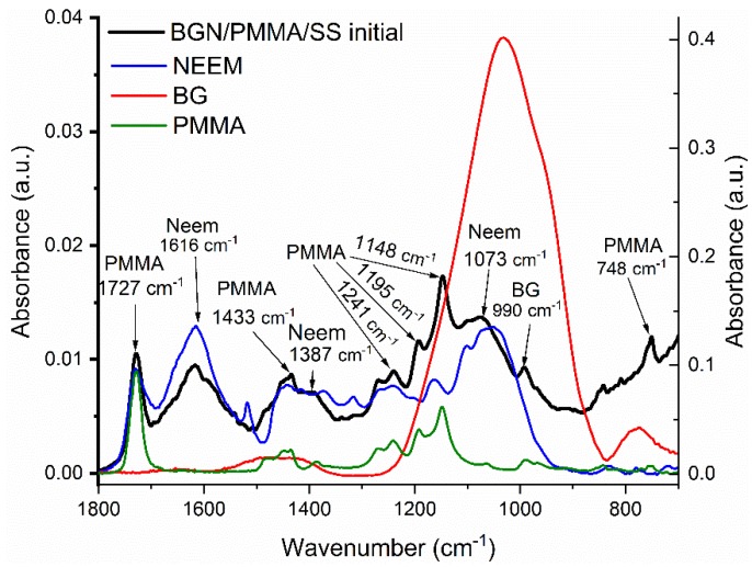
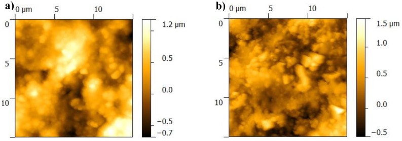
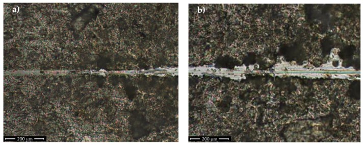
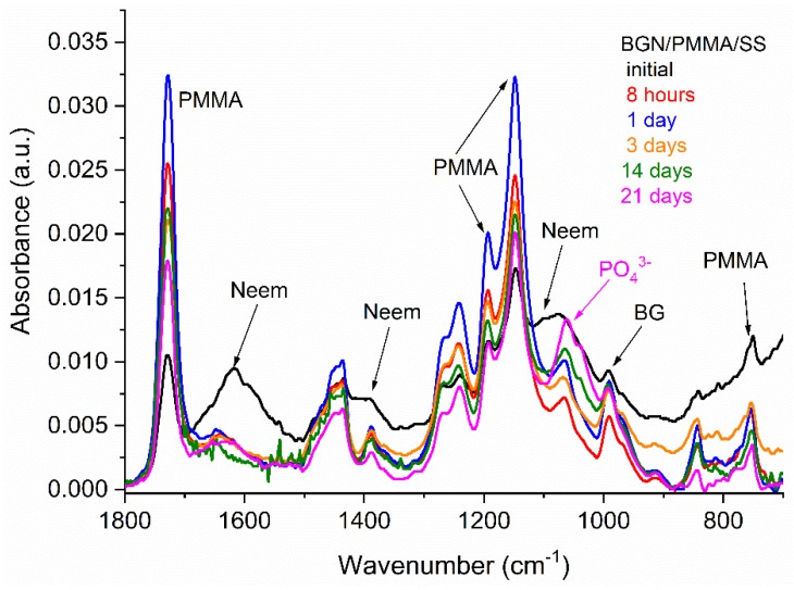
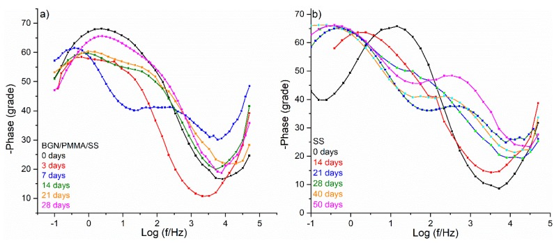
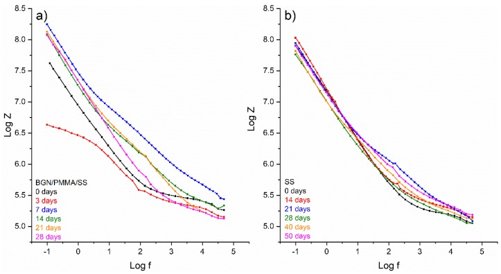
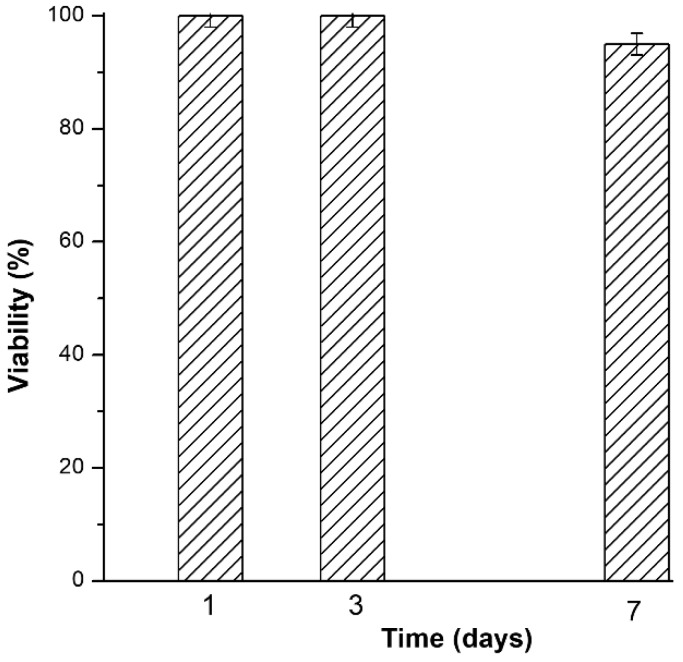
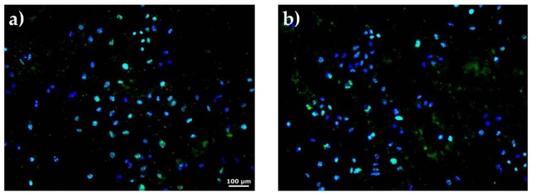
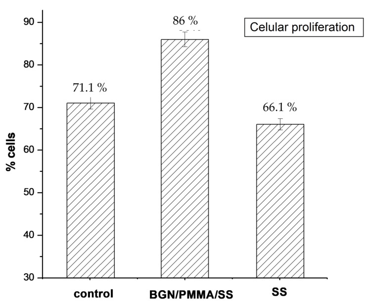
Aseptic loosening and periprosthetic infections are the main causes of implant failure. Strategies to mitigate this drawback are therefore mandatory to avoid primary and revision replacement surgeries. A functional bioapatite-biopolymer double nanostructure fabricated by matrix-assisted pulsed laser evaporation to prevent infection of orthopedic and dental implants could promote osseointegration and ensure controlled delivery of natural antimicrobial drugs. The synthesized nanostructure consists of two overlapping layers, the lower from a biocompatible polymer for anticorrosive protection, and the upper of bioactive glass incorporating antimicrobial plant extract, acting as a potential drug delivery system. Morphology, composition, adherence, ability for drug delivery and biological properties (cytotoxicity and antimicrobial effect) were studied. Structures proved compact and stable, conserving a remarkable drug delivery ability for more than 21 days, i.e., enough to ensure long-term microbes' eradication.
Keywords: antimicrobial effect; bioactive coatings; drug release; implant protection.
Conflict of interest statement
The authors declare no conflicts of interest.
Figures
References
-
- Kumar M., Kumar R., Kumar S., Prakash C. Biomechanical Properties of Orthopedic and Dental Implants: A Comprehensive Review. Handb. Res. Green Eng. Tech. Mod. Manuf. 2019:1–13. doi: 10.4018/978-1-5225-5445-5.ch001. - DOI
-
- Scholz M.S., Blanchfield J.P., Bloom L.D., Coburn B.H., Elkington M., Fuller J.D., Gilbert M.E., Muflahi S.A., Pernice M.F., Trevarthen J.A., et al. The use of composite materials in modern orthopaedic medicine and prosthetic devices: A review. Compos. Sci. Technol. 2011;71:1791–1803. doi: 10.1016/j.compscitech.2011.08.017. - DOI
-
- Floroian L., Samoila C., Badea M., Munteanu D., Ristoscu C., Sima F., Negut I., Chifiriuc M.C., Mihailescu I.N. Stainless steel surface biofunctionalization with PMMA-bioglass coatings: Compositional, electrochemical corrosion studies and microbiological assay. J. Mater. Sci. Mater. Med. 2015;26:195. doi: 10.1007/s10856-015-5527-y. - DOI - PubMed
Grants and funding
- PRO-DD (POS-CCE, O.2.2.1., ID 123, SMIS 2637, ctr. No 11/2009)/European Commission
- 2017 Fellowship/Transilvania University of Brasov, Romania
- PN-III-P2-2.1-PED-2016-0621 no.146PED/2017/Unitatea Executiva pentru Finantarea Invatamantului Superior, a Cercetarii, Dezvoltarii si Inovarii
- PN-III-P1-1.1-PD-2016-1072/Unitatea Executiva pentru Finantarea Invatamantului Superior, a Cercetarii, Dezvoltarii si Inovarii
- 16N/2019/Ministerul Educației și Cercetării Științifice
LinkOut - more resources
Full Text Sources
