Review
. 2020 Feb 22;12(2):575.
doi: 10.3390/nu12020575.

Vitamin D and Endothelial Function

Affiliations
Review

Vitamin D and Endothelial Function

Do-Houn Kim et al. Nutrients. .

Abstract

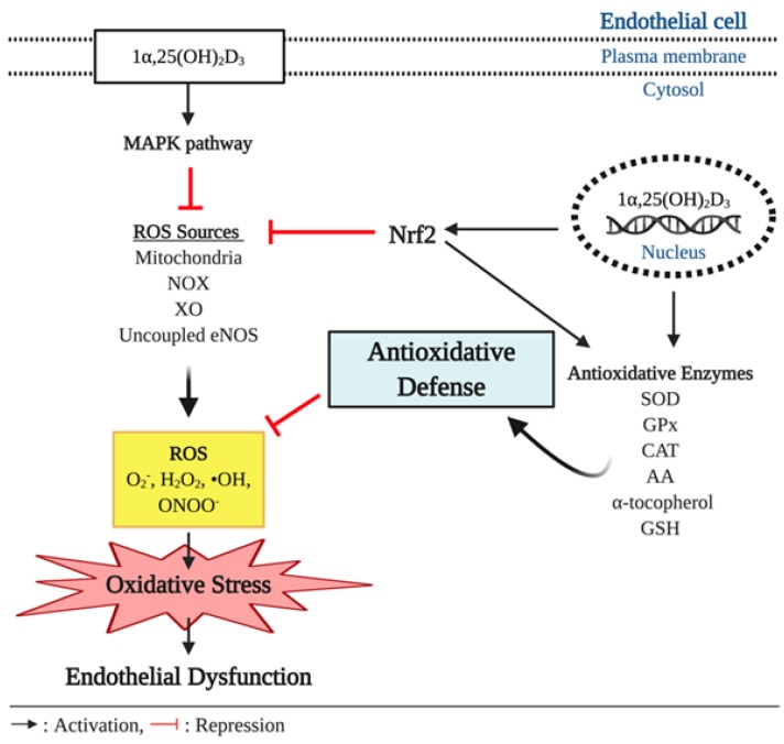
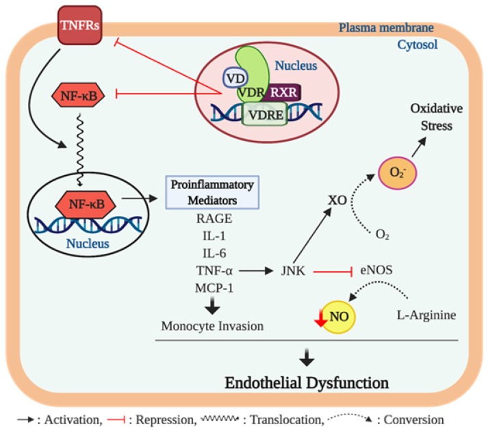
Vitamin D is known to elicit a vasoprotective effect, while vitamin D deficiency is a risk factor for endothelial dysfunction (ED). ED is characterized by reduced bioavailability of a potent endothelium-dependent vasodilator, nitric oxide (NO), and is an early event in the development of atherosclerosis. In endothelial cells, vitamin D regulates NO synthesis by mediating the activity of the endothelial NO synthase (eNOS). Under pathogenic conditions, the oxidative stress caused by excessive production of reactive oxygen species (ROS) facilitates NO degradation and suppresses NO synthesis, consequently reducing NO bioavailability. Vitamin D, however, counteracts the activity of nicotinamide adenine dinucleotide phosphate (NADPH) oxidase which produces ROS, and improves antioxidant capacity by enhancing the activity of antioxidative enzymes such as superoxide dismutase. In addition to ROS, proinflammatory mediators such as TNF-α and IL-6 are risk factors for ED, restraining NO and eNOS bioactivity and upregulating the expression of various atherosclerotic factors through the NF-κB pathway. These proinflammatory activities are inhibited by vitamin D by suppressing NF-κB signaling and production of proinflammatory cytokines. In this review, we discuss the diverse activities of vitamin D in regulating NO bioavailability and endothelial function.

Keywords: NO; NOX; ROS; calcitriol; eNOS; endothelial dysfunction; inflammation; nitric oxide; oxidative stress; vitamin D deficiency.


Conflict of interest statement

The authors declare no conflict of interest.

Figures

Figure 1
Figure 1
Basic physiology of vitamin D action. Humans obtain vitamin D mainly through endogenous production of previtamin D3 and vitamin D3 in the skin, followed by subsequent conversions in the liver and kidney. When exposed to the sun, 7-dehydrocholesterol converts to previtamin D3 and vitamin D3 by ultraviolet B (UVB) radiation and heat, respectively, in the skin. A small amount of vitamin D can be present in natural food as vitamin D2 and D3. Vitamin D2/D3 go through hydroxylation two times in the liver (vitamin D2/D3 → 25 (OH) D3) and the kidney (25 (OH) D3 → 1α,25 (OH)2D3) to become a biologically active form of vitamin D3, 1α,25 (OH)2D3;1α,25 (OH)2D3 exerts its biological actions by binding to the nuclear vitamin d receptor (VDR), which associates with a retinoid x receptor (RXR) in the nucleus. The VDR/RXR heterodimers bind to the vitamin D response element (VDRE) in the promoter region of vitamin D-regulated genes and initiate expression of various genes. VDR is also found in the plasma membrane, and the liganded plasma membrane VDR activates an intracellular signaling transduction involved in many physiological actions.
Figure 2
Figure 2
Role of vitamin D and vitamin D receptor (VDR) in regulating nitric oxide (NO) bioavailability. Ligand-bound VDR plays an important role in regulating NO synthesis via alterations in eNOS activity. Activation of plasma membrane VDR upregulates the activity of endothelial NO synthase (eNOS), a calcium dependent enzyme, by upregulating the formation of intracellular second messengers including adenylyl cyclase (AC), diacylglycerol (DAG) and inositol trisphosphate (IP3), which in turn result in calcium influx through the voltage-sensitive calcium channel (VSCC) in the plasma membrane and the sarcoplasmic reticulum and through the ip3 receptor/calcium channel (IP3CC) in the sarcoplasmic reticulum. The increased intracellular calcium concentrations facilitate the calcium-calmodulin (CaM) pathway to activate eNOS. In addition, plasma membrane VDR triggers eNOS activation through phosphorylation of serine-1779 (human serine 1177) on eNOS by activating the phosphoinositide 3-kinase (PI3K)/ protein kinase b (Akt) pathway. Furthermore, genetic action of vitamin D via the nuclear VDR promotes eNOS expression, which synthesizes NO from L-arginine, and suppresses arginase-2 (AG2) expression, which inhibits eNOS activity by hydrolyzing the substrate for NO synthesis (arginine) to ornithine and urea. Increased NO production promotes angiogenesis by upregulating gene expression of matrix metalloproteinase 2 (MMP-2) which improves endothelial cell (EC) migration and proliferation capacity. In addition, NO mediates angiogenetic activity of the cell via upregulation of vascular endothelial growth factor (VEGF) and fibroblast growth factor (FGF) and suppression of the angiogenesis inhibitor, angiostatin.
Figure 3
Figure 3
Antioxidant effect of vitamin D and endothelial function. In pathophysiological conditions, overproduction of reactive oxygen species (ROS), such as superoxide anion (O2), hydrogen peroxide (H2O2), hydroxyl radical (•OH), and peroxynitrite (ONOO), outbalances antioxidative defenses and causes oxidative stress, which is implicated to development of endothelial dysfunction. In the cell, ROS are produced by various intracellular sources including mitochondria, nicotinamide adenine dinucleotide phosphate (NADPH) oxidase (NOX), xanthine oxidase (XO), and uncoupled endothelial nitric oxide synthase (eNOS). Vitamin D elicits antioxidant effects through upregulating expression of antioxidative enzymes including superoxide dismutase (SOD), glutathione peroxidase (GPx), catalase (CAT), ascorbic acid (AA), α-tocopherol, and glutathione (GSH) that can scavenge the free radicals. In addition, the genetic action of vitamin D triggers the expression of nuclear respiratory factor 2 (Nrf2), a key transcriptional factor that suppresses ROS production from its various sources and upregulates the expression of the antioxidative enzymes.
Figure 4
Figure 4
Vitamin D, inflammation, and endothelial dysfunction. The chronic inflammation process contributes to developing endothelial dysfunction through proinflammatory cytokine activity. Ligand bound vitamin D receptor (VDR) activation suppresses gene expression of nuclear factor-κB (NF-κB) and tumor necrosis factor (TNF)-α receptors 2 and 4 (TNFRs). The NF-κB is a key transcription factor that promotes expression of various proinflammatory mediators including advanced glycation end products (AGEs), interleukin (IL)-1 and 6, TNF-α, and monocyte chemoattractant protein-(MCP)-1 which are implicated in endothelial dysfunction. Among these proinflammatory cytokines, TNF-α activates the c-Jun N-terminal kinase (JNK) pathway that inhibits endothelial NO synthase (eNOS) activity, consequently resulting in reduction of nitric oxide (NO) bioavailability. In addition, the upregulated JNK pathway induces the formation of superoxide anion O2− from oxygen (O2) through upregulating xanthine oxidase (XO) activity, which further impairs endothelial function via inducing oxidative stress in the cell. Furthermore, TNF-α bound TNFRs trigger the translocation of NF-κB to the nucleus to promote the expression of proinflammatory mediators.

References

    1. Fleming I., Busse R. Signal transduction of eNOS activation. Cardiovasc. Res. 1999;43:532–541. doi: 10.1016/S0008-6363(99)00094-2. - DOI - PubMed
    1. Dedeoglu M., Garip Y., Bodur H. Osteomalacia in Crohn’s disease. Arch. Osteoporos. 2014 doi: 10.1007/s11657-014-0177-0. - DOI - PubMed
    1. Kanikarla-Marie P., Jain S.K. 1,25(OH) 2 D 3 inhibits oxidative stress and monocyte adhesion by mediating the upregulation of GCLC and GSH in endothelial cells treated with acetoacetate (ketosis) J. Steroid Biochem. Mol. Biol. 2016 doi: 10.1016/j.jsbmb.2016.03.002. - DOI - PMC - PubMed
    1. Bischoff H., Borchers M., Gudat F., Duermueller U., Theiler R., Stähelin H., Dick W. In Situ Detection of 1,25-dihydroxyvitamin D Receptor In human Skeletal Muscle Tissue. Histochem. J. 2001;33:19–24. doi: 10.1023/A:1017535728844. - DOI - PubMed
    1. Napoli C., De Nigris F., Williams-Ignarro S., Pignalosa O., Sica V., Ignarro L.J. Nitric oxide and atherosclerosis: An update. Nitric Oxide Biol. Chem. 2006;15:265–279. doi: 10.1016/j.niox.2006.03.011. - DOI - PubMed

MeSH terms