. 2020 Feb 25;11(1):e02918-19.
doi: 10.1128/mBio.02918-19.

The Plasmodium falciparum Artemisinin Susceptibility-Associated AP-2 Adaptin μ Subunit is Clathrin Independent and Essential for Schizont Maturation

Affiliations

The Plasmodium falciparum Artemisinin Susceptibility-Associated AP-2 Adaptin μ Subunit is Clathrin Independent and Essential for Schizont Maturation

Ryan C Henrici et al. mBio. .

Abstract

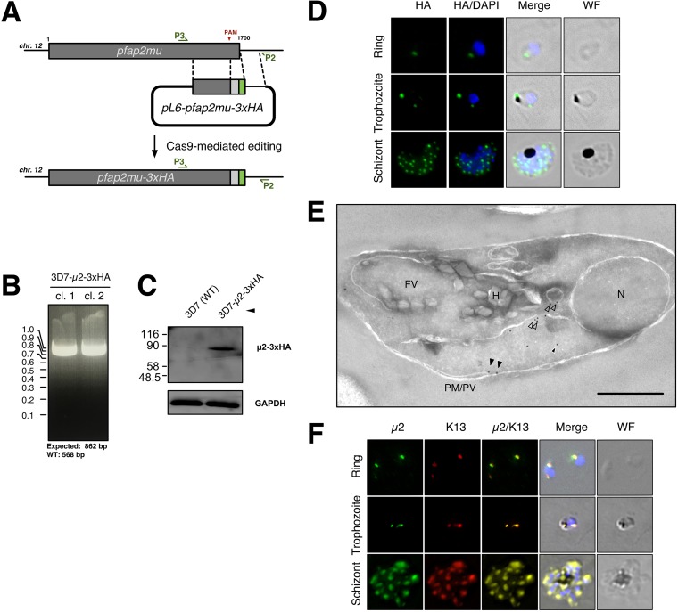
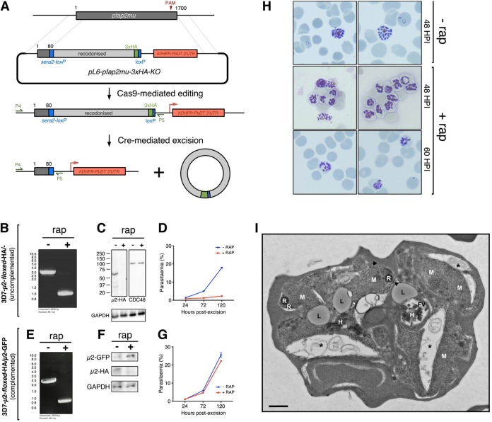
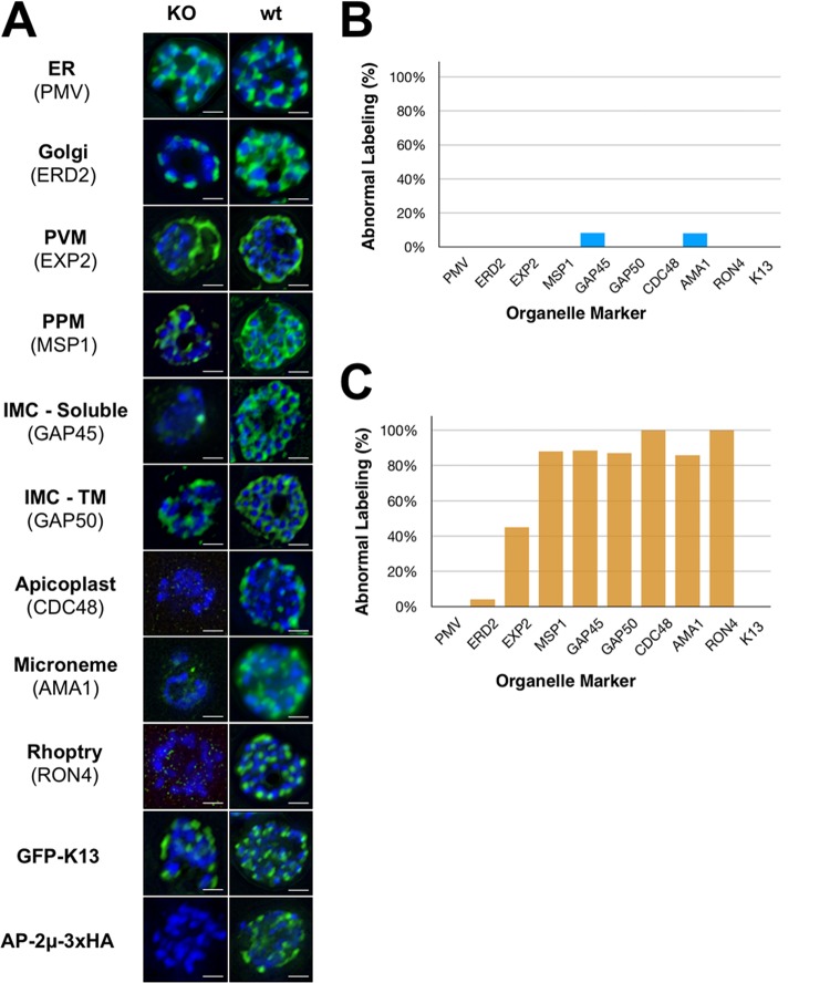
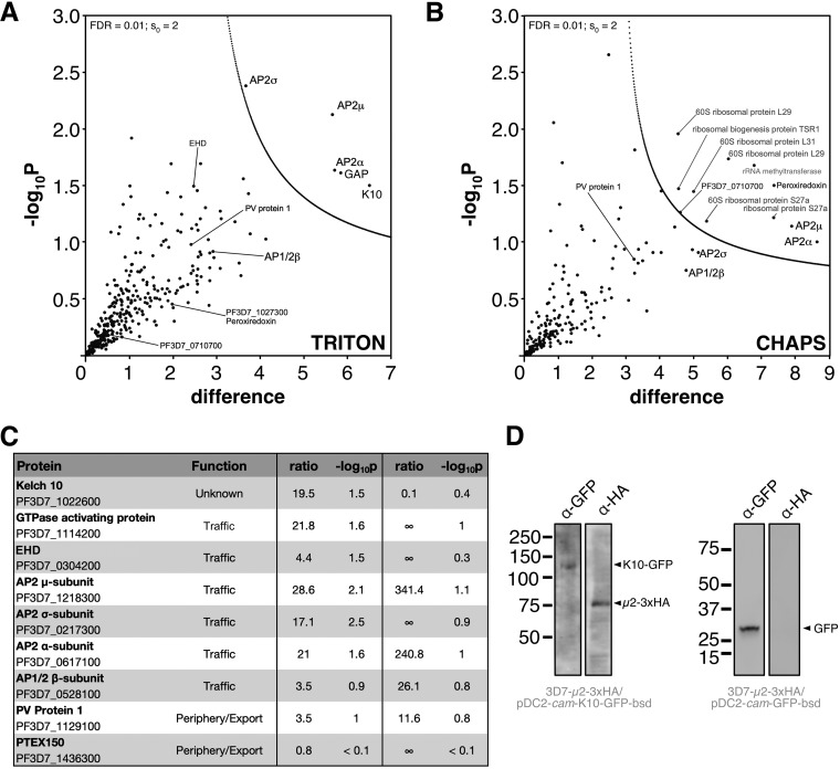
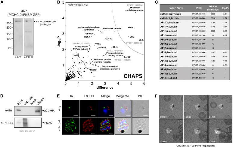
The efficacy of current antimalarial drugs is threatened by reduced susceptibility of Plasmodium falciparum to artemisinin, associated with mutations in pfkelch13 Another gene with variants known to modulate the response to artemisinin encodes the μ subunit of the AP-2 adaptin trafficking complex. To elucidate the cellular role of AP-2μ in P. falciparum, we performed a conditional gene knockout, which severely disrupted schizont organization and maturation, leading to mislocalization of key merozoite proteins. AP-2μ is thus essential for blood-stage replication. We generated transgenic P. falciparum parasites expressing hemagglutinin-tagged AP-2μ and examined cellular localization by fluorescence and electron microscopy. Together with mass spectrometry analysis of coimmunoprecipitating proteins, these studies identified AP-2μ-interacting partners, including other AP-2 subunits, the K10 kelch-domain protein, and PfEHD, an effector of endocytosis and lipid mobilization, but no evidence was found of interaction with clathrin, the expected coat protein for AP-2 vesicles. In reverse immunoprecipitation experiments with a clathrin nanobody, other heterotetrameric AP-complexes were shown to interact with clathrin, but AP-2 complex subunits were absent.IMPORTANCE We examine in detail the AP-2 adaptin complex from the malaria parasite Plasmodium falciparum In most studied organisms, AP-2 is involved in bringing material into the cell from outside, a process called endocytosis. Previous work shows that changes to the μ subunit of AP-2 can contribute to drug resistance. Our experiments show that AP-2 is essential for parasite development in blood but does not have any role in clathrin-mediated endocytosis. This suggests that a specialized function for AP-2 has developed in malaria parasites, and this may be important for understanding its impact on drug resistance.

Keywords: Plasmodium falciparum; adaptin trafficking complex; adaptor proteins; artemisinin susceptibility; endocytosis; malaria.


Figures

FIG 1
FIG 1
P. falciparum AP-2μ is localized to a noncanonical cytoplasmic compartment. (A) Homologous repair construct used to install AP-2μ variants to fuse a tandem triple hemagglutinin tag (3xHA) onto the C terminus of AP-2μ. (B) PCR-based genotyping of two parasite clones harboring AP-2μ-3xHA in place of the endogenous AP-2μ allele. Amplification of the integrated transgenic pfap2μ locus with P3 and P2 (annealing sites annotated) produces an 862-bp fragment. Genotypes were confirmed by Sanger sequencing of the PCR products shown. (C) Anti-HA Western blot confirming expression of the desired fusion protein (∼78 kDa) in mixed-stage lysates, compared to wild-type parental 3D7. Molecular weights are presented in kDa. (D) Localization of AP-2μ-3xHA (green) across the asexual life cycle by anti-HA IFA, counterstained for parasite DNA with DAPI (blue). The images shown are representative of more than 100 cells examined at each stage; merge is the superimposition of each channel on a brightfield image (WF). Maximum intensity z-projections are shown. Scale bar, 2 μm. (E) Immunoelectron micrograph of a representative young intraerythrocytic trophozoite. AP-2μ-3xHA parasites probed with an anti-HA rabbit antibody and a secondary antibody 18 nm gold conjugate. Protein disulfide isomerase (PDI), a marker for the parasite ER, is detected by an anti-PDI mouse antibody and a secondary conjugated to 12-nm gold particles (Fig. S2, S3, S4, and Table S1). N, nucleus; FV, food vacuole; H, hemazoin; PM/PV, plasma membrane/parasitophorous vacuole; empty arrows, AP-2μ associated with vesicles; black arrows, AP-2μ at the plasma membrane; white-outlined arrows, AP-2μ in the cytosol. Scale bar, 500 nm. (F) Localization of AP-2μ-3xHA (green) with respect to episomally expressed GFP-K13 (red) across the asexual life cycle by IFA. Representative images of more than 100 observed cells is shown. Maximum intensity z-projections are shown. Scale bar, 2 μm.
FIG 2
FIG 2
AP-2μ is required for asexual replication and schizont maturation. (A) Schematic for the integration of loxP recombination elements into the endogenous pfap2μ locus of a parasite line constitutively expressing a split-Cre recombinase (43). The addition of rap initiates Cre dimerization and excision of the loxP-flanked (floxed) region of pfap2μ on chromosome 12. (B) PCR confirmation of rap-induced excision of floxed region by PCR using the primers P4 and P5 (see panel A). (C) Western blot confirmation that excision of floxed pfap2μ causes a loss of AP-2μ-3xHA protein (within 24 h) but has no effect on levels of CDC48 protein. Molecular weight is presented in kDa. (D) Parasite multiplication in the 3D7-AP-2μ-floxed-3xHA line across 2.5 cell cycles, with or without rap induction of Cre. The mean parasitemia (normalized to 0.25% starting parasitemia) with the standard error is shown at each time point. Each data point represents the average of at least three biological replicates (different cultures, different days). (E) PCR confirmation of rap-mediated pfap2μ excision in 3D7-AP-2μ-floxed-3xHA parasites transfected with an episome encoding cam-AP-2μ-GFP. The construction of this complementation plasmid is described in Fig. S5. (F) Western blot confirmation that excision of chromosomal pfap2μ from 3D7-AP-2μ-floxed-3xHA/cam-AP-2μ-GFP parasites causes a loss of AP-2μ-3xHA protein, but it does not prevent episomal expression of AP-2μ-GFP. (G) Parasite multiplication in the 3D7-AP-2μ-floxed-3xHA/cam-AP-2μ-GFP line across 2.5 cell cycles, with or without induction of Cre by rap. Means and standard errors are shown as in panel D. (H) Giemsa staining of 3D7-AP-2μ-floxed-3xHA schizonts, without rap treatment at 48 h postinfection and with rap treatment at 48 and 60 h postinfection. (I) Electron micrograph of 3D7-AP-2μ-floxed-3xHA schizonts, with or without 1-h ring-stage treatment with 10 nM rap. Micronemes at the apical end of developing merozoites are labeled with arrowheads; asterisks indicate membrane separation (see Fig. S7). FV, food (digestive) vacuole; H, hemozoin; L, lipid body; M, merozoite; R, rhoptry. Scale bar, 500 nm.
FIG 3
FIG 3
AP-2μ-KO severely disrupts schizont maturation. (A) Antibodies against the ER (PMV), Golgi apparatus (ERD2), PVM (EXP2), PPM (MSP1), IMC (GAP45, GAP50), apicoplast (CDC48), micronemes (AMA1), rhoptries (RON4), episomal K13 (GFP), and AP-2μ (HA) were used to stain 3D7-AP-2μ-floxed-3xHA schizonts with rap treatment (KO) or without (wt). All organelle markers have been false colored green regardless of fluorophore-conjugated secondary antibody used for clarity. Nuclei have been false colored blue. IMC-TM, transmembrane component of inner membrane complex. Scale bar, 2 μm. (B and C) Abnormal labeling in AP-2μ KO parasites was quantitated relative to the staining observed in the majority of wild-type schizonts (B rap–; C rap+). Normal staining was defined as follows: ERD2, PMV, and CDC48, discrete punctate staining corresponding to each nucleus; EXP2, contiguous, circular, peripheral membrane staining; GAP45, GAP50, and MSP1, distinct, circular grape-like staining surrounding each daughter nucleus; AMA1 and RON4, discrete, apical punctate spots corresponding to each nucleus. At least 100 cells were scored for each marker.
FIG 4
FIG 4
Identification of AP-2μ-interacting proteins. (A) Volcano plot from P values versus the corresponding t test difference of proteins identified by immunoprecipitation (IP) in Triton buffer. Cutoff curves for statistically significant interactors (dotted curve) were calculated from the estimated false discovery rate (for details, see Materials and Methods). Selected hits are labeled (potentially nonspecific interactors are in gray). (B) Volcano plot for proteins identified by IP in CHAPS buffer. (C) Table of selected identified interactors listing functional annotation, enrichment ratios (compared to controls; see Materials and Methods) and negative log10 of corresponding P values for Triton and CHAPS buffers, respectively. Additional hits are listed in an extended table available at https://doi.org/10.17037/DATA.00001533. (D) pfk10-GFP (left panel) or GFP alone (right panel), driven by the calmodulin promoter, was expressed episomally in 3D7-AP-2μ-3xHA parasites and immunoprecipitated with α-GFP antibody-coated magnetic beads. Western blots of fractionated proteins are shown, probed with either α-GFP or α-HA antibodies. Molecular weight is presented in kDa.
FIG 5
FIG 5
P. falciparum AP-2μ does not interact with clathrin heavy chain. (A) Western blot of mixed-stage 3D7-CHC-2xFKBP-GFP lysates probed with antibodies, either α-GFP (left) or α-PfCHC (right). (B) Volcano plot from P values versus the corresponding t test difference of proteins identified by α-GFP (nanobody) immunoprecipitation in CHAPS buffer. Cutoff curves for statistically significant interactors (dotted curve) were calculated from the estimated false discovery rate. (C) Abundance/enrichment ratio table for subunits of all adaptin subunits identified in the α-CHC-GFP pulldown. Additional hits are listed in an extended table available at https://doi.org/10.17037/DATA.00001533. ND, no peptides were detected that correspond to the listed protein. (D) Western blot of α-PfCHC immunoprecipitation performed on cryomilled 3D7-AP-2μ-3xHA trophozoite lysates. Membrane was probed with α-PfCHC and α-HA antibodies. (E) Maximum intensity projection IFA of representative trophozoite and schizont stages of 3D7-AP-2μ-3xHA parasites, from among at least 100 cells examined at each stage, probed with both α-HA antibodies and α-PfCHC, which are green and red in the merge images, respectively. Scale bar, 2 μm. (F) Representative maximum intensity projection images of time-lapse live microscopic observation of CHC-2xFKBP-GFP in a trophozoite. Each frame represents the passage of 6 min.

References

    1. World Health Organization. 2017. World malaria report. World Health Organization, Geneva, Switzerland.
    1. Saunders DL, Royal Cambodian Armed Forces, Vanachayangkul P, Lon C. 2014. Dihydroartemisinin-piperaquine failure in Cambodia. N Engl J Med 371:484–485. doi:10.1056/NEJMc1403007. - DOI - PubMed
    1. Thanh NV, Thuy-Nhien N, Tuyen NTK, Tong NT, Nha-Ca NT, Dong LT, Quang HH, Farrar J, Thwaites G, White NJ, Wolbers M, Hien TT. 2017. Rapid decline in the susceptibility of Plasmodium falciparum to dihydroartemisinin-piperaquine in the south of Vietnam. Malar J 16:27. doi:10.1186/s12936-017-1680-8. - DOI - PMC - PubMed
    1. Imwong M, Hien TT, Thuy-Nhien NT, Dondorp AM, White NJ. 2017. Spread of a single multidrug-resistant malaria parasite lineage (PfPailin) to Vietnam. Lancet Infect Dis 17:1022–1023. doi:10.1016/S1473-3099(17)30524-8. - DOI - PubMed
    1. Ariey F, Witkowski B, Amaratunga C, Beghain J, Langlois A-C, Khim N, Kim S, Duru V, Bouchier C, Ma L, Lim P, Leang R, Duong S, Sreng S, Suon S, Chuor CM, Bout DM, Ménard S, Rogers WO, Genton B, Fandeur T, Miotto O, Ringwald P, Le Bras J, Berry A, Barale J-C, Fairhurst RM, Benoit-Vical F, Mercereau-Puijalon O, Ménard D. 2014. A molecular marker of artemisinin-resistant Plasmodium falciparum malaria. Nature 505:50–55. doi:10.1038/nature12876. - DOI - PMC - PubMed

Publication types

MeSH terms