. 2020 Jan 29:14:417-428.
doi: 10.2147/DDDT.S228047. eCollection 2020.

Induction of Autophagy by Baicalin Through the AMPK-mTOR Pathway Protects Human Skin Fibroblasts from Ultraviolet B Radiation-Induced Apoptosis

Affiliations

Induction of Autophagy by Baicalin Through the AMPK-mTOR Pathway Protects Human Skin Fibroblasts from Ultraviolet B Radiation-Induced Apoptosis

Jia-An Zhang et al. Drug Des Devel Ther. .

Abstract

Background: Baicalin, a natural product isolated from Scutellaria radix, has been reported to exert anti-oxidant and anti-apoptotic effects on skin, but the underlying mechanism remains poorly understood. This study aimed to investigate the possible mechanism of anti-UVB effect of baicalin in human skin fibroblasts.

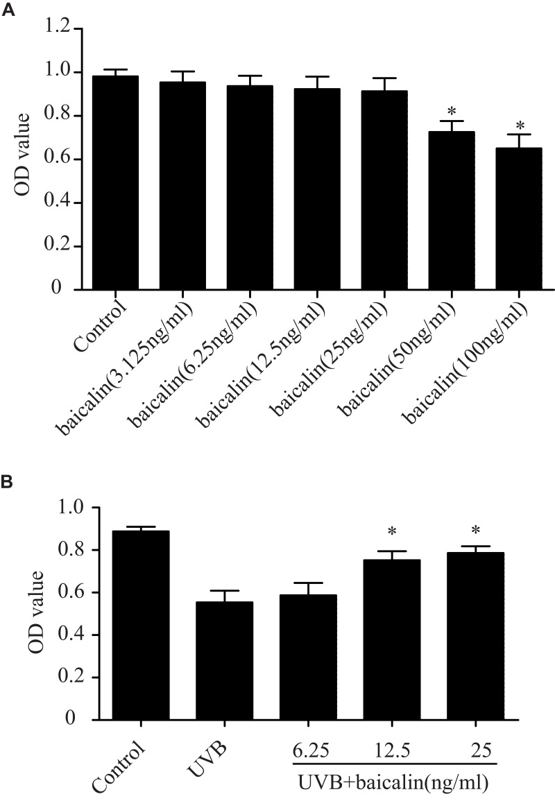
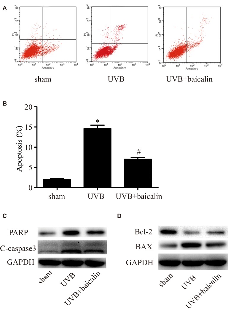
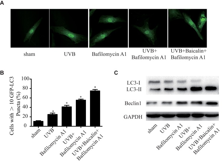
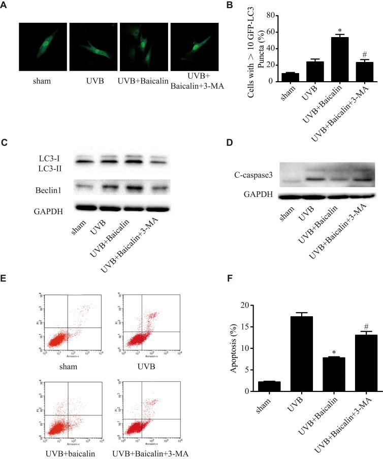
Methods: Cell proliferation was estimated by CCK-8 Kit. Apoptotic incidence was detected by flow cytometry with Annexin V-PE/PI apoptosis detection kit. Autophagy was determined by the evaluation of fluorescent LC3 puncta and Western blotting. Cell signalling was analysed by Western blotting.

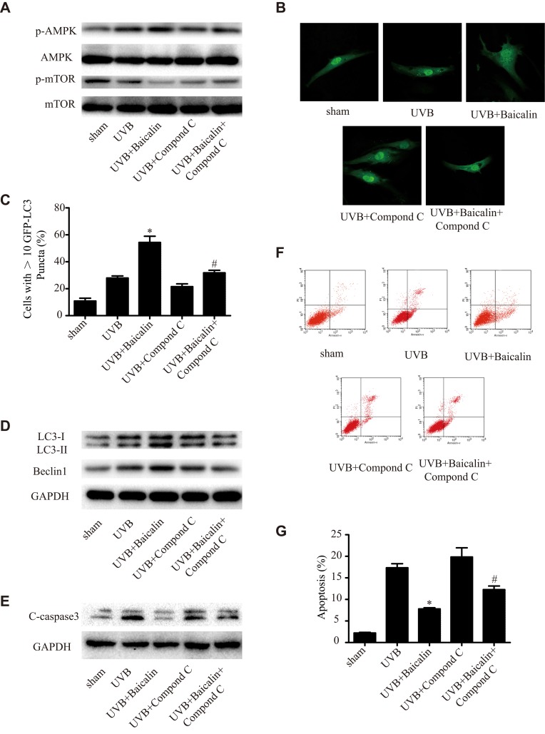
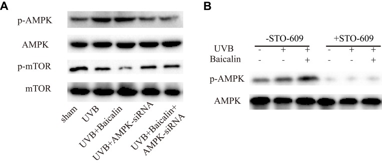
Results: Baicalin exerted cytoprotective effects in UVB-induced HSFs. Moreover, baicalin increased autophagy and suppressed UVB-induced apoptosis of HSFs. Pretreatment with 3-MA, an autophagy inhibitor, attenuated baicalin-induced HSFs autophagy and promoted apoptosis. Baicalin activated AMPK, which leads to suppression of basal mTOR activity in cultured HSFs. Administration of compound C, an AMPK inhibitor, abrogated AMPK phosphorylation and increased mTOR phosphorylation and apoptosis compared with baicalin alone.

Conclusion: Taken together, these results indicate the important role of mTOR inhibition in UVB protection by baicalin and provide a new target and strategy for better prevention of UV-induced skin disorders.

Keywords: AMPK; apoptosis; autophagy; baicalin; ultraviolet B.


Conflict of interest statement

The authors have no conflicts of interest to declare.

Figures

Figure 1
Figure 1
Baicalin protects against the UVB-induced phototoxicity in HSFs. (A) Effect of baicalin at different dosages on HSFs viability. Baicalin was applied to the HSFs at 3.125, 6.25, 12.5, 25, 50, 100 ng/mL. After 24 hrs, the cultured cell viability was assayed by using a CCK-8 assay kit. Values are given as means±SEM (n=5). *p < 0.05 versus Control. (B) Baicalin protects cultured HSFs against UVB-induced cell viability. HSFs were irradiated with 600 J/m2 of UVB, and then cultured with 6.25, 12.5, and 25 ng/mL baicalin. Twenty-four hours after UVB irradiation, the cell viability was assayed by using a CCK-8 assay kit. Values are given as means±SEM (n=6). *p < 0.05 versus UVB.
Figure 2
Figure 2
Baicalin suppresses UVB-induced apoptosis in HSFs. (A, B) Cellular apoptosis was assayed by annexin V-FITC and PI counterstaining, and analyzed with flow cytometry. The original flow cytometry figures are shown in (A) and the apoptosis rates are shown in (B). Values are given as means±SEM (n=5). *p < 0.05 versus sham, #p < 0.05 versus UVB. (C, D) HSFs were treated with indicated treatments. Then, PARP, cleaved caspase-3, Bcl-2 expression, and BAX expression were detected by Western blotting analysis. (C) Representative image of immunoblots for PARP and cleaved caspase-3. GAPDH was used as a loading control. (D) Representative image of immunoblots for Bcl-2 and BAX. GAPDH was used as a loading control. The concentration of baicalin in these studies was 25 ng/mL.
Figure 3
Figure 3
Baicalin increases UVB-induced autophagy in HSFs. HSFs were pretreated with Bafilomycin A1 (100 nM) for 2 h, then treated with 25 ng/mL baicalin. (A) Representative confocal images of GFP-LC3 in fibroblasts. Scale bars = 50μm. (B) The percentage of cells with induced autophagy was determined as the percentage of GFP-LC3 cells with greater than 10 GFP-LC3 puncta per cell (means ±SEM of the independent experiments, *p < 0.05). (C) LC3-II and Beclin1 expressions were detected by Western blotting analysis. GAPDH was used as a loading control. The concentration of baicalin in these studies was 25 ng/mL.
Figure 4
Figure 4
3-MA inhibits the effects of Baicalin on UVB-induced autophagy and apoptosis in HSFs. (A) Representative confocal images of GFP-LC3 in fibroblasts. Scale bars = 50μm. (B) The percentage of cells with induced autophagy was determined as the percentage of GFP-LC3 cells with greater than 10 GFP-LC3 puncta per cell (means ±SEM of the independent experiments, *p < 0.05 versus UVB, #p < 0.05 versus UVB+Baicalin). (C) LC3-II and Beclin1 expressions were detected by Western blotting analysis. GAPDH was used as a loading control. (D) C-caspase3 expression was detected by Western blotting analysis. GAPDH was used as a loading control. (E, F) Cellular apoptosis was assayed by annexin V-FITC and PI counterstaining, and analyzed with flow cytometry. The original flow cytometry figures are shown in (E) and the apoptosis rates are shown in (F). Values are given as means±SEM (n=5). *p < 0.05 versus UVB, #p < 0.05 versus UVB+Baicalin. The concentration of baicalin in these studies was 25 ng/mL.
Figure 5
Figure 5
(A) Western blot analysis of AMPK, mTOR, and their phosphorylated forms. (B) Representative confocal images of GFP-LC3 in fibroblasts. Scale bars = 50μm. (C) The percentage of cells with induced autophagy was determined as the percentage of GFP-LC3 cells with greater than 10 GFP-LC3 puncta per cell. *p < 0.05 versus UVB, #p < 0.05 versus UVB+Baicalin. (D) LC3-II and Beclin1 expression were detected by Western blotting analysis. GAPDH was used as a loading control. (E) C-caspase3 expression was detected by Western blotting analysis. GAPDH was used as a loading control. (F, G) Cellular apoptosis was assayed by annexin V-FITC and PI counterstaining, and analyzed with flow cytometry. The original flow cytometry figures are shown in (F), and the apoptosis rates are shown in (G). Values are given as means±SEM (n=5). *p < 0.05 versus UVB, #p < 0.05 versus UVB+Baicalin. The concentration of baicalin in these studies was 25 ng/mL.
Figure 6
Figure 6
(A) Western blot analysis of AMPK, mTOR, and their phosphorylated forms. (B) Representative Western blots showing the effect of the CaMKKb inhibitor STO-609 (10 mg/mL) on baicalin-induced AMPK and phosphorylation of AMPK protein expression.

References

    1. Pustisek N, Situm M. UV-radiationapoptosis and skin. Coll Antropol. 2011;35(Suppl 2):339–341. - PubMed
    1. P. National Toxicology. NTP 12th Report on Carcinogens, Report on carcinogens: carcinogen profiles, 12. 2011; iii–499. - PubMed
    1. Lee CH, Wu SB, Hong CH, Yu HS, Wei YH. Molecular mechanisms of UV-induced apoptosis and its effects on skin residential cells: the implication in UV-based phototherapy. Int J Mol Sci. 2013;14:6414–6435. doi: 10.3390/ijms14036414 - DOI - PMC - PubMed
    1. Raj D, Brash DE, Grossman D. Keratinocyte apoptosis in epidermal development and disease. J Invest Dermatol. 2006;126:243–257. doi: 10.1038/sj.jid.5700008 - DOI - PMC - PubMed
    1. Barrandon Y. The epidermal stem cell: an overview. Semin Dev Biol. 1993;4:209–215. doi: 10.1006/sedb.1993.1025 - DOI

MeSH terms