

Sonophoresis Enhanced Transdermal Delivery of Cisplatin in the Xenografted Tumor Model of Cervical Cancer
- PMID: 32099393
- PMCID: PMC6996214
- DOI: 10.2147/OTT.S238126
Sonophoresis Enhanced Transdermal Delivery of Cisplatin in the Xenografted Tumor Model of Cervical Cancer
Abstract
Background: Transdermal drug delivery system has been researched for a long time because of its advantage in decreasing side effects such as nausea, vomiting, and gastrointestinal disturbance. Sonophoresis has been shown to be very effective in promoting the transdermal delivery of drugs. This study is on purpose to research the feasibility of sonophoresis promoting cisplatin in the treatment of cervical cancer and the optimum drug delivery mode.
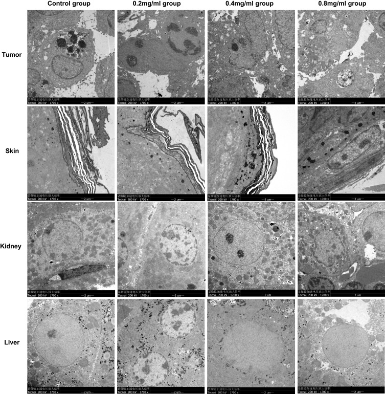
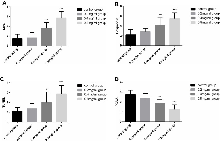
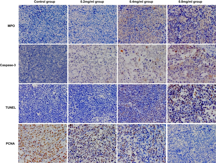
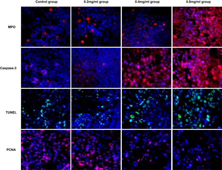
Methods: Thirty-two female nude-mice model of cervical cancer were randomly divided into 4 groups (n=8 in each group): control group without any intervention, low, medium and high concentration groups were treated with the corresponding cisplatin concentrations of 0.2mg/mL, 0.4mg/mL and 0.8mg/mL, respectively, with concurrent sonophoresis applied on the skin of local tumor, 1 mL at a time, once a day for a total of 5 days. Therapeutic pulsed ultrasound (TPU) was 1.0 MHz, 2.0 W/cm2 and 60-min duration. Weight of mice and tumor diameters were measured every day during the intervention. The concentration of cisplatin in tumors was detected by HPLC. Meanwhile, tumor, skin, liver and kidney gross structures and ultrastructure were observed in order to evaluate the effectiveness and safety of experimental conditions. In addition, apoptosis and proliferation-related factors (MPO, Caspase-3, PCNA) were detected by immunohistochemistry, immunofluorescence and TUNEL assay.
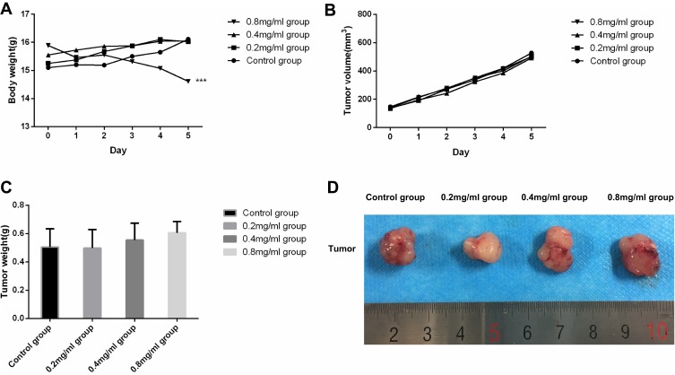
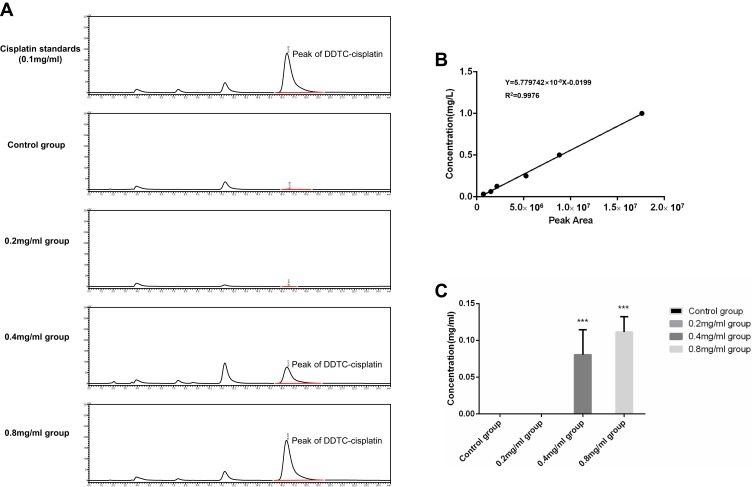
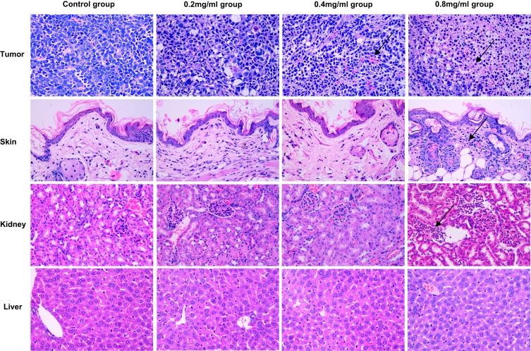
Results: The weight of nude mice in each group showed an increasing trend, except for a decrease of weight in the 0.8 mg/mL group. No obvious tumor inhibition effect was observed. Cisplatin was detected in the 0.4 mg/mL group and 0.8 mg/mL group, with relative concentrations of 0.081±0.033 mg/mL and 0.111±0.021 mg/mL, respectively. Both skin and kidney inflammation were observed in the 0.8 mg/mL group. The expression of MPO, caspase-3 and TUNEL was concentration dependent, with the highest expression in the 0.8 mg/mL group, followed by the 0.4 mg/mL group, with no significant differences between the control and the 0.2 mg/mL group. PCNA was highly expressed in both the control and 0.2 mg/mL groups but decreased in the 0.4 mg/mL and 0.8 mg/mL groups.
Conclusion: Sonophoresis enhanced transdermal delivery of cisplatin in a xenograft tumor model of cervical cancer. Considering the occurrence of skin inflammation and renal injury caused by cisplatin, the recommended concentration to be administered is 0.4mg/mL.
Keywords: cervical cancer; cisplatin; sonophoresis; transdermal drug delivery.
© 2020 Ma et al.
Conflict of interest statement
The authors report no conflicts of interest in this work.
Figures
References
-
- Forouzanfar MH, Afshin A, Alexander LT, GBD 2015 Risk Factors Collaborators. Global, regional, and national comparative risk assessment of 79 behavioural, environmental and occupational, and metabolic risks or clusters of risks, 1990-2015: a systematic analysis for the Global Burden of Disease Study 2015. Lancet. 2016;388(10053):1659–1724. doi:10.1016/S0140-6736(16)31679-8 - DOI - PMC - PubMed
-
- Woopen H, Richter R, Chekerov R, et al. Prognostic role of chemotherapy-induced nausea and vomiting in recurrent ovarian cancer patients: results of an individual participant data meta-analysis in 1213. Support Care Cancer. 2019;28:73–78. - PubMed
-
- Tianze Z, Guangwei X, Yi W, et al. Oncology [M]. Tianjin: Tianjin Science and Technology Press. 2005;535–930. - PubMed
LinkOut - more resources
Full Text Sources
Research Materials
Miscellaneous
