

Case Reports
doi: 10.1016/j.radcr.2020.01.025.
eCollection 2020 Apr.
Endometrial cancer arising from adenomyosis: Case report and literature review of MRI findings
Affiliations
- PMID: 32099587
- PMCID: PMC7031131
- DOI: 10.1016/j.radcr.2020.01.025
Item in Clipboard
Case Reports
Endometrial cancer arising from adenomyosis: Case report and literature review of MRI findings
Radiol Case Rep.
.
Abstract
Endometrial cancer arising from adenomyosis (EC-AIA) is extremely rare, and the typical magnetic resonance imaging (MRI) findings of EC-AIA have not been established. We report a case of EC-AIA that was detected preoperatively on MRI and conduct a literature review of the MRI findings of EC-AIA.
Keywords: Adenomyosis; Endometrial cancer; Magnetic resonance imaging; Uterine cancer.
© 2020 The Authors. Published by Elsevier Inc. on behalf of University of Washington.
Figures
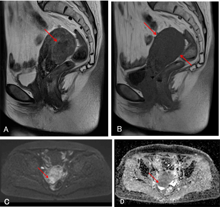
Initial magnetic resonance images (MRI) of the pelvis. (A) T2-weighted sagittal MRI demonstrates swelling of the posterior uterine wall and ill-defined signal intensity higher than that of myometrium (arrow). (B) T1-weighted sagittal MRI demonstrates uniform low signal intensity of the uterine corpus (arrows). (C) Diffusion weighted imaging (DWI) demonstrates high signal intensity in the posterior uterine wall (arrow). (D) Apparent diffusion coefficient (ADC) map demonstrates low signal intensity in the posterior uterine wall (arrow).
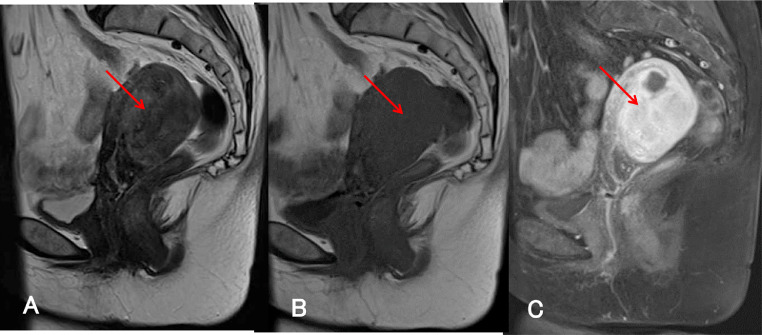
MRI acquired 4 months after the initial MRI. (A) T2-weighted sagittal and (B) T1-weighted sagittal MRI demonstrate increased thickening of the posterior uterine wall (arrow). (C) Fat-saturated T1-weighted sagittal MRI with gadolinium-based contrast material demonstrates strong and uniform enhancement of the posterior uterine wall (arrow).
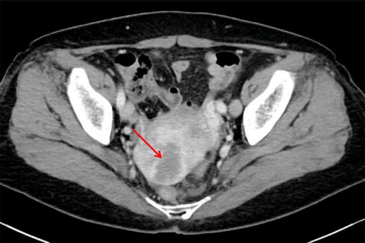
Contrast-enhanced computed tomography shows an ill-defined area of low density in the posterior uterine wall (arrow).
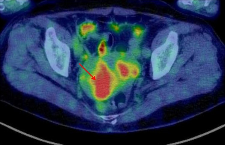
Fluorodeoxyglucose-positron emission tomography reveals a hypermetabolic area in the posterior uterine wall (arrow).
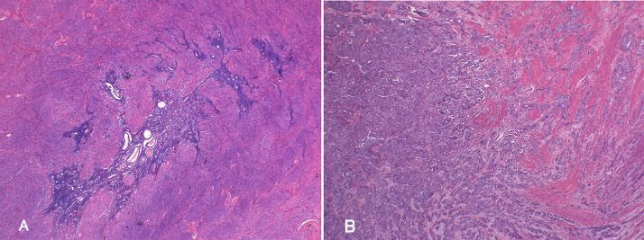
Histopathological analysis of the uterine specimen. (A) Adenomyotic glands are diffusely distributed within the posterior uterine wall. Hematoxylin and eosin (H&E) staining: low power field. (B) Acinar and papillary proliferation of atypical duct resembling endometrial gland is seen in the posterior uterine wall. H&E staining: low power field.
References
-
- Bray F, Ferlay J, Soerjomataram I, Siegel RL, Torre LA, Jemal A. Global cancer statistics 2018: GLOBOCAN estimates of incidence and mortality worldwide for 36 cancers in 185 countries. CA Cancer J Clin. 2018;68:394–424. - PubMed
-
- Vercellini P, Vigan´ø P, Somigliana E, Daguati R, Abbiati A, Fedele L. Adenomyosis: epidemiological factors. Best Pract Res Clin Obstet Gynaecol. 2006;20:465–477. - PubMed
-
- Colman HI, Rosenthal AH. Carcinoma developing in areas of adenomyosis. Obstet Gynecol. 1959;14:342–348. - PubMed
-
- Motohara K., Tashiro H., Ohtake H., Saito F., Ohba T., Katabuchi H. Endometrioid adenocarcinoma arising in adenomyosis: elucidation by periodic magnetic resonance imaging evaluations. Int J Clin Oncol. 2008;13:266–270. - PubMed
Publication types
LinkOut - more resources
Full Text Sources
