. 2020 Jan 31:18:323-331.
doi: 10.1016/j.csbj.2020.01.008. eCollection 2020.

Genetic heterogeneity and clonal evolution during metastasis in breast cancer patient-derived tumor xenograft models

Affiliations

Genetic heterogeneity and clonal evolution during metastasis in breast cancer patient-derived tumor xenograft models

Kathleen Sprouffske et al. Comput Struct Biotechnol J. .

Abstract

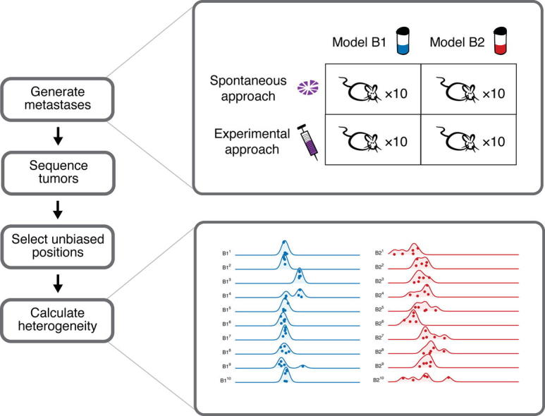
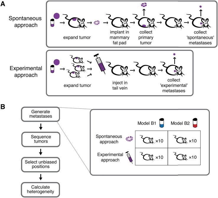
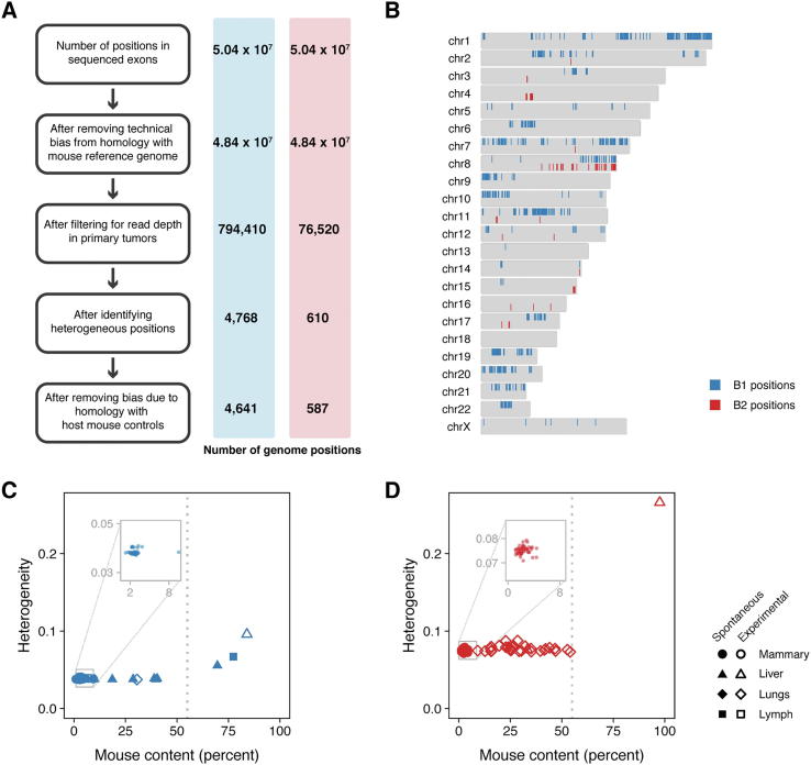
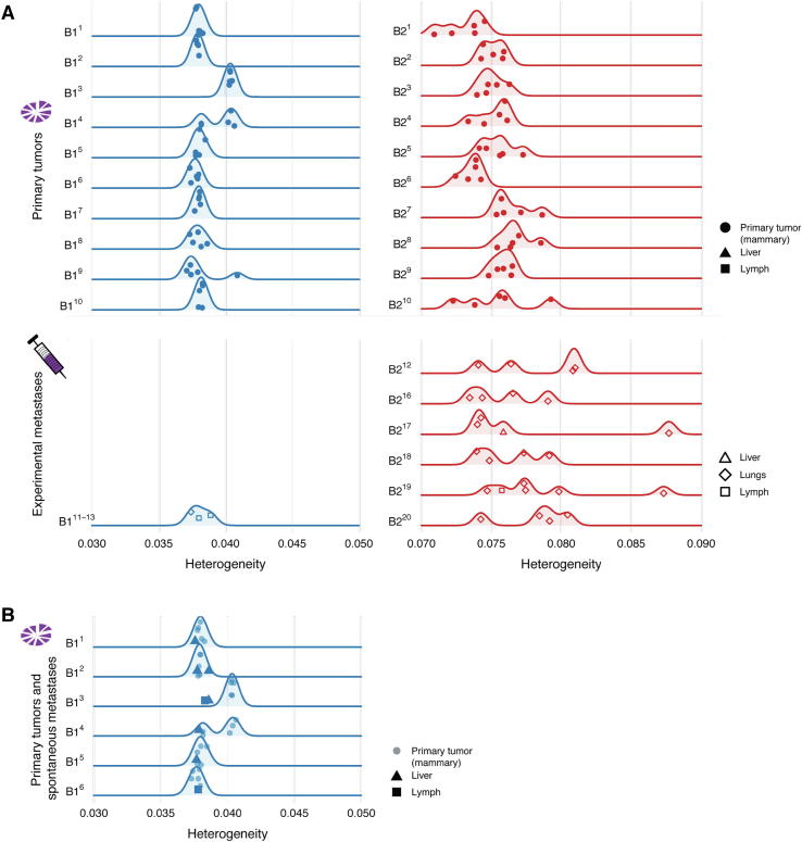
Genetic heterogeneity within a tumor arises by clonal evolution, and patients with highly heterogeneous tumors are more likely to be resistant to therapy and have reduced survival. Clonal evolution also occurs when a subset of cells leave the primary tumor to form metastases, which leads to reduced genetic heterogeneity at the metastatic site. Although this process has been observed in human cancer, experimental models which recapitulate this process are lacking. Patient-derived tumor xenografts (PDX) have been shown to recapitulate the patient's original tumor's intra-tumor genetic heterogeneity, as well as its genomics and response to treatment, but whether they can be used to model clonal evolution in the metastatic process is currently unknown. Here, we address this question by following genetic changes in two breast cancer PDX models during metastasis. First, we discovered that mouse stroma can be a confounding factor in assessing intra-tumor heterogeneity by whole exome sequencing, thus we developed a new bioinformatic approach to correct for this. Finally, in a spontaneous, but not experimental (tail-vein) metastasis model we observed a loss of heterogeneity in PDX metastases compared to their orthotopic "primary" tumors, confirming that PDX models can faithfully mimic the clonal evolution process undergone in human patients during metastatic spreading.

Keywords: Breast cancer; Clonal evolution; Heterogeneity; Metastasis; Patient derived xenograft models.


Figures

None
Graphical abstract
Fig. 1
Fig. 1
(A) Two common approaches were used to obtain either spontaneous (top) or experimental (bottom) metastases. For spontaneous metastasis (top), a fragment of the patient-derived tumor xenograft was implanted in the mammary fat pad of a mouse to generate the ‘primary orthotopic’ tumor. This primary tumor was removed, and divided into five fragments for sequencing. Spontaneous metastases are any metastases that subsequently arose in the mouse. Experimental metastases (bottom) are any metastases that arose in a mouse after the tail vein injection of a patient-derived tumor xenograft cell solution. (B) Metastases were obtained from two PDX models of breast cancer (B1 and B2) using the spontaneous and experimental approaches with 10 mice per condition. The primary tumors from the mice in the spontaneous metastasis experiments and all metastases were collected. Large tumors were dissected into several pieces and sequenced.
Fig. 2
Fig. 2
(A, B) The number of tumor samples sequenced for each of the 40 mice (rows) implanted with B1 (blue) or B2 (red) PDX tissue using the spontaneous (panel A) or the experimental (panel B) approaches. An empty box indicates that no metastasis was found in that mouse’s organ. For example in mouse B11, no metastases were found in its lungs or lymph nodes, but five samples from its primary tumor and one spontaneously-arising metastasis from its liver was sequenced. Dashes indicate that a mouse did not survive the implantation procedure (e.g., B118, B119, B120). (C) Primary orthotopic tumors were collected from 20 mice (10 B1 and 10 B2), and each primary tumor was dissected into five pieces for sequencing, as depicted in a representative image from mouse B25. (D) Large metastases were also divided into smaller pieces for sequencing. The lungs often contained multiple metastases that were dissected into multiple pieces for sequencing, as shown here for mouse B219. (E) The mouse content for all sequenced PDX tumor samples for the orthotopic primary tumors (mammary gland) and the metastases for models B1 (blue) and B2 (red) was estimated from the number of sequencing reads that uniquely mapped to the mouse and human genomes. (For interpretation of the references to colour in this figure legend, the reader is referred to the web version of this article.)
Fig. 3
Fig. 3
(A) Sequenced samples from PDX models contain sequences from the host mouse that can bias heterogeneity estimates. Unbiased genomic positions were selected using the steps outlined in the boxes. Each step is described in full detail in the Supplementary Methods. The number of genomic positions remaining for model B1 and model B2 after each step are indicated in the blue and red bars. (B) The distribution of the unbiased genomic positions across the genome for models B1 (blue) and B2 (red). The heterogeneity for each (C) model B1 and (D) model B2 sample was estimated using the unbiased genomic positions selected for each model (vertical axes) and plotted against the amount of mouse contamination in the sample (horizontal axes). Heterogeneity and mouse contamination are uncorrelated for the primary samples depicted in the inset panels (Spearman’s rank correlation; B1: ρ = 0.013, p = 0.93, n = 50; B2: ρ = 0.25, p = 0.08, n = 50). However, heterogeneity increased with mouse content for samples with more than 55% mouse content; therefore, these samples were removed from downstream analyses. (For interpretation of the references to colour in this figure legend, the reader is referred to the web version of this article.)
Fig. 4
Fig. 4
The heterogeneity score (horizontal axes) of the samples obtained from each mouse (vertical axes) for PDX model B1 (blue) and B2 (red). Five samples were collected from each primary tumor (one solid circle is shown for each of the five sequenced regions from a primary tumor), in addition to metastases from the liver (triangles), lungs (diamonds), and lymph nodes (squares). (A) The primary tumors collected during the spontaneous metastasis experiments are depicted in the top panels, while the metastases collected during the experimental metastasis experiments are depicted in the bottom panels. Comparing heterogeneity between primary tumors and experimental metastases was inconclusive for model B1 (primary tumors B11 to B110 vs. experimental metastases B111 to B113, Mann-Whitney, p = 0.95) and increased for model B2 (primary tumors B21 to B210 vs. experimental metastases B211 to B220, Mann-Whitney, p = 0.01). (B) Primary tumors and their metastases collected during spontaneous metastasis experiments. Heterogeneity decreased in the mice (B11 to B16) when comparing matched primary tumors and spontaneous metastases (linear mixed effects analysis, ??2(1) = 6.2, p = 0.013, n = 38). (For interpretation of the references to colour in this figure legend, the reader is referred to the web version of this article.)

References

    1. Hidalgo M., Amant F., Biankin A.V., Budinska E., Byrne A.T. Patient-derived xenograft models: an emerging platform for translational cancer research. Cancer Discov. 2014;4:998–1013. - PMC - PubMed
    1. Byrne A.T., Alférez D.G., Amant F., Annibali D., Arribas J. Interrogating open issues in cancer precision medicine with patient-derived xenografts. Nat Rev Cancer. 2017;17:254–268. - PubMed
    1. Nowell P. The clonal evolution of tumor cell populations. Sci N Y NY. 1976;194:23–28. - PubMed
    1. Merlo L.M.F., Pepper J., Reid B., Maley C.C. Cancer as an evolutionary and ecological process. Nat Rev Cancer. 2006;6:924–935. - PubMed
    1. Marusyk A., Polyak K. Tumor heterogeneity: causes and consequences. Biochim Biophys Acta. 2010;1805:105–117. - PMC - PubMed

LinkOut - more resources