Review
. 2020 Feb 1:2020:3491764.
doi: 10.1155/2020/3491764. eCollection 2020.

Lipoprotein(a) the Insurgent: A New Insight into the Structure, Function, Metabolism, Pathogenicity, and Medications Affecting Lipoprotein(a) Molecule

Affiliations
Review

Lipoprotein(a) the Insurgent: A New Insight into the Structure, Function, Metabolism, Pathogenicity, and Medications Affecting Lipoprotein(a) Molecule

Motasim M Jawi et al. J Lipids. .

Abstract

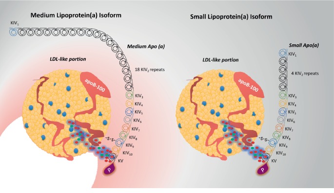
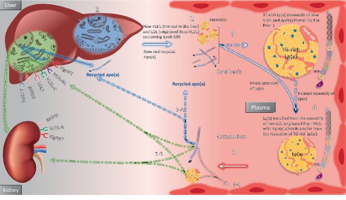
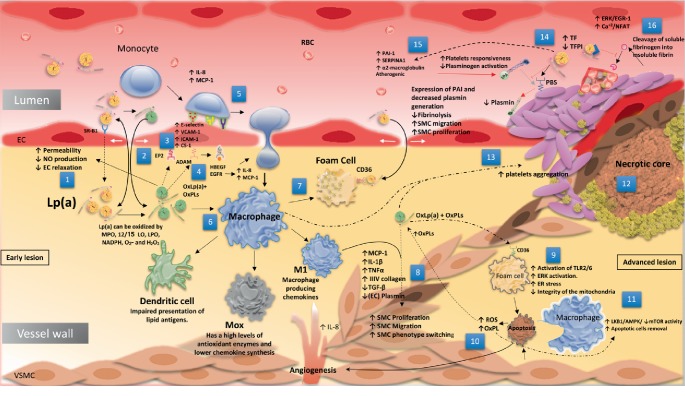
Lipoprotein(a) [Lp(a)], aka "Lp little a", was discovered in the 1960s in the lab of the Norwegian physician Kåre Berg. Since then, we have greatly improved our knowledge of lipids and cardiovascular disease (CVD). Lp(a) is an enigmatic class of lipoprotein that is exclusively formed in the liver and comprises two main components, a single copy of apolipoprotein (apo) B-100 (apo-B100) tethered to a single copy of a protein denoted as apolipoprotein(a) apo(a). Plasma levels of Lp(a) increase soon after birth to a steady concentration within a few months of life. In adults, Lp(a) levels range widely from <2 to 2500 mg/L. Evidence that elevated Lp(a) levels >300 mg/L contribute to CVD is significant. The improvement of isoform-independent assays, together with the insight from epidemiologic studies, meta-analyses, genome-wide association studies, and Mendelian randomization studies, has established Lp(a) as the single most common independent genetically inherited causal risk factor for CVD. This breakthrough elevated Lp(a) from a biomarker of atherosclerotic risk to a target of therapy. With the emergence of promising second-generation antisense therapy, we hope that we can answer the question of whether Lp(a) is ready for prime-time clinic use. In this review, we present an update on the metabolism, pathophysiology, and current/future medical interventions for high levels of Lp(a).


Conflict of interest statement

The authors declare that they have no conflicts of interest regarding the publication of this paper.

Figures

Figure 1
Figure 1
Lp(a) structure, composition, and physicochemical properties. A—Lp(a) is composed of apo-B100 covalently fastened together with apo(a), which originates from kringle IV (KIV) and KV, and the inactive protease domain of PLG. Apo(a) has important differences compared with PLG. (1) Apo(a) has an unpaired cysteine and forms a disulfide bond with apoB to generate the lipoprotein particle Lp(a). (2) Apo(a) has an inactive protease domain. (3) Apo(a) includes 10 subtypes of KIV repeats, composed of 1 copy each of KIV1, multiple copies of KIV2, and 1 copy of KIV310, KV, and an inactive protease-like domain. (4) Apo(a) lacks kringles 1–3 of PLG but has kringles 5 and 10 of KIV, of which KIV2 is present in numerous repeats. OxPLs exist covalently bonded to the apo(a) component and are suspended in the lipid phase of apo-B100. B—Comparison between Lp(a) and LDL with regard to their composition and physicochemical properties.
Figure 2
Figure 2
Comparison between different Lp(a) isoform sizes. In these 2 illustrations, apo(a) molecules of 4 (right) and 18 (left) KIV2 repeats are presented, representing 13 and 27 total KIV repeats.
Figure 3
Figure 3
Model for the metabolism of apo(a). 1—Lipoprotein (a) production (hepatocyte level). Four stages are likely responsible for apo(a) Lp(a) production in liver cells: (A) transcription of the apo(a) gene and apo(a) mRNA stability in the nucleus; (B) influence of apo(a) translation on the production rate; (c) in the ER, posttranslation modifications and folding of apo(a) kringles; (D) Golgi-specific addition and modification of apo(a) carbohydrates; and (E) transport to the cell surface. 2—Assembly of Lp(a): The site of Lp(a) assembly is controversial. (A) cell surface. (B) The space of Disse. (C) Plasma. 3—Apo(a) associates with a recently made TG-abundant molecule to form Lp(a) with VLDL properties and/or with a cholesterol-abundant molecule with LDL properties. 4—TG-abundant Lp(a) may be transformed into a cholesterol-abundant molecule with LDL properties. 5—Catabolism and clearance: The two Lp(a) components become separated. The generation of apo(a) fragments is most likely from proteolytic cleavage by elastases or metalloproteinases secreted by cells in the arterial wall. (5—A) This permits apo(a) to unite the apo(a) pool recently produced by the hepatocytes. (5-B) Hepatocyte internalization and uptake by megalin, gp330 receptor, macrophage scavenger receptor-BI, lipoprotein receptor, VLDL receptor, PlgRKT receptor, asialoglycoprotein receptor (ASGPR), and LDLR. (5-B) Kidney cellular internalization and uptake. (5-C) Vascular wall deposition. Solid lines represent metabolic pathways; dotted lines represent hypothesized metabolic pathways.
Figure 4
Figure 4
Different theories of how Lp(a) causes atherosclerosis. Early lesions (1) Lp(a) enters the vascular wall and is oxidized by MPO, 12/15 LO, LPO, NADPH, O2, and H2O2. (2) OxLp(a) and OxPLs constitute a substantial increase in monolayer permeability, resulting in increased Lp(a) and LDL entry into the vascular wall. (3) OxLp(a) and OxPLs bind to the E-type prostaglandin receptor (EP2) receptor, causing deposition of connecting segment 1 (CS-1). Additionally, OxLp(a) stimulates the expression of cell adhesion molecules (ICAM, VCAM E-selectin) that bind to monocytes on the endothelial cell surface. (4) OxLp(a) may also activate specific disintegrin and metalloproteinases (ADAMs) to cause the release of active heparin binding epidermal growth factor (HBEGF) and activation of epidermal growth factor receptor (EGFR) and vascular endothelial growth factor receptor 2 (VEGFR2), causing IL-8 and monocyte chemotactic protein (MCP)-1 production. (5) These chemokines simplify access of the attracted monocytes to the artery wall. (6) OxPL build-up causes monocytes to differentiate into M1, dendritic cells, Mox cells, and foam cells. Advanced lesions (7) macrophages engulf OxLp(a) through its scavenger receptor CD36 to form the foam cell. (8) Ox-Lp(a) and Lp(a) also induce aberrant proliferation, migration, and phenotype switching of smooth muscle cells (SMCs). (9) OxLp(a) stimulates CD36 to activate TLR2/6, which activates ERK and results in ER stress-induced loss of integrity of the mitochondria, which eventually leads to apoptosis. (10) Apoptotic cells provide more OxPLs and stimulate angiogenesis. (11) OxLp(a) may stimulate LKB1/AMPK/ mTOR activity and induce apoptotic cell removal by macrophages. (12) Necrotic core formation and vessel wall rupture. (13) Macrophage and OxLp(a) cause increased platelet aggregation. (14) Apo(a) binding to PLG binding sites blocks the interaction between PLG and tissue PLG activator (tPA). (15) Lp(a) increases the production and activity of tPA inhibitor-1 (PAI-1), which eventually leads to a decrease in fibrinolysis (16). Lp(a) increases the expression of TF and inhibits the potent inhibitory effect of tissue factor pathway inhibitor (TFPI), which leads to thrombosis. Dotted lines: hypothesized pathways.

References

    1. Finney A. C., Stokes K. Y., Pattillo C. B., Orr A. W. Integrin signaling in atherosclerosis. Cellular and Molecular Life Sciences. 2017;74(12):2263–2282. doi: 10.1007/s00018-017-2490-4. - DOI - PMC - PubMed
    1. Luna-Luna M., Medina-Urrutia A., Vargas-Alarcon G., Coss-Rovirosa F., Vargas-Barron J., Perez-Mendez O. Adipose tissue in metabolic syndrome: onset and progression of atherosclerosis. Archives of Medical Research. 2015;46(5):392–407. doi: 10.1016/j.arcmed.2015.05.007. - DOI - PubMed
    1. Giannini C., de Giorgis T., Scarinci A., et al. Obese related effects of inflammatory markers and insulin resistance on increased carotid intima media thickness in pre-pubertal children. Atherosclerosis. 20081;197(1):448–456. doi: 10.1016/j.atherosclerosis.2007.06.023. - DOI - PubMed
    1. Bergheanu S. C., Bodde M. C., Jukema J. W. Pathophysiology and treatment of atherosclerosis. Netherlands Heart Journal. 2017;25(4):231–242. doi: 10.1007/s12471-017-0959-2. - DOI - PMC - PubMed
    1. Kamstrup P. R. Lipoprotein (a) and ischemic heart disease—a causal association? A Review. Atherosclerosis. 2010;211(1):15–23. doi: 10.1016/j.atherosclerosis.2009.12.036. - DOI - PubMed

LinkOut - more resources