. 2019 Dec 21;7(4):1900949.
doi: 10.1002/advs.201900949. eCollection 2020 Feb.

Circ-MALAT1 Functions as Both an mRNA Translation Brake and a microRNA Sponge to Promote Self-Renewal of Hepatocellular Cancer Stem Cells

Affiliations

Circ-MALAT1 Functions as Both an mRNA Translation Brake and a microRNA Sponge to Promote Self-Renewal of Hepatocellular Cancer Stem Cells

Liang Chen et al. Adv Sci (Weinh). .

Erratum in

Abstract

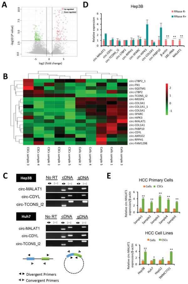
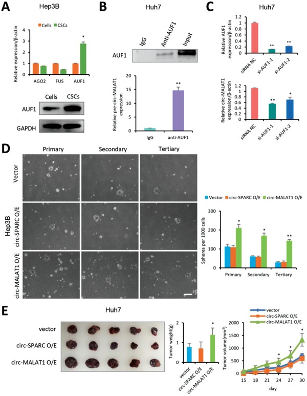
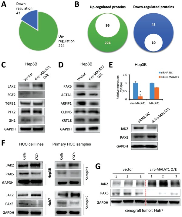
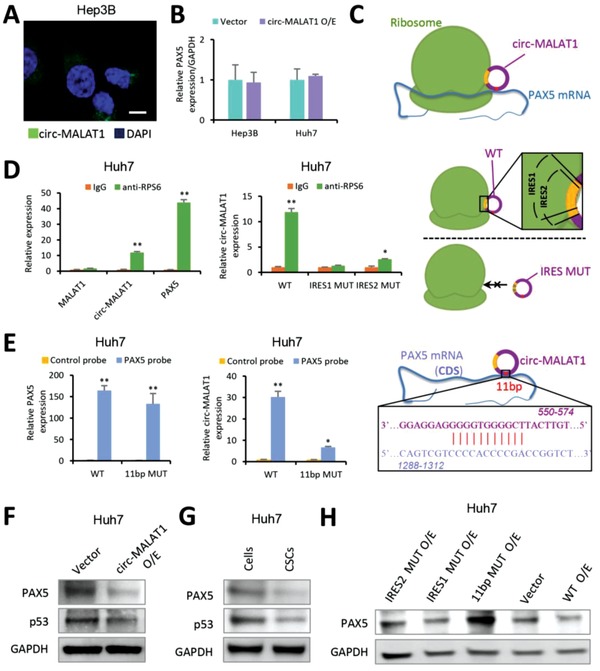
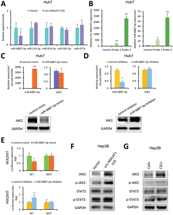
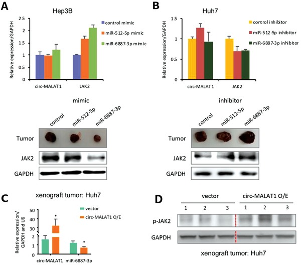
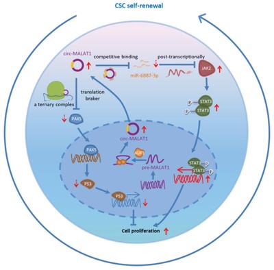
Both circular RNAs (circRNAs) and cancer stem cells (CSCs) are separately known to be involved in cancer, but their interaction remains unclear. Here, the regulation of hepatocellular CSC self-renewal is discovered by a circRNA, circ-MALAT1, which is produced by back-splicing of a long noncoding RNA, MALAT1. Circ-MALAT1 is highly expressed in CSCs from clinical hepatocellular carcinoma samples under the mediation of an RNA-binding protein, AUF1. Surprisingly, circMALAT1 functions as a brake in ribosomes to retard PAX5 mRNA translation and promote CSCs' self-renewal by forming an unprecedented ternary complex with both ribosomes and mRNA. The discovered braking mechanism of a circRNA, termed mRNA braking, along with its more traditional role of miRNA sponging, uncovers a dual-faceted pattern of circRNA-mediated post-transcriptional regulation for maintaining a specific cell state.

Keywords: PAX5; circular RNA; hepatocellular cancer stem cells; miR‐6887‐3p; ribosomes; self‐renewal.


Conflict of interest statement

The authors declare no conflict of interest.

Figures

Figure 1
Figure 1
Circ‐MALAT1 is highly expressed in Hepatocellular CSCs. A) Volcano plot of differential circRNA expression between adherent cells and matched CSCs from five pairs of HCC primary samples. Points‐of‐interest (fold‐change > 2 or < 0.5 and p < 0.001) were represented in red (upregulated) and green (downregulated). Those points having no significant difference were shown in gray. B) Heat map of 18 differentially expressed circRNAs in at least three of the five pairs from circRNA‐seq data. Each column represented a sample of HCC adherent cells (Cells sample) or matched CSCs (CSCs sample), while each row represented an individual circRNA. The stripe color indicated the level of expression. C) Divergent primers amplified circR‐MALAT1, circ‐CDYL, and circ‐TCONS_I2 in cDNA but not genomic DNA (gDNA) or RNA (No RT). Convergent Primers could amplify linear RNA and circRNA in cDNA and parental gene in gDNA. D) CircRNAs were insensitive to RNase R treatment. Total RNA samples were split into two aliquots; one aliquot was treated with the RNase R exonuclease (RNase R+) and the other was subjected to a mock treatment (RNase R−). Linear RNAs such as β‐actin, 18S rRNA and MALAT1 were substantially digested with RNase R, while circRNAs were almost unaffected and even enriched (such as circ‐MALAT1, circ‐HIPK3 and circ‐FKBP10) due to degradation of linear RNAs. E) Circ‐MALAT1 expression was increased in CSCs of HCC primary cells (top panel) and cell lines (bottom panel). CSCs were enriched by the tumorsphere assay. Control primers, β‐actin. Columns, means from three independent experiments; bars, SD. ** p < 0.01, * p < 0.05.
Figure 2
Figure 2
Circ‐MALAT1 promotes the self‐renewal of hepatocellular CSCs. A) Top panel—mRNA of RNA‐binding protein AUF1 (not AGO2 or FUS) was upregulated in Hep3B CSCs. Control primers, β‐actin. Bottom panel, AUF1 protein was analyzed in Hep3B cells and CSCs by western blot assay. CSCs were enriched by the tumorsphere assay. B) RIP was performed using Huh7 cell lysate and either anti‐AUF1 or IgG as IP antibody. Top panel, AUF1 antibody was confirmed by western blot. AUF1 protein was observed in the anti‐AUF1 RIP (lane 2) and 10% Input (lane 3) but not in the IgG RIP (Lane1). Bottom panel, purified RNA from RIP was analyzed by qRT‐PCR using primers specific for the pre‐circ‐MALAT1. C) Top panel, AUF1 was silenced in Huh7 cells by two independent siRNAs (si‐AUF1‐1 and si‐AUF1‐2). Bottom panel, circ‐MALAT1 expression was decreased after silencing AUF1 in Huh7 cells. Control primers, β‐actin. D) Left panel, bright images of spheres of circ‐MALAT1 overexpressed (circ‐MALAT1 O/E), circ‐SPARC overexpressed (circ‐SPARC O/E), or vector control (Vector) Hep3B cells. Right panel, quantitative statistics of the numbers of the primary, secondary, and tertiary spheres per 1000 cells. Scale bar, 200 µm. E) Both Circ‐MALAT1 overexpressed (circ‐MALAT1 O/E), circ‐SPARC overexpressed (circ‐SPARC O/E), and vector control (Vector) Huh7 cells were diluted and subcutaneously implanted into five BALB/c nude mice, respectively. Tumors were observed every 3 d for a period of one month 14 d post‐tumor inoculation. Similar results were obtained in three to six independent experiments. ** p < 0.01, * p < 0.05.
Figure 3
Figure 3
Circ‐MALAT1 expression results in JAK2 and PAX5 Differential Expression. A) Pie chart showed that 224 proteins were upregulated and 43 were downregulated from protein microarray. Ratio of comparison at ≥1.8 or ≤1/1.8. B) Left panel, of the 224 upregulated proteins, 96 proteins were associated with CSCs self‐renewal or tumor metastasis and proliferation. Right panel, 10 of the 43 downregulated proteins were associated with tumor suppression. C) Five upregulated proteins from antibody‐array results were analyzed in Hep3B cells transfected with circ‐MALAT1 overexpression (circ‐MALAT1 O/E) or empty vector (Vector) by western blot analysis. D) Five downregulated proteins from antibody‐array results were analyzed in circ‐MALAT1 overexpression (circ‐MALAT1 O/E) or empty vector (Vector) cells by western blot analysis. E) Top panel, expression of circ‐MALAT1 and MALAT1 was analyzed by qRT‐PCR in Hep3B cells transfected with sicirc‐MALAT1 and siRNA NC. Bottom panel, protein expression of JAK2 and PAX5 was detected by western blot in circ‐MALAT1‐silenced‐Hep3B cells. siRNA NC, scramble control siRNA; sicirc‐MALAT1, siRNA against circ‐MALAT1. Columns, means from three independent experiments; bars, SD. * p < 0.05. F) Expression of JAK2 and PAX5 of CSCs and adherent cells of two HCC cell lines (left panel) Hep3B and Huh7 and two HCC primary cells (right panel) were detected by western blot. CSCs were enriched by the tumorsphere assay. G) JAK2 and PAX5 expressions were determined in the xenograft tumors by western blot.
Figure 4
Figure 4
Circ‐MALAT1 obstructs PAX5 translation by binding to PAX5 coding sequence and ribosomes. A) RNA fluorescence in situ hybridization for circ‐MALAT1. Nuclei were stained with DAPI. Green staining signal against circ‐MALAT1 was localized in cytoplasm. Scale bar, 10 µm. B) The level of PAX5 mRNA was analyzed by qRT‐PCR. Circ‐MALAT1 overexpression (circ‐MALAT1 O/E) led to no significant difference in PAX5 mRNA level compared to the control (Vector). Control primers, GAPDH. C) An interaction model showing that circ‐MALAT1 recognized PAX5 mRNA via 11 bp bases paring (red site) and competitively inhibited PAX5 translation through IRESs (yellow sites). D) Left panel, RIP lysates prepared from circ‐MALAT1 overexpressed Huh7 cells were subjected to immunoprecipitation using either a normal rabbit IgG or anti‐RPS6 antibody. Purified RNA was analyzed by qRT‐PCR using primers specific for MALAT1, circ‐MALAT1 and PAX5, respectively. Middle panel, RIP was performed using lysates prepared from Huh7 cells overexpressing wild type (WT), IRES1‐mutated (IRES1 MUT) and IRES2‐mutated (IRES2 MUT) circ‐MALAT1, respectively. Purified RNA was analyzed by qRT‐PCR using primers specific for circ‐MALAT1. Right panel, the model above the dashed line (indicating the result in the left panel of (D) showed that circ‐MALAT1 with wild type of IRES (WT, “1” and “2” overlapped yellow sites) could bind to ribosome while the model below the dashed line (indicating the result in the middle panel of (D) showed that circ‐MALAT1 with mutated IRES (MUT) could not bind to ribosome. E) In vivo RNA pull‐down using PAX5 specific probes was performed in circ‐MALAT1 WT or 11 bp MUT overexpressed Huh7 cells, followed by qRT‐PCR to detect PAX5 (left panel) and circ‐MALAT1 (middle panel). Right panel, the interaction model between circ‐MALAT1 and the CDS region of PAX5 mRNA via 11 bp bases paring (red site). F) PAX5 and its downstream protein p53 were detected by western blot. Vector, empty vector overexpression; circ‐MALAT1 O/E, circ‐MALAT1 overexpression. G) PAX5 and p53 were analyzed in CSCs and adherent cells of Hep3B cell line by western blot. CSCs were enriched by the tumorsphere assay. H) PAX5 was detected in Huh7 cells overexpressing circ‐MALAT1 wild (WT O/E) or mutant type (11 bp MUT O/E, IRES1 MUT O/E, and IRES2 MUT O/E) or none (Vector) by western blot. Columns, means from three independent experiments; bars, SD. ** p < 0.01, * p < 0.05.
Figure 5
Figure 5
Circ‐MALAT1 acts as miR‐6887‐3p sponge to up‐regulate JAK2. A) Top five miRNAs with the highest binding free energy were analyzed in circ‐MALAT1 overexpressed (circ‐MALAT1 O/E) or empty vector (Vector) Huh7 cells by qRT‐PCR. Control primers, U6. B) In vivo circ‐MALAT1 pull‐down using circ‐MALAT1 specific probes (Probe 1 and Probe 2) was performed in circ‐MALAT1 overexpressed Huh7 cells, followed by qRT‐PCR to detect circ‐MALAT1 (left panel) and miR‐6887‐3p (right panel). C) After treatment with miR‐6887‐3p mimic or its scrambled version (control mimic), both miR‐6887‐3p level and JAK2 expression at mRNA were detected by qRT‐PCR (top panel), and JAK2 expression at protein level was detected by western blot (bottom panel), respectively. U6 and GAPDH were used as control primers. D) After treatment with the miR‐6887‐3p inhibitor or its scrambled version (the control inhibitor), the level of both the miR‐6887‐3p and JAK2 mRNA was detected by qRT‐PCR (top panel), and JAK2 expression at the protein level was detected by western blot (bottom panel). U6 and GAPDH were used as control primers. E) The luciferase activities of pMIR‐JAK2‐3'UTR wild type (WT) and mutated type (MUT) were detected when cells were cotransfected with miR‐6887‐3p mimic (top panel) or miR‐6887‐3p inhibitor (bottom panel). F) The key molecules of JAK2/STAT3 pathway were analyzed in circ‐MALAT1 overexpressed (circ‐MALAT1 O/E) or empty vector (Vector) cells by western blot. p‐JAK2, phospho‐JAK2; p‐STAT3, phospho‐STAT3. G) The key protein molecules in JAK2/STAT3 signaling pathway were analyzed in CSCs and adherent cells of Hep3B cell line by western blot. p‐JAK2, phospho‐JAK2; p‐STAT3, phospho‐STAT3. CSCs were enriched by the tumorsphere assay. Columns, means from three independent experiments; bars, SD. ** p < 0.01, * p < 0.05.
Figure 6
Figure 6
Circ‐MALAT1 absorbs miR‐6887‐3p to upregulate JAK2 in vivo. A) After treatment with miR‐6887‐3p mimic, miR‐512‐5p mimic, or its scrambled version (control mimic), both circ‐MALAT1 and JAK2 mRNA of xenograft tumors were detected by qRT‐PCR (top panel), and JAK2 expression at the protein level was detected by western blot (bottom panel). miR‐512‐5p was used as a control miRNA. GAPDH was used as a control primer. B) After treatment with miR‐6887‐3p inhibitor, miR‐512‐5p inhibitor or its scrambled version (control inhibitor), both circ‐MALAT1 and JAK2 mRNA of xenograft tumors were detected by qRT‐PCR (top panel), and JAK2 expression at the protein level was detected by western blot (bottom panel). miR‐512‐5p was used as a control miRNA. GAPDH was used as a control primer. C) MiR‐6887‐3p expression was analyzed in the xenograft tumors generated by circ‐MALAT1 overexpressed (circ‐MALAT1 O/E) or empty vector (Vector) Huh7 cells. Control primers, GAPDH and U6. D) p‐JAK2 was determined in the xenograft tumors generated by circ‐MALAT1 overexpressed (circ‐MALAT1 O/E) or empty vector (Vector) Huh7 cells. Columns, means from three independent experiments; bars, SD. * p < 0.05.
Figure 7
Figure 7
Schematic diagram for the mechanisms of circ‐MALAT1 functioning as both an mRNA translation brake and an miRNA sponge to promote self‐renewal of hepatocellular CSCs.

References

    1. Memczak S., Jens M., Elefsinioti A., Torti F., Krueger J., Rybak A., Maier L., Mackowiak S. D., Gregersen L. H., Munschauer M., Loewer A., Ziebold U., Landthaler M., Kocks C., le Noble F., Rajewsky N., Nature 2013, 495, 333. - PubMed
    1. Chen L. L., Nat. Rev. Mol. Cell Biol. 2016, 17, 205. - PubMed
    1. Ashwal‐Fluss R., Meyer M., Pamudurti N. R., Ivanov A., Bartok O., Hanan M., Evantal N., Memczak S., Rajewsky N., Kadener S., Mol. Cell 2014, 56, 55. - PubMed
    1. Hansen T. B., Jensen T. I., Clausen B. H., Bramsen J. B., Finsen B., Damgaard C. K., Kjems J., Nature 2013, 495, 384. - PubMed
    1. Pamudurti N. R., Bartok O., Jens M., Ashwal‐Fluss R., Stottmeister C., Ruhe L., Hanan M., Wyler E., Perez‐Hernandez D., Ramberger E., Shenzis S., Samson M., Dittmar G., Landthaler M., Chekulaeva M., Rajewsky N., Kadener S., Mol. Cell 2017, 66, 9. - PMC - PubMed

LinkOut - more resources