. 2020 Jan 7;9(2):e205-e212.
doi: 10.1016/j.eats.2019.09.024. eCollection 2020 Feb.

Bone-on-Bone Anatomic Patellar Tendon Graft Anterior Cruciate Ligament Reconstruction: A Reproducible Technique Combining Press-Fit and Extracortical Fixation

Affiliations

Bone-on-Bone Anatomic Patellar Tendon Graft Anterior Cruciate Ligament Reconstruction: A Reproducible Technique Combining Press-Fit and Extracortical Fixation

Georg Brandl et al. Arthrosc Tech. .

Abstract

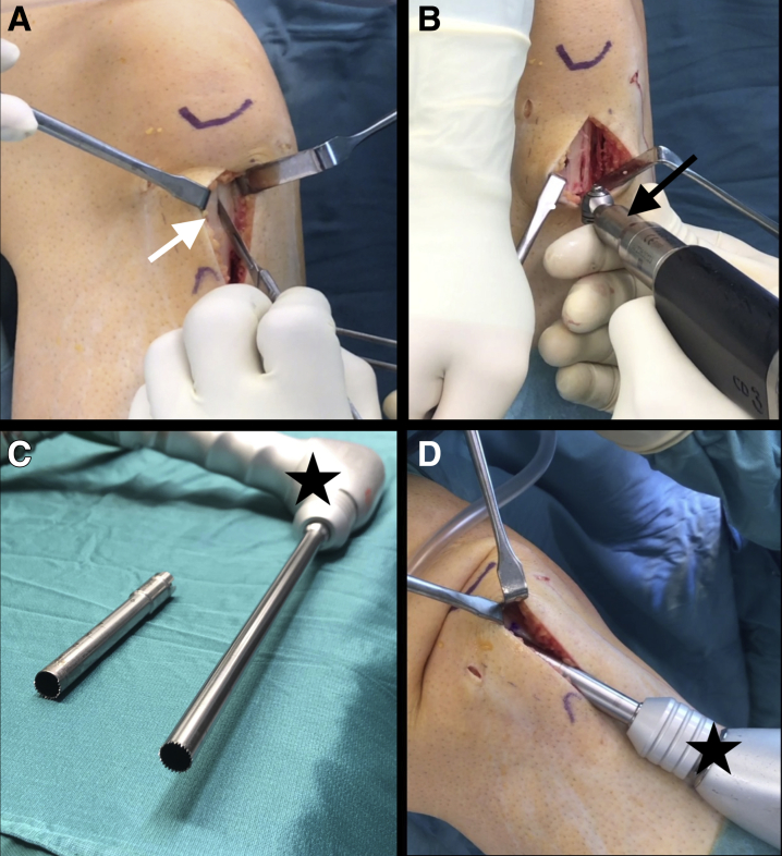
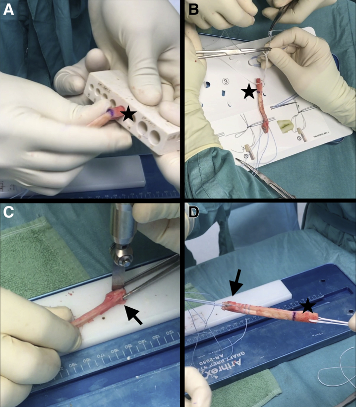
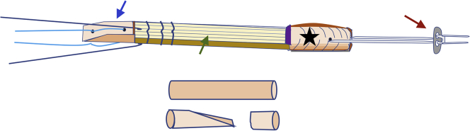
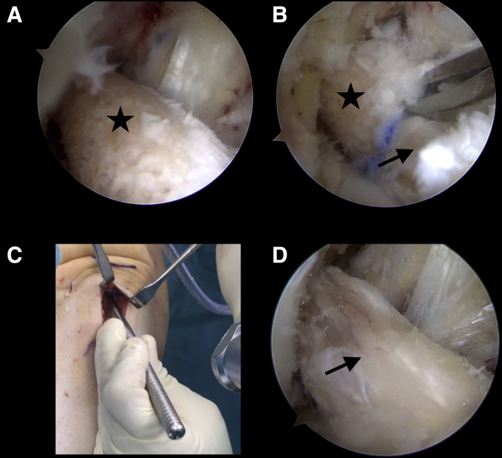
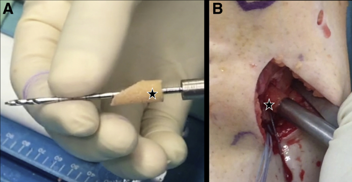
Scientific interest in optimizing outcomes after anterior cruciate ligament (ACL) reconstruction is ongoing, and some recent developments have focused on graft shape as one of the most important factors of anatomic graft placement. The double-bundle or fanlike structure of the native ACL seems to more closely restore normal function and control of rotational stability of the knee with implant-free or press-fit techniques, creating a favorable situation in terms of direct graft-tunnel healing. The ACL reconstruction technique presented in this article enables safe, reproducible, and anatomic fixation of the patellar tendon autograft, providing the biological and biomechanical benefits of direct bony integration of the ribbonlike bone-patellar tendon-bone graft. Because press-fit fixation represents a technically challenging surgical procedure, the goal of the described technique is to enable direct bone-to-bone healing by using secondary extracortical femoral and tibial fixation without the need for a true press-fit situation. Safe and anatomic femoral tunnel drilling is achieved with an outside-in technique (retrograde drilling), hence providing advantages in the routine clinical setting in terms of applicability and time effort. To reduce donor-site morbidity caused by bone block harvesting, refilling of the harvest sites with autologous material is performed.


Figures

Fig 1
Fig 1
Graft harvest in right knee, with patient in supine position. The right leg is hanging with the thigh fixed in a tourniquet. (A) A central 9- to 10-mm strip is created from the patellar tendon (white arrow). (B) At the height of the tibial tuberosity, a 1-cm-wide calibrated oscillating saw blade is used to create the vertical bone cuts, and a smaller, 0.5-cm saw blade (black arrow) is used for the distal horizontal cut about 15 to 20 mm distal to the tendon insertion to create a bone block of sufficient length. (C) A hollow saw (Wolf) with a 9.5-mm diameter for creating the tibial tunnel is shown on the right (black star); a 9.4-mm (two-thirds toothed) hollow saw used to harvest the patellar bone block is shown on the left. (D) A cylindrical bone block, approximately 15 to 20 mm in length, is harvested from the patella, after insertion of the patellar tendon strip with the tibial bone block into the 9.4-mm trephine (black star).
Fig 2
Fig 2
External view of bone–patellar tendon–bone graft during preparation. (A) The cylindrical patellar bone block, 15 to 20 mm in length, is shaped with a rongeur and rasp so that the tip just fits the entrance of a 9-mm template (black star) (or an 8-mm template when the 8.4-mm trephine has been used). (B) For further femoral backup fixation, a BTB TightRope is brought through a 2-mm drill hole at the center of the proximal bone block (black star). (C) Narrowing of the free end of the distal bone block (from the tibial tuberosity) is performed with an oscillation saw (black arrow) so that a trapezoid bone block is shaped (Fig 3). (D) The total length of the graft is determined after placement of 2 No. 2 FiberWire threads within the distal ligament and the trapezoid bone block (black arrow). The cylindrical femoral bone block is shown with the BTB TightRope (black star) in place.
Fig 3
Fig 3
To perform graft preparation, a BTB TightRope (red arrow) is brought through a 2-mm drill hole at the center of the proximal bone block (black star). A No. 2 FiberWire suture (light blue) is brought through each of two 2-mm drill holes in the distal bone block (blue arrow). A No. 2 FiberLoop (dark blue) is used to place a whipstitch through the distal part of the patellar tendon itself. One suture end is again brought through the graft so that tilting of the bone block is avoided when pulling on the sutures. The bone cylinder harvested from creating the tibial tunnel is prepared with a small oscillating saw blade to refill the harvest site at the distal patella (short bone cylinder) and the tibial tunnel in a press-fit fashion. Therefore, the cylinder is trapezoidal to enable tapping in the bone material in a press-fit manner, ensuring direct bone-to-bone healing of the graft in the tibial tunnel. The green arrow indicates the patellar tendon graft.
Fig 4
Fig 4
Arthroscopic view of femoral tunnel drilling from anteromedial portal (A, B) and tibial tunnel creation from anterolateral portal (C, D). (A) At 90° of knee flexion (a right knee in this case), the femoral anterior cruciate ligament (ACL) guide is inserted into the anterolateral portal and placed at the anatomic ACL footprint on the lateral intercondylar wall to ideally cover the anteromedial- and posterolateral-bundle area (black star). (B) With a FlipCutter (fold-out drill), an anatomic femoral socket (black arrow) is placed with an outside-in, retrograde drilling technique. (C) A 2.5-mm guidewire is introduced at the center of the tibial ACL footprint with a 60° aimer or an adjustable aimer (Wolf, Tuttlingen, Germany) (red star). (D) The hollow saw (9.5-mm outer diameter) is used with a centering device. The cylindrical bone block from the tibial tunnel can then be removed, after cutting of the remnant ACL fibers with electrocautery (blue arrow). The red arrow indicates the fully toothed 9.5-mm trephine.
Fig 5
Fig 5
Graft insertion and fixation with arthroscopic view from anterolateral portal (A, (B, D) and external view of distal pole of patella (C). (A) The graft is introduced into the tibial tunnel with correct angulation of the bone block (black star) and advanced into the joint by pulling all 4 sutures coming from the BTB TightRope. (B) The bone block (black star) is guided into the femoral tunnel between the posterior cruciate ligament and lateral intercondylar wall with a suture grasper (Kingfisher). The bone-tendon transition is marked with a skin marker (black arrow). In this step, correct angulation in line with the femoral tunnel is crucial. The bone block can usually then be pulled into the socket without the need for an arthroscopic tappet. (C) The harvest site at the distal pole of the patella is refilled with bone material (Fig 3) using a tappet. An absorbable No. 0 Vicryl suture is then used to close the defect. (D) Final arthroscopic evaluation is performed, and proof of graft tension (black arrow) is confirmed.
Fig 6
Fig 6
Refilling of tibial tunnel with external view of bone cylinder (stars) on 2-mm guidewire (A) and tibial tunnel at anteromedial tibial surface (B). The inversely shaped bone cylinder (harvested from creating the tibial tunnel with the 9.5-mm trephine) is placed on a 2-mm guidewire (A) and is then placed over the graft in the tibial tunnel and tapped into the tunnel with the use of a centering device or a tappet (B).

References

    1. Wipfler B., Donner S., Zechmann C.M., Springer J., Siebold R., Paessler H.H. Anterior cruciate ligament reconstruction using patellar tendon versus hamstring tendon: A prospective comparative study with 9-year follow-up. Arthroscopy. 2011;27:653–665. - PubMed
    1. Sheean A.J., Musahl V., Slone H.S. Quadriceps tendon autograft for arthroscopic knee ligament reconstruction: Use it now, use it often. Br J Sports Med. 2018;52:698–701. - PubMed
    1. Gifstad T., Foss O.A., Engebretsen L. Lower risk of revision with patellar tendon autografts compared with hamstring autografts: A registry study based on 45,998 primary ACL reconstructions in Scandinavia. Am J Sports Med. 2014;42:2319–2328. - PubMed
    1. Boszotta H. Arthroscopic anterior cruciate ligament reconstruction using a patellar tendon graft in press-fit technique: Surgical technique and follow-up. Arthroscopy. 1997;13:332–339. - PubMed
    1. Robin B.N., Jani S.S., Marvil S.C., Reid J.B., Schillhammer C.K., Lubowitz J.H. Advantages and disadvantages of transtibial, anteromedial portal, and outside-in femoral tunnel drilling in single-bundle anterior cruciate ligament reconstruction: A systematic review. Arthroscopy. 2015;31:1412–1417. - PubMed

LinkOut - more resources