

doi: 10.1016/j.eats.2019.10.001.
eCollection 2020 Feb.
Arthroscopic, Nonrigid Fixation of a Displaced Glenoid Fracture After Anterior Shoulder Dislocation
Affiliations
- PMID: 32099777
- PMCID: PMC7029179
- DOI: 10.1016/j.eats.2019.10.001
Item in Clipboard
Arthroscopic, Nonrigid Fixation of a Displaced Glenoid Fracture After Anterior Shoulder Dislocation
Arthrosc Tech.
.
Abstract
Glenoid rim fractures are recognized as a risk factor for recurrent instability after anterior shoulder dislocation. In addition to traditional open treatments of bony Bankart lesions, several arthroscopic techniques of fixation and reconstruction recently have been described. We present a technique of arthroscopic nonrigid fixation for large glenoid rim fractures, as an alternative to existing procedures.
© 2019 by the Arthroscopy Association of North America. Published by Elsevier.
Figures
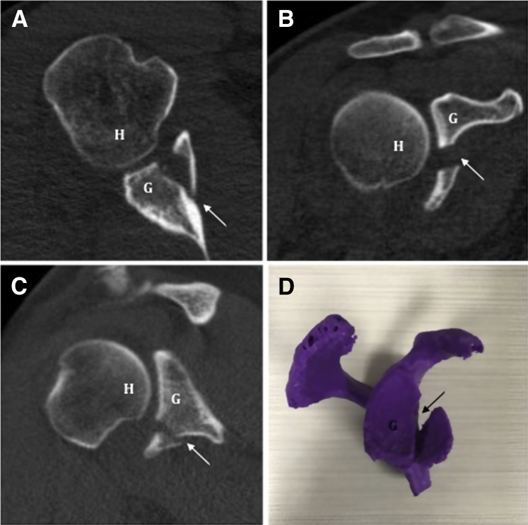
(A-C) Axial, coronal, and sagittal computed tomography images of a right anteroinferior glenoid rim fracture are shown. This fragment represents 30% loss of glenoid width. (D) A 3-dimensional printed model assists with preoperative planning. Arrows demonstrate the glenoid rim fracture. (G, stable glenoid; H, humeral head.)
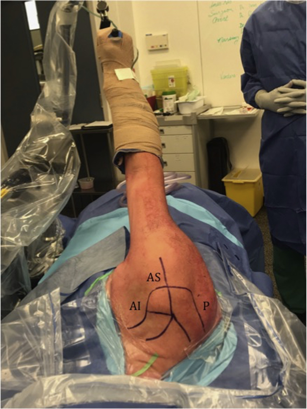
Lateral decubitus positioning and skin markings for standard shoulder arthroscopy portals (right shoulder). (AI, anteroinferior portal; AS, anterosuperior portal; P, posterior portal.)
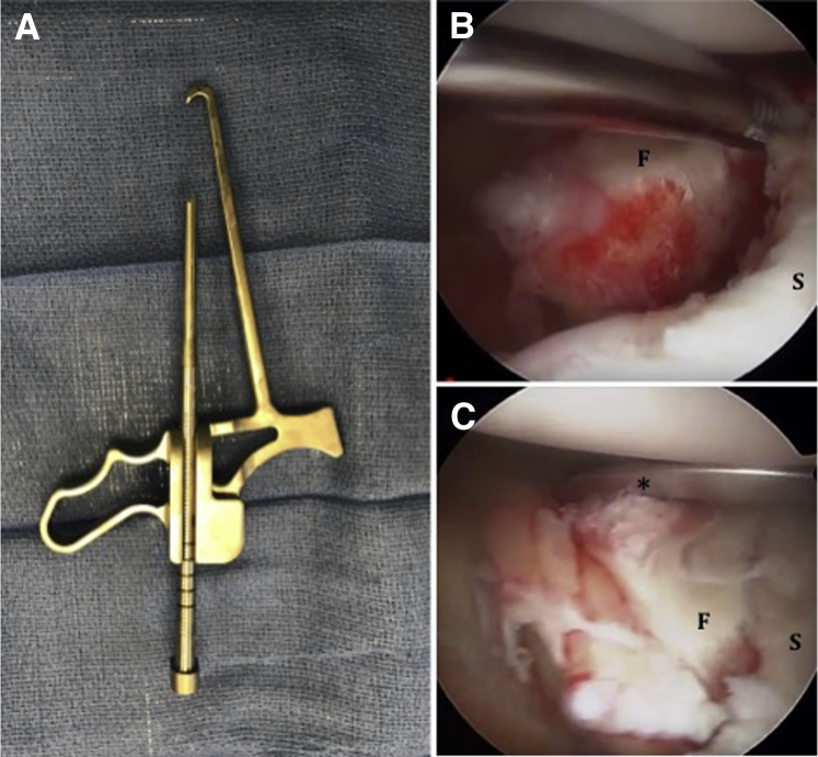
(A) Viewing the right shoulder through an anterosuperior portal, the bullet drill guide is used to manipulate (B) the glenoid fracture fragment into (C) its reduced position. *The bullet drill guide. (F, glenoid fracture fragment; S, stable glenoid.)
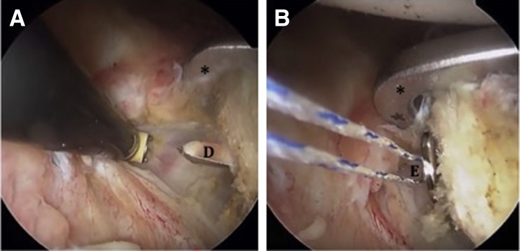
(A) Viewing the right shoulder through an anterosuperior portal, a 2.8-mm drill is advanced through the anterior cortex under direct visualization, establishing a tunnel through which (B) the ENDOBUTTON is then passed. *The bullet drill guide. (D, 2.8-mm drill; E, ENDOBUTTON device.)
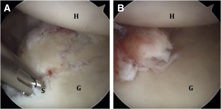
(A) Viewing the right shoulder through an anterosuperior portal, a 1.8-mm suture anchor is positioned just superior to the reduced glenoid fragment. (B) This enables our final Bankart repair, which creates a concentrically reduced glenohumeral joint. (G, repaired glenoid; H, humeral head; S, placement of the suture anchor.)
References
-
- Griffith J.F., Antonio G.E., Yung P.S. Prevalence, pattern, and spectrum of glenoid bone loss in anterior shoulder dislocation: CT analysis of 218 patients. AJR Am J Roentgenol. 2008;190:1247–1254. - PubMed
-
- Burkhart S.S., De Beer J.F. Traumatic glenohumeral bone defects and their relationship to failure of arthroscopic Bankart repairs: Significance of the inverted-pear glenoid and the humeral engaging Hill–Sachs lesion. Arthroscopy. 2000;16:677–694. - PubMed
-
- Sheibel M., Hug K., Gerhardt C., Kruger D. Arthroscopic reduction and fixation of large solitary and multifragmented anterior glenoid rim fractures. J Shoulder Elbow Surg. 2016;25:781–790. - PubMed
LinkOut - more resources
Full Text Sources
