

Knee Arthroscopy: The "Crevice Sign," a New Pathognomonic Sign for Unstable Posterior Medial Meniscal Tear in Anterior Cruciate Ligament-Deficient Knees
- PMID: 32099780
- PMCID: PMC7029181
- DOI: 10.1016/j.eats.2019.10.004
Knee Arthroscopy: The "Crevice Sign," a New Pathognomonic Sign for Unstable Posterior Medial Meniscal Tear in Anterior Cruciate Ligament-Deficient Knees
Abstract
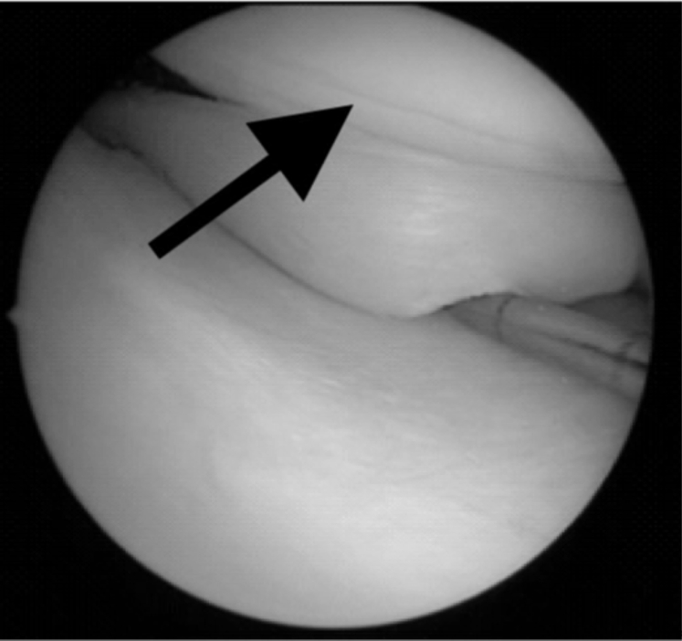
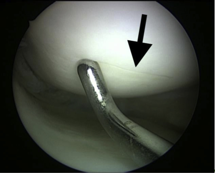
There has been increased emphasis on medial meniscus repair in the anterior cruciate ligament-reconstructed knee, as this improves stability. We describe an arthroscopic sign of an unstable medial meniscal tear that is diagnostic. The "crevice sign" is a longitudinal fissure located on the distal medial femoral condyle. In the anterior cruciate ligament-deficient knee, there is increased strain on the medial meniscus. A posterior longitudinal medial meniscal tear can occur at the time of the index injury or with subsequent instability events. During this injury, the knee pivots and the anterior edge of the unstable medial meniscus digs into the articular cartilage of the medial femoral condyle, resulting in a longitudinal split of the distal femoral condyle articular cartilage. If this sign is observed during arthroscopy, it is recommended that surgeons thoroughly probe the medial meniscus to ensure no pathology is missed.
© 2019 by the Arthroscopy Association of North America. Published by Elsevier.
Figures
References
-
- Neuman P., Englund M., Kostogiannis I., Friden T., Roos H., Dahlberg L.E. Prevalence of tibiofemoral osteoarthritis 15 years after nonoperative treatment of anterior cruciate ligament injury: A prospective cohort study. Am J Sports Med. 2008;36:1717–1725. - PubMed
-
- Pujol N., Beaufils P. Save the meniscus again! Knee Surg Sports Traumatol Arthrosc. 2019;27:341–342. - PubMed
-
- Muriuki M.G., Tuason D.A., Tucker B.G., Harner C.D. Changes in tibiofemoral contact mechanics following radial split and vertical tears of the medial meniscus an in vitro investigation of the efficacy of arthroscopic repair. J Bone Joint Surg Am. 2011;93:1089–1095. - PubMed
-
- Inoue H., Furumatsu T., Miyazawa S., Fujii M., Kodama Y., Ozaki T. Improvement in the medial meniscus posterior shift following anterior cruciate ligament reconstruction. Knee Surg Sports Traumatol Arthrosc. 2018;26:434–441. - PubMed
-
- Okazaki Y., Furumatsu T., Miyazawa S. Meniscal repair concurrent with anterior cruciate ligament reconstruction restores posterior shift of the medial meniscus in the knee-flexed position. Knee Surg Sports Traumatol Arthrosc. 2019;27:361–368. - PubMed
LinkOut - more resources
Full Text Sources
