. 2020 Feb 20;4(2):bvz039.
doi: 10.1210/jendso/bvz039. eCollection 2020 Feb 1.

Intestinal Barrier Dysfunction, LPS Translocation, and Disease Development

Affiliations

Intestinal Barrier Dysfunction, LPS Translocation, and Disease Development

Siddhartha S Ghosh et al. J Endocr Soc. .

Abstract

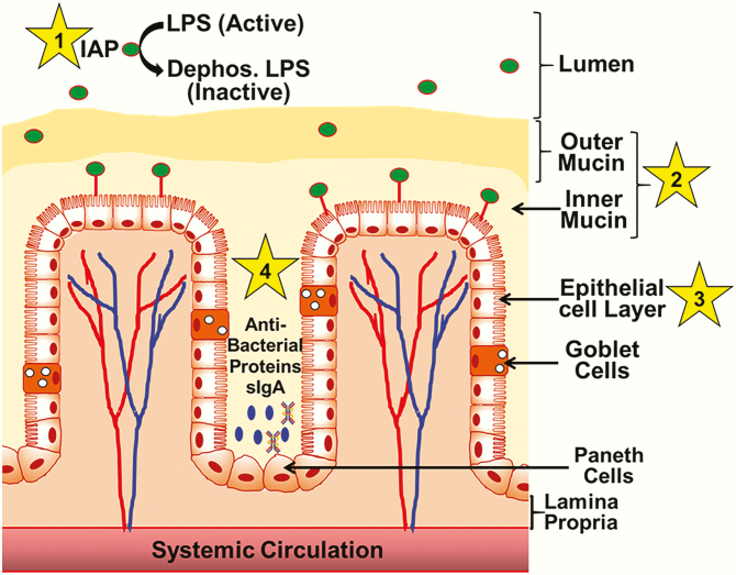
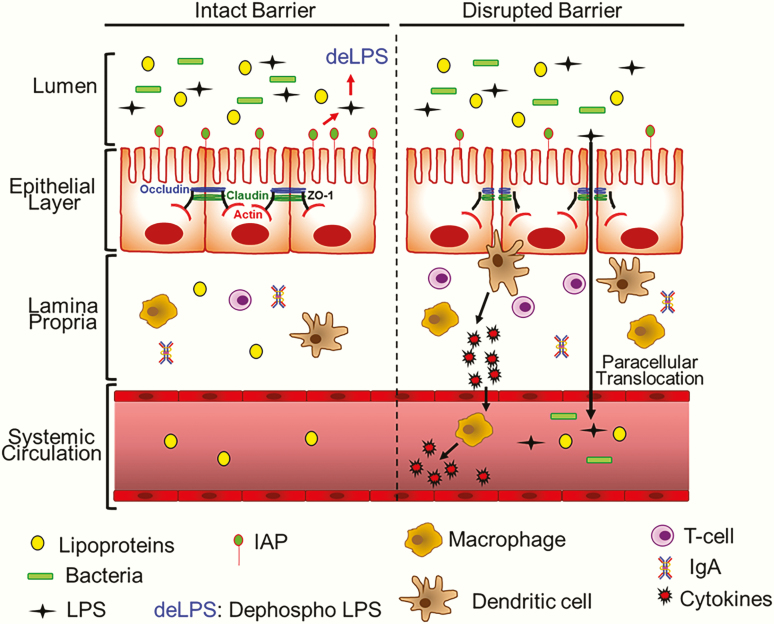
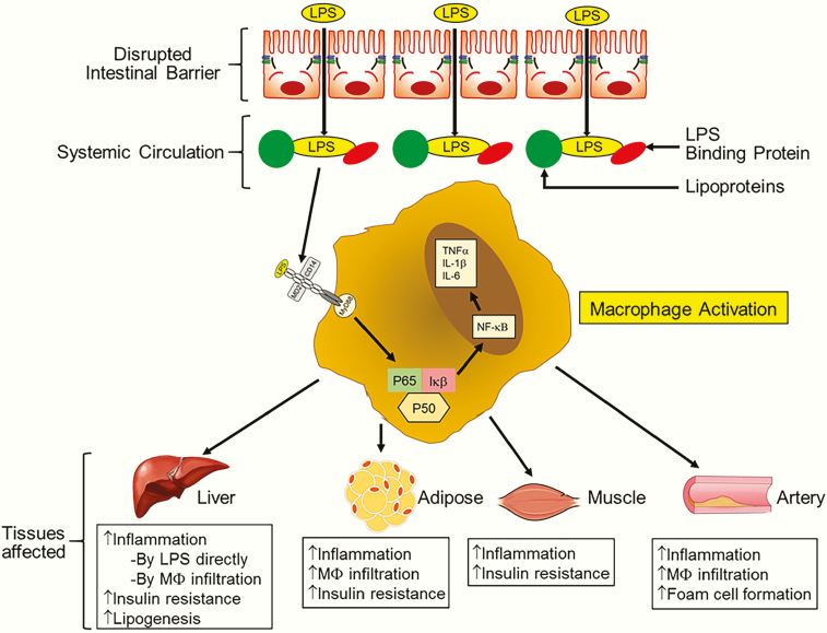
The intestinal barrier is complex and consists of multiple layers, and it provides a physical and functional barrier to the transport of luminal contents to systemic circulation. While the epithelial cell layer and the outer/inner mucin layer constitute the physical barrier and are often referred to as the intestinal barrier, intestinal alkaline phosphatase (IAP) produced by epithelial cells and antibacterial proteins secreted by Panneth cells represent the functional barrier. While antibacterial proteins play an important role in the host defense against gut microbes, IAP detoxifies bacterial endotoxin lipopolysaccharide (LPS) by catalyzing the dephosphorylation of the active/toxic Lipid A moiety, preventing local inflammation as well as the translocation of active LPS into systemic circulation. The causal relationship between circulating LPS levels and the development of multiple diseases underscores the importance of detailed examination of changes in the "layers" of the intestinal barrier associated with disease development and how this dysfunction can be attenuated by targeted interventions. To develop targeted therapies for improving intestinal barrier function, it is imperative to have a deeper understanding of the intestinal barrier itself, the mechanisms underlying the development of diseases due to barrier dysfunction (eg, high circulating LPS levels), the assessment of intestinal barrier function under diseased conditions, and of how individual layers of the intestinal barrier can be beneficially modulated to potentially attenuate the development of associated diseases. This review summarizes the current knowledge of the composition of the intestinal barrier and its assessment and modulation for the development of potential therapies for barrier dysfunction-associated diseases.

Keywords: chronic inflammation; diabetes; intestinal alkaline phosphatase; layers of intestinal barrier; macrophage activation; metabolic diseases.


Figures

Figure 1.
Figure 1.
The “layers” of the intestinal barrier. Functional intestinal barrier consists of four “layers” (shown by numbers 1–4) extending from the lumen that contains gut bacteria and bacterial endotoxin LPS. Layer 1, or intestinal alkaline phosphatase (IAP), released from the intestinal epithelial cells, dephosphorylates luminal LPS inactive, producing dephospho-LPS. Mucin layer (layer 2), consisting of a firmly attached inner layer and a loose outer layer provide the first physical barrier restricting the interaction between luminal bacteria and epithelial cells. A single layer of epithelial cells (layer 3) separates the lumen from systemic circulation. Specialized secretory cells of the epithelial layer, namely goblet and Paneth cells, contribute to the formation of the mucin layer and the production of antibacterial proteins, respectively. Panneth cell-derived antibacterial proteins/peptides, along with secreted IgA (sIgA) from plasma cells present in the lamina propria, restrict bacterial growth and represent the fourth layer (layer 4) of the intestinal barrier.
Figure 2.
Figure 2.
Disruption of the intestinal barrier function. Under normal conditions with an intact barrier, while intestinal epithelial cells facilitate the transcellular movement of ions and nutrients, paracellular transport of bacteria/bacterial products such as LPS is restricted. Cells within the epithelial layer are sealed by tight junction proteins such as Occludin, Claudin, and ZO-1, preventing paracellular transport. In addition, appropriate/homeostatic expression of intestinal alkaline phosphatase (IAP) continuously dephosphorylates and detoxifies LPS in the luminal space. Lamina propria, below the epithelial layer, contains immune cells, both of the innate immune system (eg, macrophages, dendritic cells) and the adaptive immune system (eg, T-cells and IgA-producing plasma celsl [not shown here]). When the intestinal barrier is disrupted (eg, by a Western diet, pathogenic bacteria, LPS due to inadequate detoxification by reduced IAP levels, etc.), the tight junctions are disordered, allowing for paracellular transport of LPS as well as luminal bacteria. In response to these stimuli, dendritic cells and/or macrophages are activated to produce proinflammatory cytokines that not only enhance further infiltration of immune cells into the lamia propria but also activate macrophages in circulation. Paracellularly transported bacteria and LPS also enter systemic circulation, resulting in increased systemic inflammation.
Figure 3.
Figure 3.
Consequences of disrupted intestinal barrier. Increased LPS in systemic circulation is causally linked to the development of multiple diseases. LPS associates with circulating lipoproteins and also interacts with LPS binding protein (LBP). LPS acts as a trigger for macrophage activation via LBP-dependent binding to TLR4. In co-ordination with MD2 and CD14, this results in the homodimerization of TLR4 and the initiation of intracellular signaling. Activation and nuclear translocation of proinflammatory transcription factor NF-κB leads to the eventual production of proinflammatory cytokines (eg, TNFα, IL-1β, IL-6), resulting in increased tissue inflammation. In the liver, LPS reaching through portal blood also activates resident macrophages or Kupffer cells, and increased inflammation underlies increased hepatic insulin resistance and lipogenesis. Activated macrophages infiltrate adipose tissue and the resulting inflamed adipose tissue is also insulin resistant and underlies the development of diabetes. Increased inflammation also leads to insulin resistance in skeletal muscles. Increased infiltration of LPS-activated macrophages into the artery wall initiates foam cell formation, resulting in atherosclerotic plaque development.

References

    1. Bäckhed F, Ley RE, Sonnenburg JL, Peterson DA, Gordon JI. Host-bacterial mutualism in the human intestine. Science. 2005;307(5717):1915–1920. - PubMed
    1. Gill SR, Pop M, Deboy RT, et al. Metagenomic analysis of the human distal gut microbiome. Science. 2006;312(5778):1355–1359. - PMC - PubMed
    1. Sender R, Fuchs S, Milo R. Revised estimates for the number of human and bacteria cells in the body. Plos Biol. 2016;14(8):e1002533. - PMC - PubMed
    1. den Besten G, van Eunen K, Groen AK, Venema K, Reijngoud DJ, Bakker BM. The role of short-chain fatty acids in the interplay between diet, gut microbiota, and host energy metabolism. J Lipid Res. 2013;54(9):2325–2340. - PMC - PubMed
    1. Bäumler AJ, Sperandio V. Interactions between the microbiota and pathogenic bacteria in the gut. Nature. 2016;535(7610):85–93. - PMC - PubMed

LinkOut - more resources