Anti-HMGCR Antibody-Positive Myopathy Shows Bcl-2-Positive Inflammation and Lymphocytic Accumulations
- PMID: 32100014
- PMCID: PMC7092361
- DOI: 10.1093/jnen/nlaa006
Anti-HMGCR Antibody-Positive Myopathy Shows Bcl-2-Positive Inflammation and Lymphocytic Accumulations
Abstract
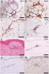
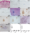
Anti-3-hydroxy-3-methylglutaryl-coenzyme A reductase (HMGCR) and antisignal recognition particle (SRP) antibodies are frequently associated with immune-mediated necrotizing myopathy (IMNM). However, the difference in clinical manifestations between anti-HMGCR and anti-SRP antibodies is unclear. HMGCR is an essential enzyme for cholesterol biosynthesis and is inhibited by statins that regulate apoptosis of Bcl-2-positive and beta chemokine receptor 4 (CCR4)-positive lymphoma cells. In this study, we aimed to clarify Bcl-2 and CCR4 expressions of lymphocytes in anti-HMGCR antibody-positive IMNM and explore the difference between anti-HMGCR antibody-positive myopathy and other inflammatory myopathies. We retrospectively examined Bcl-2- and CCR4-positive lymphocyte infiltrations in muscle and skin biopsy specimens from 19 anti-HMGCR antibody-positive patients and 75 other idiopathic inflammatory myopathies (IIMs) patients. A higher incidence of Bcl-2- and CCR4-positive lymphocytes was detected in the muscle and skin of anti-HMGCR antibody-positive IMNM patients (p < 0.001). In 5 patients with anti-HMGCR antibodies, Bcl-2-positive lymphocytes formed lymphocytic accumulations, which were not observed in other IIMs. Low-density lipoprotein cholesterol levels were not increased except for patients with Bcl-2-positive lymphocytic accumulations (p = 0.010). Bcl-2 and CCR4 lymphocyte infiltrations could be a pathological characteristic of anti-HMGCR antibody-positive IMNM.
Keywords: 3-hydroxy-3-methylglutaryl-coenzyme A reductase (HMGCR); Bcl-2; Hyperlipidemia; Immune-mediated necrotizing myopathy.
© 2020 American Association of Neuropathologists, Inc.
Figures


Similar articles
-
Atypical skin conditions of the neck and back as a dermal manifestation of anti-HMGCR antibody-positive myopathy.BMC Immunol. 2024 May 11;25(1):30. doi: 10.1186/s12865-024-00622-2. BMC Immunol. 2024. PMID: 38734636 Free PMC article.
-
Pediatric Immune-Mediated Necrotizing Myopathy: A Single-Center Retrospective Cohort Study.Pediatr Neurol. 2025 Jun;167:33-41. doi: 10.1016/j.pediatrneurol.2025.03.002. Epub 2025 Mar 14. Pediatr Neurol. 2025. PMID: 40203548
-
Clinical features and prognosis in anti-SRP and anti-HMGCR necrotising myopathy.J Neurol Neurosurg Psychiatry. 2016 Oct;87(10):1038-44. doi: 10.1136/jnnp-2016-313166. Epub 2016 May 4. J Neurol Neurosurg Psychiatry. 2016. PMID: 27147697
-
Anti-HMGCR antibodies as a biomarker for immune-mediated necrotizing myopathies: A history of statins and experience from a large international multi-center study.Autoimmun Rev. 2016 Oct;15(10):983-93. doi: 10.1016/j.autrev.2016.07.023. Epub 2016 Aug 1. Autoimmun Rev. 2016. PMID: 27491568 Review.
-
Immune-mediated necrotizing myopathy (IMNM): A myopathological challenge.Autoimmun Rev. 2022 Feb;21(2):102993. doi: 10.1016/j.autrev.2021.102993. Epub 2021 Nov 16. Autoimmun Rev. 2022. PMID: 34798316 Review.
Cited by
-
A case of anti-HMGCR myopathy in a patient with breast cancer and anti-Th/To antibodies.Oxf Med Case Reports. 2023 Sep 25;2023(9):omad097. doi: 10.1093/omcr/omad097. eCollection 2023 Sep. Oxf Med Case Reports. 2023. PMID: 37771688 Free PMC article.
-
Idiopathic inflammatory myopathy and non-coding RNA.Front Immunol. 2023 Sep 6;14:1227945. doi: 10.3389/fimmu.2023.1227945. eCollection 2023. Front Immunol. 2023. PMID: 37744337 Free PMC article. Review.
-
Juvenile idiopathic inflammatory myopathies with anti-3-hydroxy-3-methylglutaryl-coenzyme A reductase antibodies in a Chinese cohort.CNS Neurosci Ther. 2021 May 1;27(9):1041-7. doi: 10.1111/cns.13658. Online ahead of print. CNS Neurosci Ther. 2021. PMID: 33932258 Free PMC article.
-
Pathologic Features of Anti-Ku Myositis.Neurology. 2024 Apr 23;102(8):e209268. doi: 10.1212/WNL.0000000000209268. Epub 2024 Mar 28. Neurology. 2024. PMID: 38547417 Free PMC article.
-
Challenges in the diagnosis and management of immune-mediated necrotising myopathy (IMNM) in a patient on long-term statins.Rheumatol Int. 2023 Feb;43(2):383-390. doi: 10.1007/s00296-022-05230-0. Epub 2022 Oct 19. Rheumatol Int. 2023. PMID: 36260115 Free PMC article. Review.
References
-
- Bohan A, Peter JB.. Polymyositis and dermatomyositis (first of two parts). N Engl J Med 1975;292:344–7 - PubMed
-
- Hoogendijk JE, Amato AA, Lecky BR, et al. 119th ENMC international workshop: Trial design in adult idiopathic inflammatory myopathies, with the exception of inclusion body myositis, 10-12 October 2003, Naarden, The Netherlands. Neuromuscul Disord 2004;14:337–45 - PubMed
-
- Love LA, Leff RL, Fraser DD, et al. A new approach to the classification of idiopathic inflammatory myopathy: Myositis-specific autoantibodies define useful homogeneous patient groups. Medicine (Baltimore) 1991;70:360–74 - PubMed
-
- Mescam-Mancini L, Allenbach Y, Hervier B, et al. Anti-Jo-1 antibody-positive patients show a characteristic necrotizing perifascicular myositis. Brain 2015;138:2485–92 - PubMed
MeSH terms
Substances
LinkOut - more resources
Full Text Sources
Medical

