. 2020 Jun;36(6):625-638.
doi: 10.1007/s12264-020-00471-0. Epub 2020 Feb 25.

Aloin Protects Against Blood-Brain Barrier Damage After Traumatic Brain Injury in Mice

Affiliations

Aloin Protects Against Blood-Brain Barrier Damage After Traumatic Brain Injury in Mice

Yao Jing et al. Neurosci Bull. 2020 Jun.

Abstract

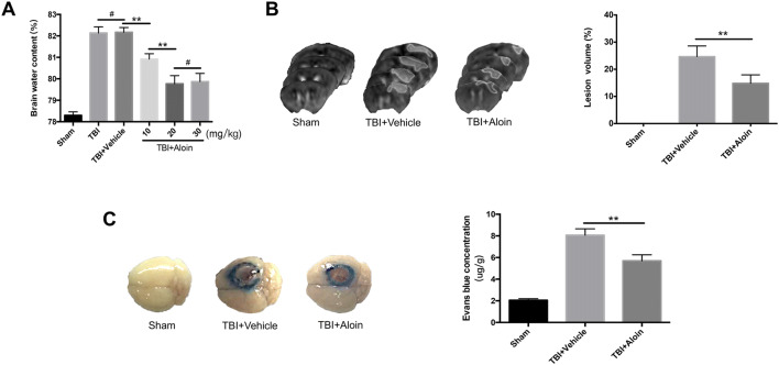
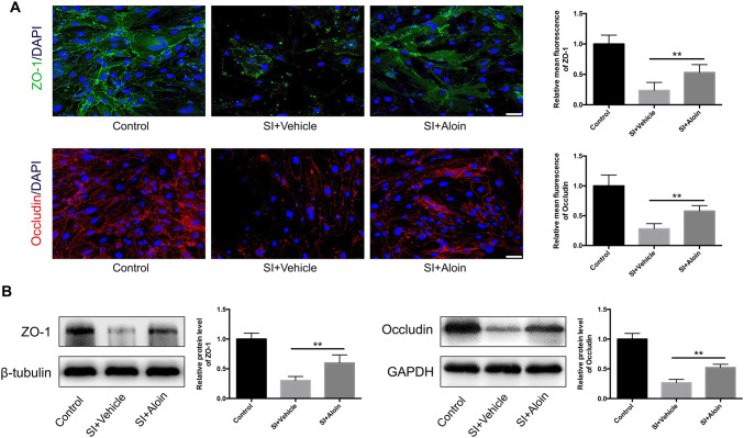
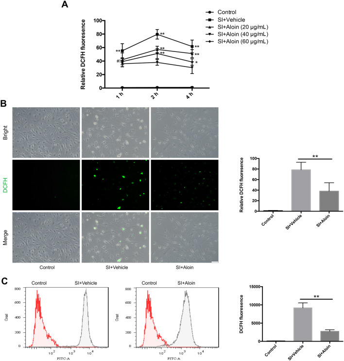
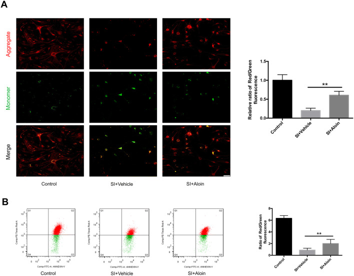
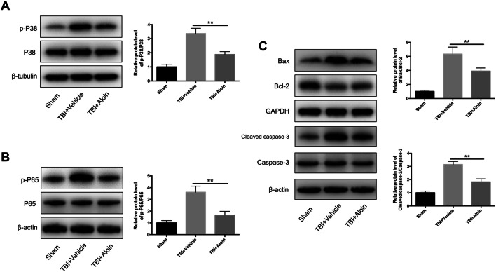
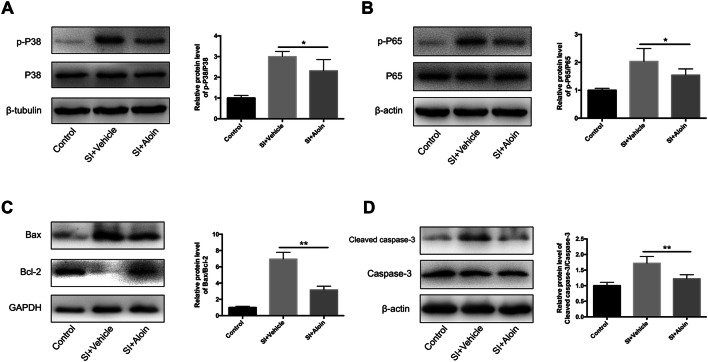
Aloin is a small-molecule drug well known for its protective actions in various models of damage. Traumatic brain injury (TBI)-induced cerebral edema from secondary damage caused by disruption of the blood-brain barrier (BBB) often leads to an adverse prognosis. Since the role of aloin in maintaining the integrity of the BBB after TBI remains unclear, we explored the protective effects of aloin on the BBB using in vivo and in vitro TBI models. Adult male C57BL/6 mice underwent controlled cortical impact injury, and mouse brain capillary endothelial bEnd.3 cells underwent biaxial stretch injury, then both received aloin treatment. In the animal experiments, we found 20 mg/kg aloin to be the optimum concentration to decrease cerebral edema, decrease disruption of the BBB, and improve neurobehavioral performance after cortical impact injury. In the cellular studies, the optimum concentration of 40 μg/mL aloin reduced apoptosis and reversed the loss of tight junctions by reducing the reactive oxygen species levels and changes in mitochondrial membrane potential after stretch injury. The mechanisms may be that aloin downregulates the phosphorylation of p38 mitogen-activated protein kinase, the activation of p65 nuclear factor-kappa B, and the ratios of B cell lymphoma (Bcl)-2-associated X protein/Bcl-2 and cleaved caspase-3/caspase-3. We conclude that aloin exhibits these protective effects on the BBB after TBI through its anti-oxidative stress and anti-apoptotic properties in mouse brain capillary endothelial cells. Aloin may thus be a promising therapeutic drug for TBI.

Keywords: Aloin; Apoptosis; Blood–brain barrier; Oxidative stress; Traumatic brain injury.


Conflict of interest statement

The authors declare no competing interests.

Figures

Fig. 1
Fig. 1
Aloin attenuates TBI-induced brain edema caused by damage to the BBB in mice. A Brain water content in different groups at 3 days post-TBI. B Representative coronal T2-weighted MRI scans and brain lesion volumes in sham, TBI+vehicle, and TBI+aloin groups at 3 days after TBI (white contours indicate areas of edema). C Representative images of EB extravasation and statistics for EB content in the three groups at 3 days after TBI (blue areas indicate EB extravasation). n = 6/group, data are presented as the mean ± SD, **P < 0.01, #P < 0.05.
Fig. 2
Fig. 2
Aloin alleviates the loss of TJ proteins in the BBB in the pericontusional area 3 days after experimental TBI in mice. A Representative western blots and levels of ZO-1 and occludin in the sham, TBI+vehicle, and TBI+aloin groups. B Representative co-stained immunofluorescence and levels of ZO-1/CD31 and occludin/CD31 in the three groups (scale bars, 75 μm). n = 6/group, data are presented as the mean ± SD. **P < 0.01, aloin vs vehicle group.
Fig. 3
Fig. 3
Aloin improves neurological functions after experimental TBI in mice. A, B mNSS (A), and rotarod latency (B) before TBI and at 1, 3, 7, and 14 days after TBI in the TBI + vehicle and TBI + aloin groups. C Morris water maze training results during 14–18 days after TBI in the sham, TBI + vehicle, and TBI + aloin groups. DF Time of first arrival at the platform (D), number of times crossing the platform (E), and percentage of time in the platform quadrant (F) in the Morris water maze 19 days after TBI in the three groups. n = 12/group, data are presented as the mean ± SD, *P < 0.05, **P < 0.01, aloin vs vehicle group.
Fig. 4
Fig. 4
Aloin reduces the damage to bEnd.3 cells by experimental SI. A Cell viability at different concentrations of aloin after 4.5 h assessed by CCK-8. B Effects of different concentrations of aloin on cells 4 h after SI assessed by LDH release. C Representative TUNEL staining of apoptotic cells and quantified apoptosis rate at 4 h after SI in control, SI+vehicle, and SI+aloin groups (scale bar, 75 μm). n = 6/group, data are presented as the mean ± SD, *P < 0.05, **P < 0.01, #P > 0.05.
Fig. 5
Fig. 5
Aloin alleviates the loss of TJ proteins after experimental SI in bEnd.3 cells. A Representative immunofluorescence images and quantification of ZO-1 and occludin proteins 4 h after SI in the three groups (scale bars, 30 μm). B Representative western blots and quantification of ZO-1 and occludin proteins 4 h after SI in the three groups. n = 6/group, data are presented as the mean ± SD, **P < 0.01, aloin vs vehicle group.
Fig. 6
Fig. 6
Aloin decreases intracellular ROS generation after experimental SI in bEnd.3 cells, as detected by DCFH-DA. A ROS levels at different time points post-SI after treatment with different concentrations of aloin. B Representative intracellular ROS fluorescence images with bright field and quantification 2 h after SI in the control, SI+vehicle, and SI+aloin groups (scale bar, 75 μm). C Representative fluorescence intensity using flow cytometry and quantification 2 h after SI in the three groups. n = 6/group, data are presented as the mean ± SD, *P < 0.05, **P < 0.01, #P > 0.05.
Fig. 7
Fig. 7
Aloin protects against the changes in ΔΨm after SI in bEnd.3 cells. A Representative ΔΨm fluorescence images and red/green fluorescence ratios 2 h after SI in the control, SI + vehicle, and SI + aloin groups (scale bar, 75 μm). B Representative fluorescence intensity using flow cytometry and red/green fluorescence ratios 2 h after SI in the three groups. n = 6/group, data are presented as the mean ± SD, **P < 0.01, aloin vs vehicle group.
Fig. 8
Fig. 8
Aloin regulates MAPK, NF-kB, and apoptosis-associated pathways in mouse TBI models. AC Representative western blots of p-P38 and P38 (A), p-P65 and P65 (B), and Bax, Bcl-2, cleaved caspase-3, and caspase-3 proteins (C), along with their quantifications 3 days after TBI in the sham, TBI+vehicle, and TBI+aloin groups. n = 6/group, data are presented as the mean ± SD, **P < 0.01, aloin vs vehicle group.
Fig. 9
Fig. 9
Aloin regulates MAPK, NF-kB, and apoptosis-associated pathways after experimental stretch injury in bEnd.3 cells. AD Representative western blots of p-P38 and P38 (A), p-P65 and P65 (B), and Bax, Bcl-2 (C), cleaved caspase-3, and caspase-3 proteins (D), along with quantifications at 2 h after SI in the control, SI+vehicle, and SI+aloin groups. n = 6/group, data are presented as the mean ± SD, *P < 0.05, **P < 0.01, aloin vs vehicle group.

Similar articles

Cited by

References

    1. Needham EJ, Helmy A, Zanier ER, Jones JL, Coles AJ, Menon DK. The immunological response to traumatic brain injury. J Neuroimmunol. 2019;332:112–125. - PubMed
    1. Yang DX, Jing Y, Liu YL, Xu ZM, Yuan F, Wang ML, et al. Inhibition of transient receptor potential vanilloid 1 attenuates blood–brain barrier disruption after traumatic brain injury in mice. J Neurotrauma. 2019;36:1279–1290. - PubMed
    1. Jiang JY, Gao GY, Feng JF, Mao Q, Chen LG, Yang XF, et al. Traumatic brain injury in China. Lancet Neurol. 2019;18:286–295. - PubMed
    1. Zhang L, Wang H, Zhou X, Mao L, Ding K, Hu Z. Role of mitochondrial calcium uniporter-mediated Ca(2+) and iron accumulation in traumatic brain injury. J Cell Mol Med. 2019;23:2995–3009. - PMC - PubMed
    1. Liu YL, Yuan F, Yang DX, Xu ZM, Jing Y, Yang GY, et al. Adjudin attenuates cerebral edema and improves neurological function in mice with experimental traumatic brain injury. J Neurotrauma. 2018;35:2850–2860. - PubMed