Review
. 2020 Apr;250(5):480-495.
doi: 10.1002/path.5405. Epub 2020 Mar 16.

Long non-coding RNAs in development and disease: conservation to mechanisms

Affiliations
Review

Long non-coding RNAs in development and disease: conservation to mechanisms

Ioannis Tsagakis et al. J Pathol. 2020 Apr.

Abstract

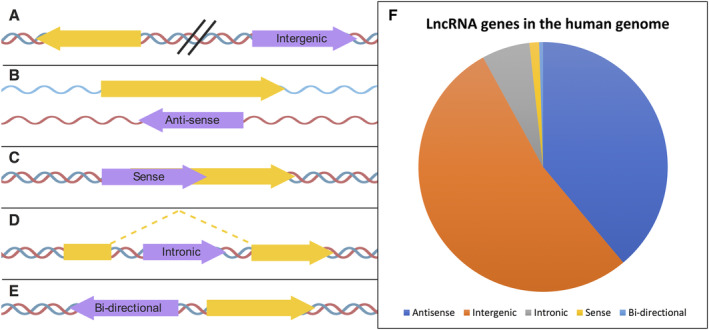
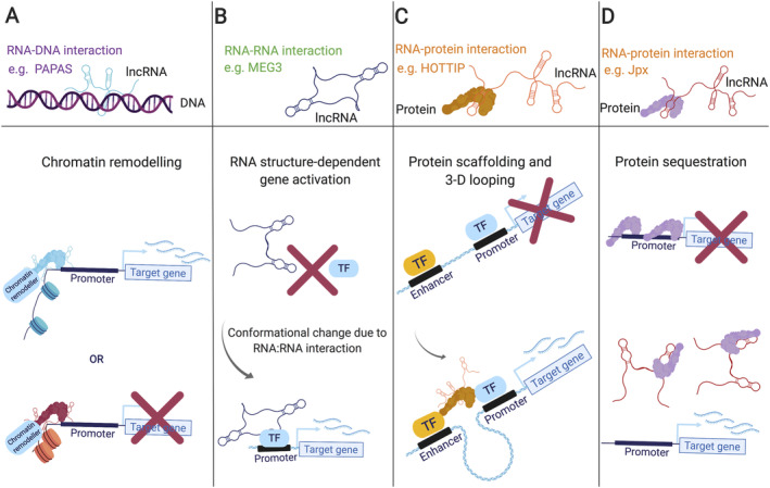
Our genomes contain the blueprint of what makes us human and many indications as to why we develop disease. Until the last 10 years, most studies had focussed on protein-coding genes, more specifically DNA sequences coding for proteins. However, this represents less than 5% of our genomes. The other 95% is referred to as the 'dark matter' of our genomes, our understanding of which is extremely limited. Part of this 'dark matter' includes regions that give rise to RNAs that do not code for proteins. A subset of these non-coding RNAs are long non-coding RNAs (lncRNAs), which in particular are beginning to be dissected and their importance to human health revealed. To improve our understanding and treatment of disease it is vital that we understand the molecular and cellular function of lncRNAs, and how their misregulation can contribute to disease. It is not yet clear what proportion of lncRNAs is actually functional; conservation during evolution is being used to understand the biological importance of lncRNA. Here, we present key themes within the field of lncRNAs, emphasising the importance of their roles in both the nucleus and the cytoplasm of cells, as well as patterns in their modes of action. We discuss their potential functions in development and disease using examples where we have the greatest understanding. Finally, we emphasise why lncRNAs can serve as biomarkers and discuss their emerging potential for therapy. © 2020 The Authors. The Journal of Pathology published by John Wiley & Sons Ltd on behalf of Pathological Society of Great Britain and Ireland.

Keywords: X chromosome inactivation; anti-sense lncRNAs; cancer; conservation; development; diabetes; long intergenic non-coding RNA; long non-coding RNA; neurodegenerative disease; stem cells; translation.


Figures

Figure 1
Figure 1
Numbers of different types of genes in humans and selected other eukaryotes. Data from Homo sapiens: GENCODE Release (version 30) , Mus musculus : GENCODE Release (version M21) , Rattus norvegicus : Ensembl RGSC assembly (v6.0) , Danio rerio: Ensembl (GRCz11) , Drosophila melanogaster : FlyBase (FB2019_02 R6.27) .
Figure 2
Figure 2
Categories of lncRNA. Types of lncRNAs based on their genomic position, orientation, and relative location to nearby protein‐coding genes. (A) intergenic, (B) anti‐sense, (C) sense, (D) intronic, and (E) bi‐directional. lncRNA genes are marked in purple and protein‐coding sequences in yellow. (F) Proportion of lncRNAs present in the human genome by location. Annotation from Gencode January 2019 (release 29, GRch38) . Created using BioRender.
Figure 3
Figure 3
The principle mechanisms governing lncRNA interactions. lncRNAs have been found to interact with (A) DNA, via Hoogstein bonding to form triple helical structures, (B) RNA, via Watson–Crick–Franklin (hydrogen) base‐pairing, or (C) proteins. These interactions underlie all effector functions elicited by lncRNAs studied to date. Created using BioRender.
Figure 4
Figure 4
Localisation of nuclear lncRNAs to specific nuclear regions and their dynamic nature. lncRNAs generated in the nucleus can be anchored at specific locations (e.g. XIST) or dynamically shift their intra‐ and inter‐cellular localisation in response to environmental cues such as heat shock (e.g. PAPAS) or metabolic stress (e.g. UCHL1‐AS). Created using BioRender.
Figure 5
Figure 5
Molecular functions of nuclear lncRNAs. (A) lncRNAs can guide chromatin remodelling complexes to transcription sites, which can either deposit active or repressive chromatin marks (e.g. Xist). (B) lncRNA:RNA interactions can cause a shift in tertiary structure, activating transcription factors which regulate gene expression (e.g. MEG3). (C) By bridging protein interactions or scaffolding the assembly of multi‐protein complexes, lncRNAs can facilitate enhancer and promoter element interactions, critical for gene activation (e.g. HOTTIP). (D) IncRNAs bind specific proteins with high affinity to titrate proteins away from typical occupancy sites, impacting gene activity and/or 3‐D genome compaction (e.g. Jpx). Created using BioRender.
Figure 6
Figure 6
Molecular functions of cytoplasmic lncRNAs. (A) lncRNAs interact with proteins and/or mRNAs to form RNP complexes, which regulate post‐transcriptional gene regulation. (B) lncRNAs act as molecular sponges for miRNAs, thus stabilising and protecting mRNAs from degradation. (C) lncRNAs associate with the translation machinery and regulate the translation of mRNAs. (D) lncRNAs can be actively engaged by translating ribosomes. (E) lncRNAs have been found in extracellular vesicles. Created using BioRender.
Figure 7
Figure 7
lncRNAs can promote or protect from cancer and neurodegeneration. (A) NBAT1‐lncRNA promotes neuronal differentiation and prevents neuroblastoma. (B) LINK‐A interacts with PIP3, PC, and AKT, resulting in AKT hyperactivity and drug resistance in breast cancer. (C) BC200 disrupts mRNA delivery and translation at the synapse and leads to neurodegeneration. (D) HTT‐AS controls HTT expression levels. Downregulation of HTT‐AS has been linked to the occurrence of Huntington's disease. Created using BioRender.

References

    1. Brannan CI, Dees EC, Ingram RS, et al. The product of the H19 gene may function as an RNA. Mol Cell Biol 1990; 10 : 28–36. - PMC - PubMed
    1. Brown CJ, Ballabio A, Rupert JL, et al. A gene from the region of the human X inactivation centre is expressed exclusively from the inactive X chromosome. Nature 1991; 349 : 38–44. - PubMed
    1. Frankish A, Diekhans M, Ferreira AM, et al. GENCODE reference annotation for the human and mouse genomes. Nucleic Acids Res 2019; 47 : D766–D773. - PMC - PubMed
    1. Consortium GT. The genotype‐tissue expression (GTEx) project. Nat Genet 2013; 45 : 580–585. - PMC - PubMed
    1. Lizio M, Harshbarger J, Shimoji H, et al. Gateways to the FANTOM5 promoter level mammalian expression atlas. Genome Biol 2015; 16 : 22. - PMC - PubMed

Publication types