

Risk factors for progression of age-related macular degeneration
- PMID: 32100327
- PMCID: PMC7155063
- DOI: 10.1111/opo.12675
Risk factors for progression of age-related macular degeneration
Abstract
Purpose: Age-related macular degeneration (AMD) is a degenerative disease of the macula, often leading to progressive vision loss. The rate of disease progression can vary among individuals and has been associated with multiple risk factors. In this review, we provide an overview of the current literature investigating phenotypic, demographic, environmental, genetic, and molecular risk factors, and propose the most consistently identified risk factors for disease progression in AMD based on these studies. Finally, we describe the potential use of these risk factors for personalised healthcare.
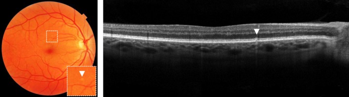
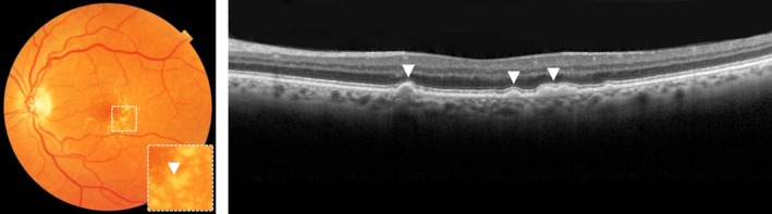
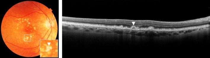
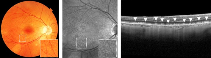
Recent findings: While phenotypic risk factors such as drusen and pigment abnormalities become more important to predict disease progression during the course of the disease, demographic, environmental, genetic and molecular risk factors are more valuable at earlier disease stages. Demographic and environmental risk factors such as age and smoking are consistently reported to be related to disease progression, while other factors such as sex, body mass index (BMI) and education are less often associated. Of all known AMD variants, variants that are most consistently reported with disease progression are rs10922109 and rs570618 in CFH, rs116503776 in C2/CFB/SKIV2L, rs3750846 in ARMS2/HTRA1 and rs2230199 in C3. However, it seems likely that other AMD variants also contribute to disease progression but to a lesser extent. Rare variants have probably a large effect on disease progression in highly affected families. Furthermore, current prediction models do not include molecular risk factors, while these factors can be measured accurately in the blood. Possible promising molecular risk factors are High-Density Lipoprotein Cholesterol (HDL-C), Docosahexaenoic acid (DHA), eicosapentaenoic acid (EPA), zeaxanthin and lutein.
Summary: Phenotypic, demographic, environmental, genetic and molecular risk factors can be combined in prediction models to predict disease progression, but the selection of the proper risk factors for personalised risk prediction will differ among individuals and is dependent on their current disease stage. Future prediction models should include a wider set of genetic variants to determine the genetic risk more accurately, and rare variants should be taken into account in highly affected families. In addition, adding molecular factors in prediction models may lead to preventive strategies and personalised advice.
Keywords: age-related macular degeneration; epidemiology; genetics.
© 2020 The Authors. Ophthalmic and Physiological Optics published by John Wiley & Sons Ltd on behalf of College of Optometrists.
Conflict of interest statement
The author(s) have no proprietary or commercial interest in any materials discussed in this article.
Figures
References
-
- Wong WL, Su X, Li X, et al. Global prevalence of age‐related macular degeneration and disease burden projection for 2020 and 2040: a systematic review and meta‐analysis. Lancet Glob Health 2014; 2: e106–116. - PubMed
-
- Tomany SC, Wang JJ, Van Leeuwen R, et al. Risk factors for incident age‐related macular degeneration: pooled findings from 3 continents. Ophthalmology 2004; 111: 1280–1287. - PubMed
-
- Schaal KB, Rosenfeld PJ, Gregori G, Yehoshua Z & Feuer WJ. Anatomic clinical trial endpoints for nonexudative age‐related macular degeneration. Ophthalmology 2016; 123: 1060–1079. - PubMed
Publication types
MeSH terms
Substances
LinkOut - more resources
Full Text Sources
Medical
Research Materials
Miscellaneous
