Case Reports
. 2020 Apr;21(4):501-504.
doi: 10.3348/kjr.2020.0112. Epub 2020 Feb 26.

2019 Novel Coronavirus (COVID-19) Pneumonia: Serial Computed Tomography Findings

Affiliations
Case Reports

2019 Novel Coronavirus (COVID-19) Pneumonia: Serial Computed Tomography Findings

Jiangping Wei et al. Korean J Radiol. 2020 Apr.

Abstract

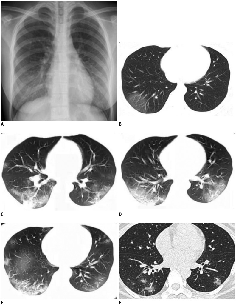
From December 2019, Coronavirus disease 2019 (COVID-19) pneumonia (formerly known as the 2019 novel Coronavirus [2019-nCoV]) broke out in Wuhan, China. In this study, we present serial CT findings in a 40-year-old female patient with COVID-19 pneumonia who presented with the symptoms of fever, chest tightness, and fatigue. She was diagnosed with COVID-19 infection confirmed by real-time reverse-transcriptase-polymerase chain reaction. CT showed rapidly progressing peripheral consolidations and ground-glass opacities in both lungs. After treatment, the lesions were shown to be almost absorbed leaving the fibrous lesions.

Keywords: 2019-nCoV; COVID-19; Coronavirus; Pneumonia; Tomography, X-ray computed.


Conflict of interest statement

The authors have no potential conflicts of interest to disclose.

Figures

Fig. 1
Fig. 1. 40-year-old female patient with Coronavirus disease 2019 pneumonia.
A, B. Initial posteroanterior chest radiograph and chest CT scan were performed on day of admission (3 days after onset of fever). Chest radiograph (A) shows no thoracic abnormalities. Axial CT scan (B) shows GGOs in subpleural area of right lower lobe. Left lung is normal. C–E. Follow-up chest CT scans taken 6 days after onset of fever show increased density of GGOs in right lower lobe, which then progressed into consolidations with perilobular thickening. Multifocal peripheral patchy areas of nodular consolidations and nodular GGO lesions are newly developed in subpleural areas of both lower lobes. F. Progressive resolution of parenchymal lesions is seen in follow-up high-resolution CT scan obtained on day 12. Patchy consolidations and GGOs in both lungs were almost absorbed leaving a few fibrous lesions that may represent residual organizing pneumonia. Repeat real-time reverse-transcriptase-polymerase chain reaction was negative and patient was discharged. CT = computer tomography, GGO = ground-glass opacity

References

    1. Rubin EJ, Baden LR, Morrissey S, Campion EW. Medical journals and the 2019-nCoV outbreak. N Engl J Med. 2020 Jan 27; doi: 10.1056/NEJMe2001329. [Epub] - DOI - PubMed
    1. The Lancet. Emerging understandings of 2019-nCoV. Lancet. 2020;395:311. - PMC - PubMed
    1. Carlos WG, Dela Cruz CS, Cao B, Pasnick S, Jamil S. Novel Wuhan (2019-nCoV) Coronavirus. Am J Respir Crit Care Med. 2020;201:P7–P8. - PubMed
    1. Lei J, Li J, Li X, Qi X. CT imaging of the 2019 Novel Coronavirus (2019-nCoV) pneumonia. Radiology. 2020 Jan 31; doi: 10.1148/radiol.2020200236. [Epub] - DOI - PMC - PubMed
    1. Zhu N, Zhang D, Wang W, Li X, Yang B, Song J, et al. China Novel Coronavirus Investigating and Research Team. A novel coronavirus from patients with pneumonia in China, 2019. N Engl J Med. 2020 Jan 24; doi: 10.1056/NEJMoa2001017. [Epub] - DOI - PMC - PubMed

Publication types