. 2020 Mar 17;59(10):1093-1103.
doi: 10.1021/acs.biochem.0c00043. Epub 2020 Mar 3.

Epigallocatechin-3-gallate Inhibits Cu(II)-Induced β-2-Microglobulin Amyloid Formation by Binding to the Edge of Its β-Sheets

Affiliations

Epigallocatechin-3-gallate Inhibits Cu(II)-Induced β-2-Microglobulin Amyloid Formation by Binding to the Edge of Its β-Sheets

Tyler M Marcinko et al. Biochemistry. .

Abstract

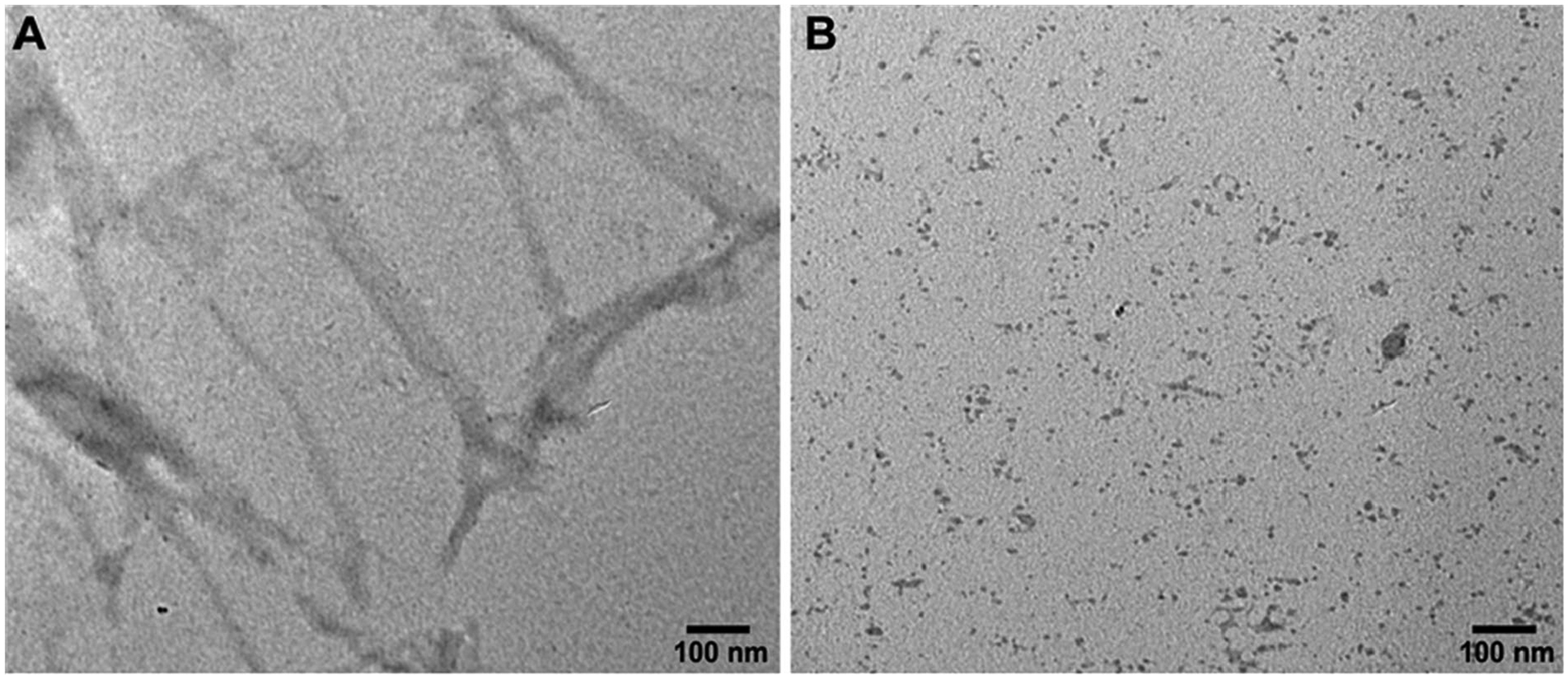
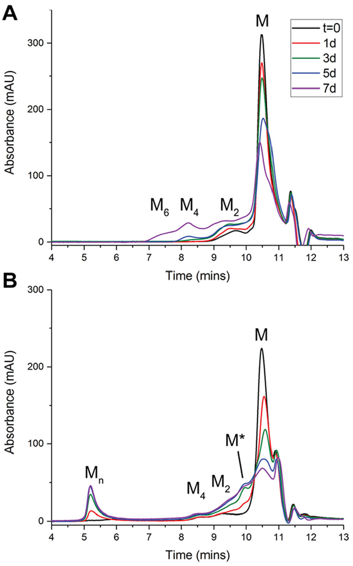
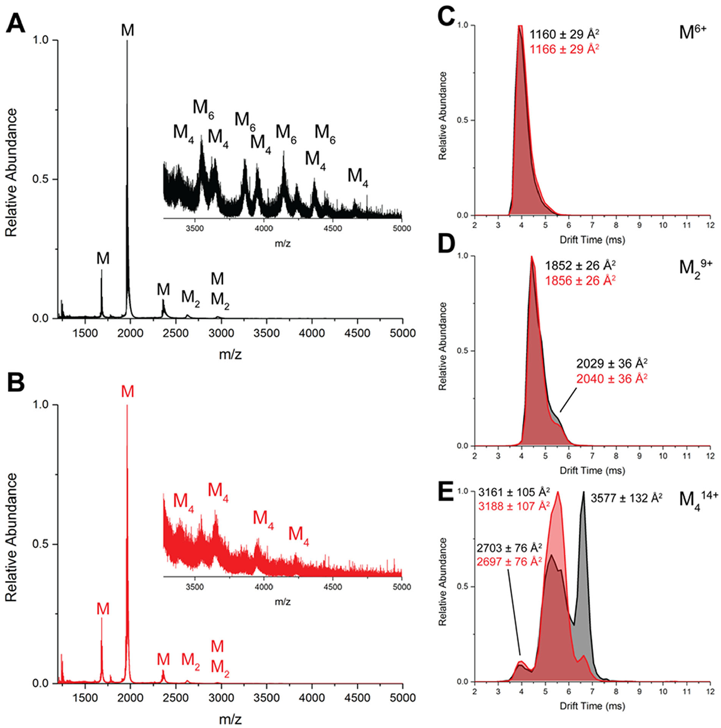
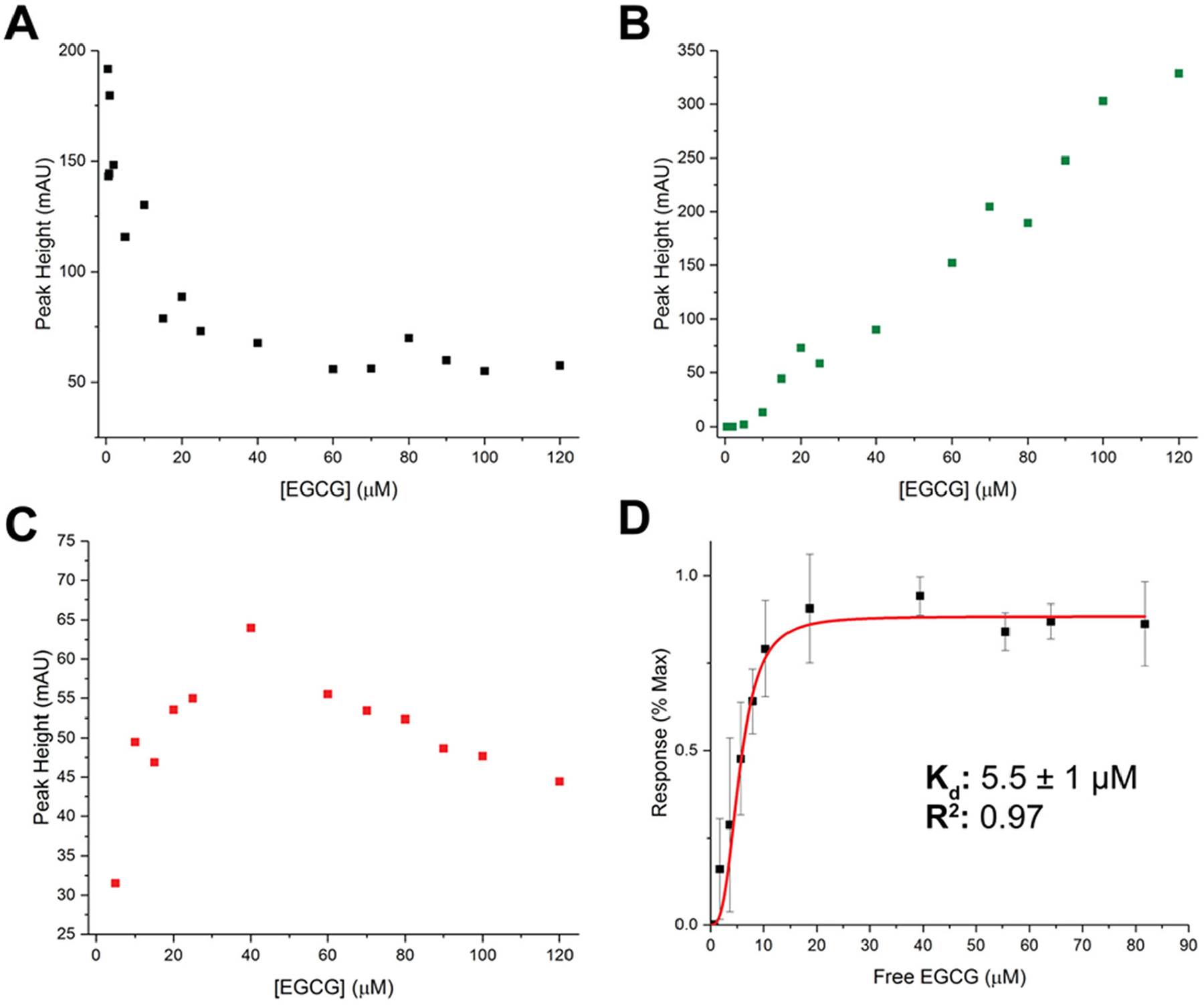
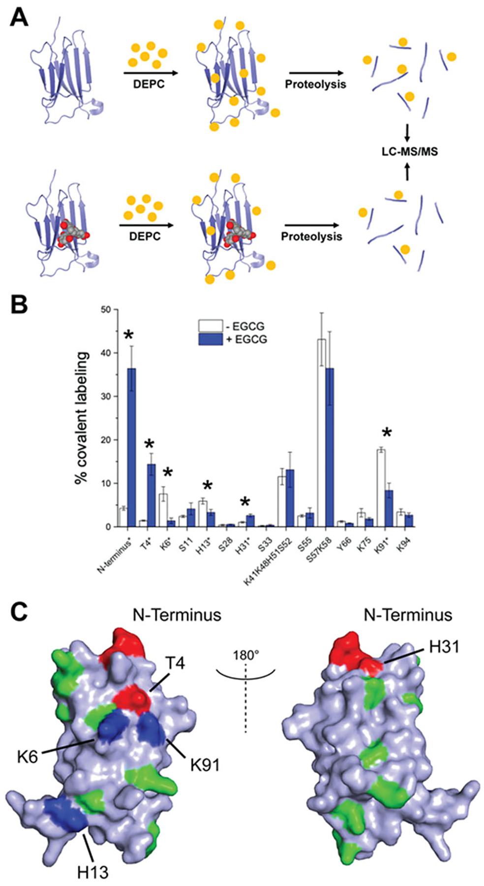
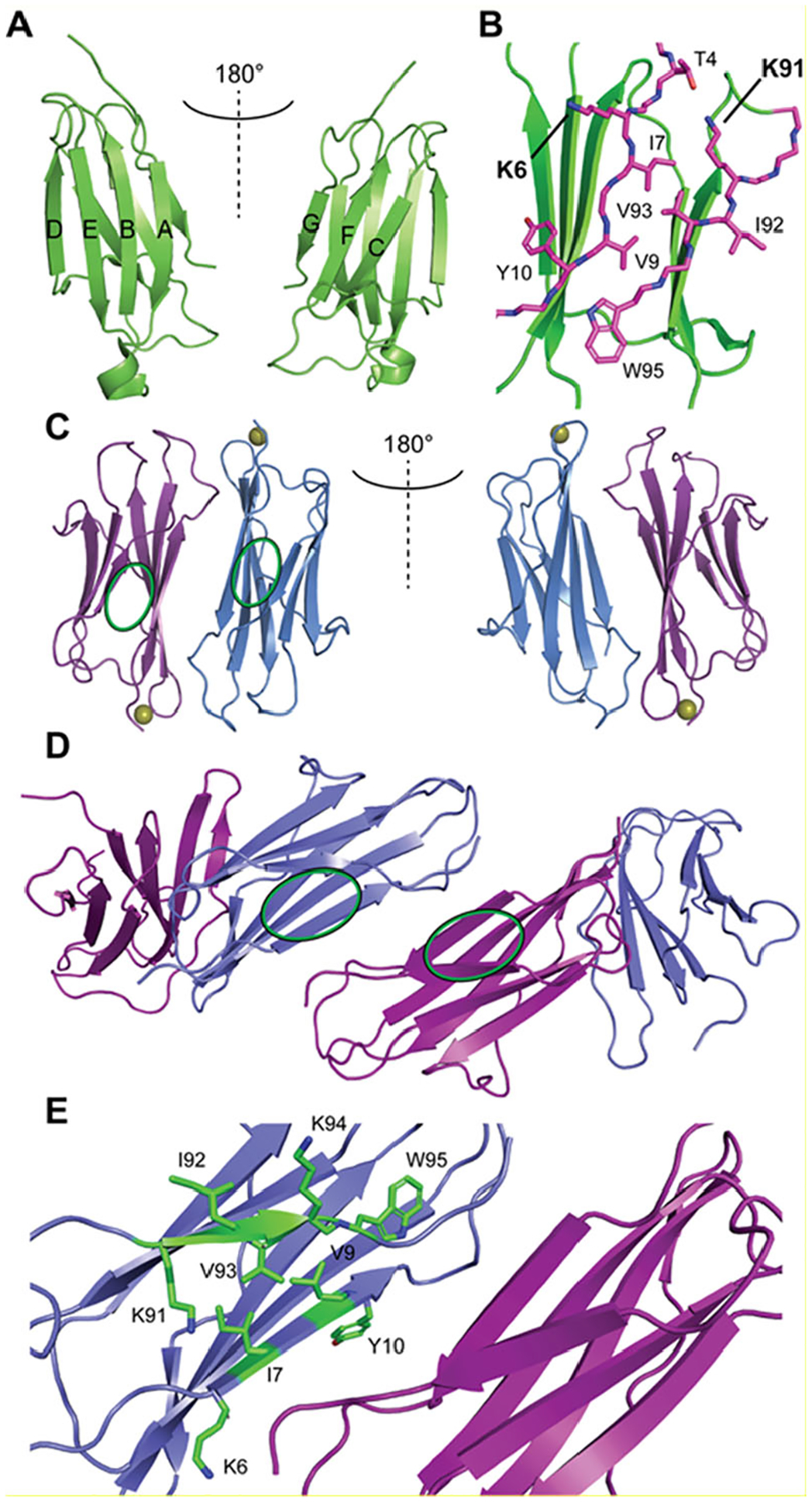
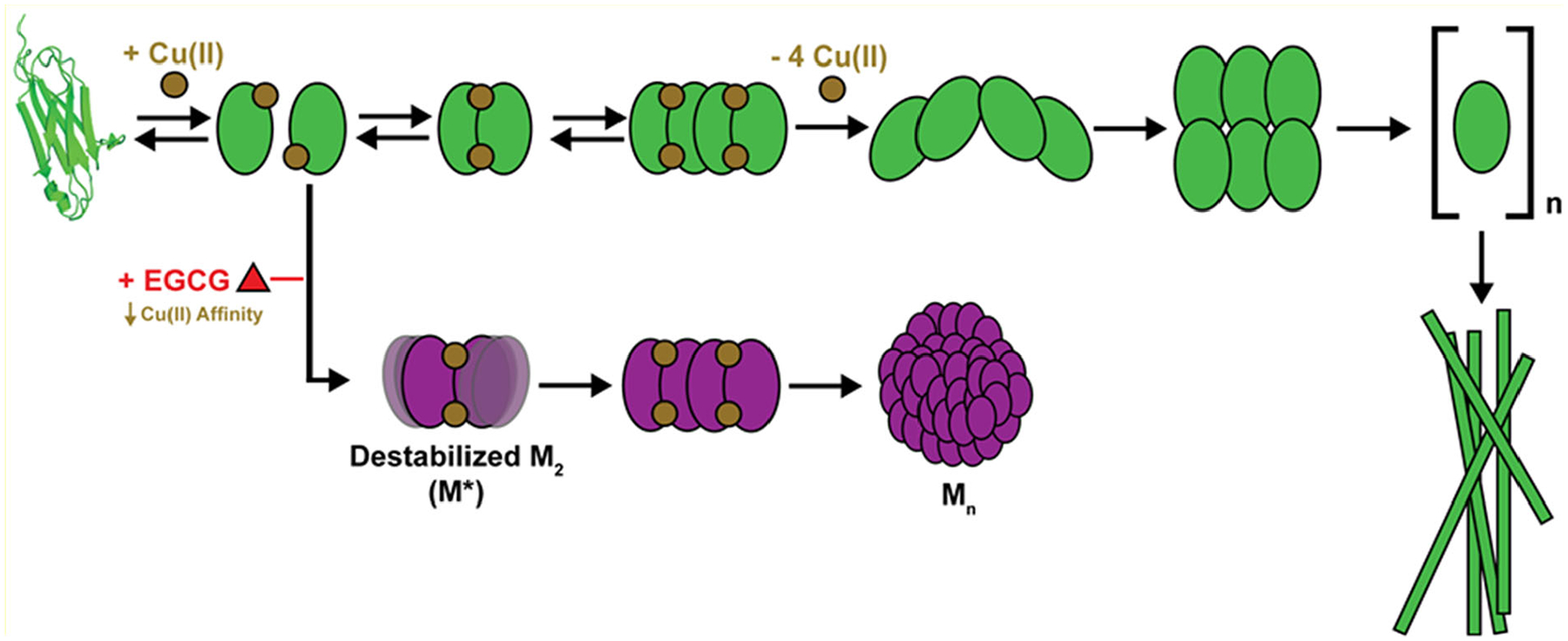
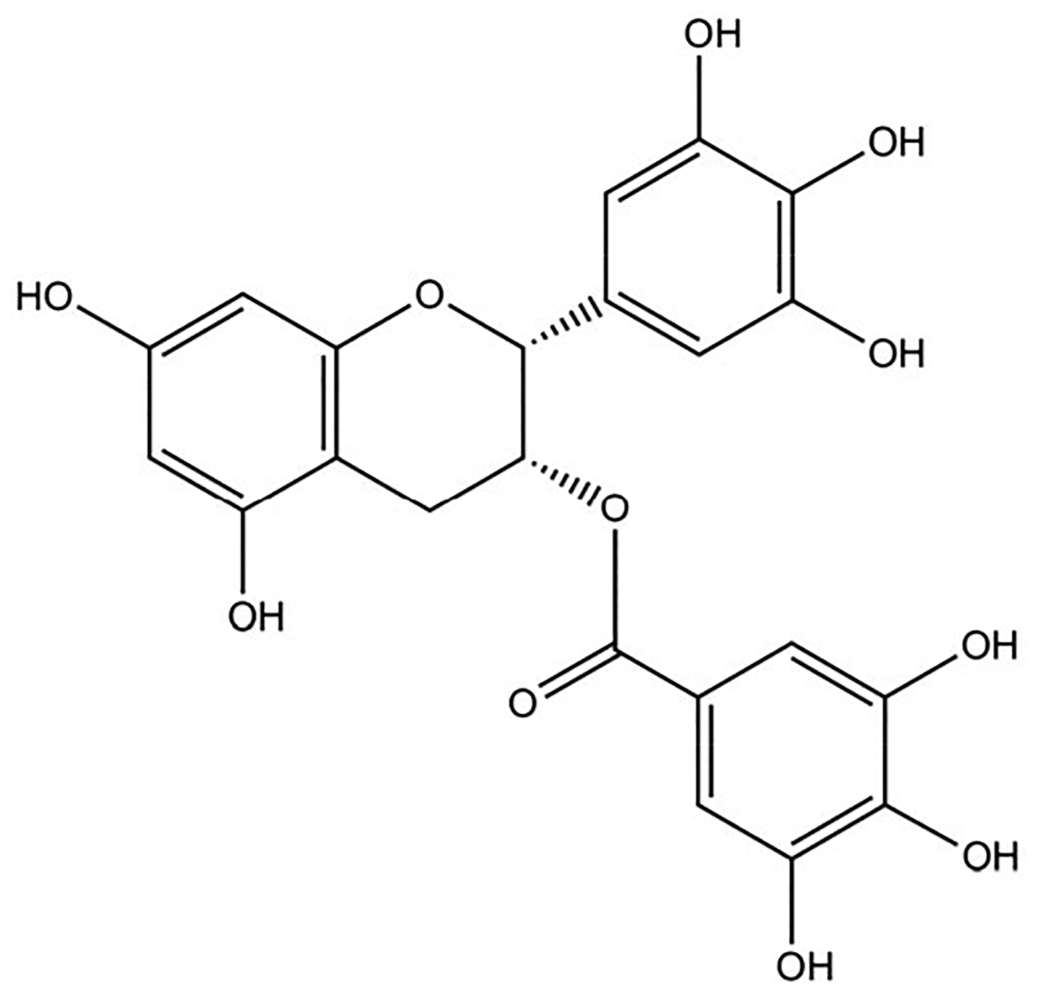
Epigallocatechin-3-gallate (EGCG) is a catechin found in green tea that can inhibit the amyloid formation of a wide variety of proteins. EGCG's ability to prevent or redirect the amyloid formation of so many proteins may reflect a common mechanism of action, and thus, greater molecular-level insight into how it exerts its effect could have broad implications. Here, we investigate the molecular details of EGCG's inhibition of the protein β-2-microglobulin (β2m), which forms amyloids in patients undergoing long-term dialysis treatment. Using size-exclusion chromatography and a collection of mass spectrometry-based techniques, we find that EGCG prevents Cu(II)-induced β2m amyloid formation by diverting the normal progression of preamyloid oligomers toward the formation of spherical, redissolvable aggregates. EGCG exerts its effect by binding with a micromolar affinity (Kd ≈ 5 μM) to the β2m monomer on the edge of two β-sheets near the N-terminus. This interaction destabilizes the preamyloid dimer and prevents the formation of a tetramer species previously shown to be essential for Cu(II)-induced β2m amyloid formation. EGCG's binding at the edge of the β-sheets in β2m is consistent with a previous hypothesis that EGCG generally prevents amyloid formation by binding cross-β-sheet aggregation intermediates.


Figures

Figure 1.
Figure 1.
Transmission electron microscopy (TEM) images after 1 month of β2m incubation at 37°C. Panel A shows a representative image of non-EGCG treated control amyloid fibrils that are formed in the presence of Cu(II). Panel B shows a representative image of the aggregates resulting from incubation under the same conditions except with EGCG present.
Figure 2.
Figure 2.
Size exclusion chromatography (SEC) analysis of β2m oligomers over the course of 7 days, with insets showing an expanded region where oligomers typically elute. Panel A includes chromatograms from samples incubated under amyloid-forming conditions that include 1:2 β2m:Cu(II) but no EGCG. Panel B includes chromatograms incubated under the same conditions but with EGCG present at a ratio of 1:2:0.3 β2m:Cu(II):EGCG. β2m monomer is denoted as M, and the dimer, tetramer, and hexamer are indicated as M2, M4, and M6, respectively. The identities of M* and Mn are described in the text.
Figure 3.
Figure 3.
Representative native ESI-IM-MS data for β2m (A) under amyloid-forming conditions without EGCG and (B) under the same conditions with EGCG (250 μM). A 1:2 β2m:Cu(II) ratio was used in both cases, and the spectra are from samples incubated for 6 days. Panels C-E show extracted arrival time distributions and determined collision cross sections for select oligomers for the control (black) and the EGCG-containing samples (red).
Figure 4.
Figure 4.
Abundances of β2m monomer and oligomers from SEC separations in the presence of increasing concentrations of EGCG under conditions that would normally induce amyloid formation. Panels A-C show abundances of the monomer, Mn, and M*, respectively, as determined from SEC chromatographic peaks after 5 days of incubation. Panel D shows results for fitting the normalized M*/monomer absorbance ratio as function of free EGCG concentration to obtain an effective EGCG-β2m Kd. Error bars are from three replicates on different days of analysis and represent the standard deviation.
Figure 5.
Figure 5.
EGCG binding site determination via covalent labeling-mass spectrometry. Panel A shows the CL-MS experimental workflow, using DEPC as the covalent labeling reagent. Panel B shows CL-MS data, where the covalent labeling percentage has been measured for the displayed residues. Statistically significant differences in covalent labeling percentage are denoted by an asterisk (n = 3). Panel C shows a surface rendered structure of monomeric β2m, with covalently labeled residues colored (PDB: 1LDS). Residues that underwent an increase or a decrease in covalent labeling upon addition of EGCG are colored red and blue, respectively. Residues that were labeled but did not exhibit significant differences are colored in green.
Figure 6.
Figure 6.
Structures of the β2m monomer and oligomers. (A) Cartoon structure of monomeric β2m (PDB: 1LDS) with β-strand notations. (B) View of monomeric β2m A and G β-strands, with notable residues proposed to be near the EGCG binding site displayed as purple sticks, with K6 and K91 highlighted in bold. (C) Cu(II)-bound pre-amyloid dimer structural model,, with the proposed EGCG binding site on each subunit highlighted by a green oval. (D) Extended Cu(II)-free tetramer model, with the proposed EGCG binding site on each subunit highlighted by a green oval. (E) Expanded view of the central interface of the extended tetramer shown in panel D, with selected A and G β-strand residues shown as green sticks. The same residues are found on the opposing subunit, albeit they are arranged in an antiparallel fashion.
Figure 7.
Figure 7.
Proposed model for EGCG inhibition of Cu(II)-catalyzed amyloid formation with β2m. Under amyloid-forming conditions, Cu(II) binding initiates the formation of dimers and tetramers before being released to allow the formation of a Cu(II)-free tetramer, a Cu(II)-free hexamer, and Cu(II)-free amyloid fibrils (top pathway in green). With EGCG present, the initially formed dimer is destabilized and the extended Cu(II)-free tetramer is not formed, leading to the formation of a large aggregate, depicted as Mn without Cu(II) for simplicity (bottom purple pathway).
Scheme 1:
Scheme 1:
Chemical structure of Epigallocatechin-3-gallate (EGCG).

Similar articles

Cited by

References

    1. Ozdal T, Capanoglu E, and Altay F (2013) A review on protein–phenolic interactions and associated changes. Food Res. Int 51, 954–970.
    1. Tanaka T, Ishii T, Mizuno D, Mori T, Yamaji R, Nakamura Y, Kumazawa S, Nakayama T, and Akagawa M (2011) (−)-Epigallocatechin-3-gallate suppresses growth of AZ521 human gastric cancer cells by targeting the DEAD-box RNA helicase p68. Free Radic. Biol. Med 50, 1324–1335. - PubMed
    1. Tachibana H, Koga K, Fujimura Y, and Yamada K (2004) A receptor for green tea polyphenol EGCG. Nat. Struct. Mol. Biol 11, 380–381. - PubMed
    1. Andrich K, and Bieschke J (2015) The Effect of (−)-Epigallo-catechin-(3)-gallate on Amyloidogenic Proteins Suggests a Common Mechanism. Adv. Exp. Med. Biol 863, 139–161. - PMC - PubMed
    1. Cheng B, Gong H, Xiao H, Petersen RB, Zheng L, and Huang K (2013) Inhibiting toxic aggregation of amyloidogenic proteins: A therapeutic strategy for protein misfolding diseases. Biochim. Biophys. Acta (BBA) - Gen. Subj 1830, 4860–4871. - PubMed

Publication types

MeSH terms