. 2020 Mar 18;12(11):12510-12524.
doi: 10.1021/acsami.9b23100. Epub 2020 Mar 9.

Boosting the Osteogenic and Angiogenic Performance of Multiscale Porous Polycaprolactone Scaffolds by In Vitro Generated Extracellular Matrix Decoration

Affiliations

Boosting the Osteogenic and Angiogenic Performance of Multiscale Porous Polycaprolactone Scaffolds by In Vitro Generated Extracellular Matrix Decoration

Betül Aldemir Dikici et al. ACS Appl Mater Interfaces. .

Abstract

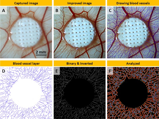
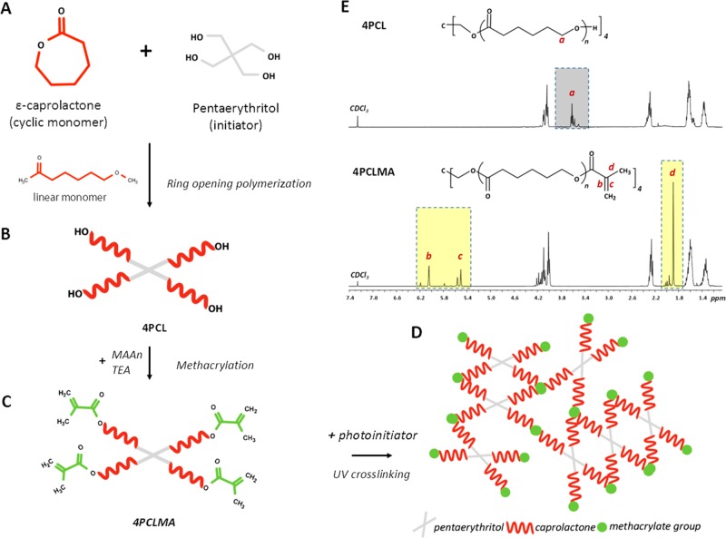
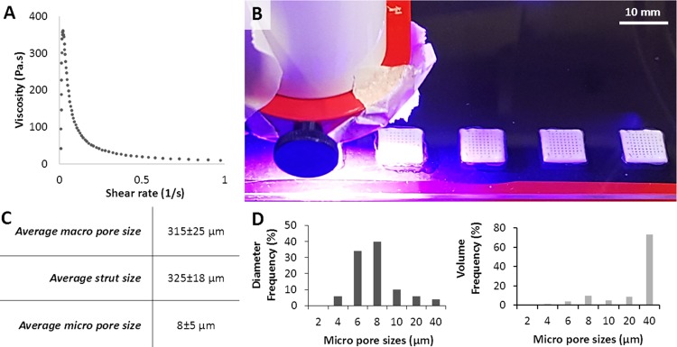
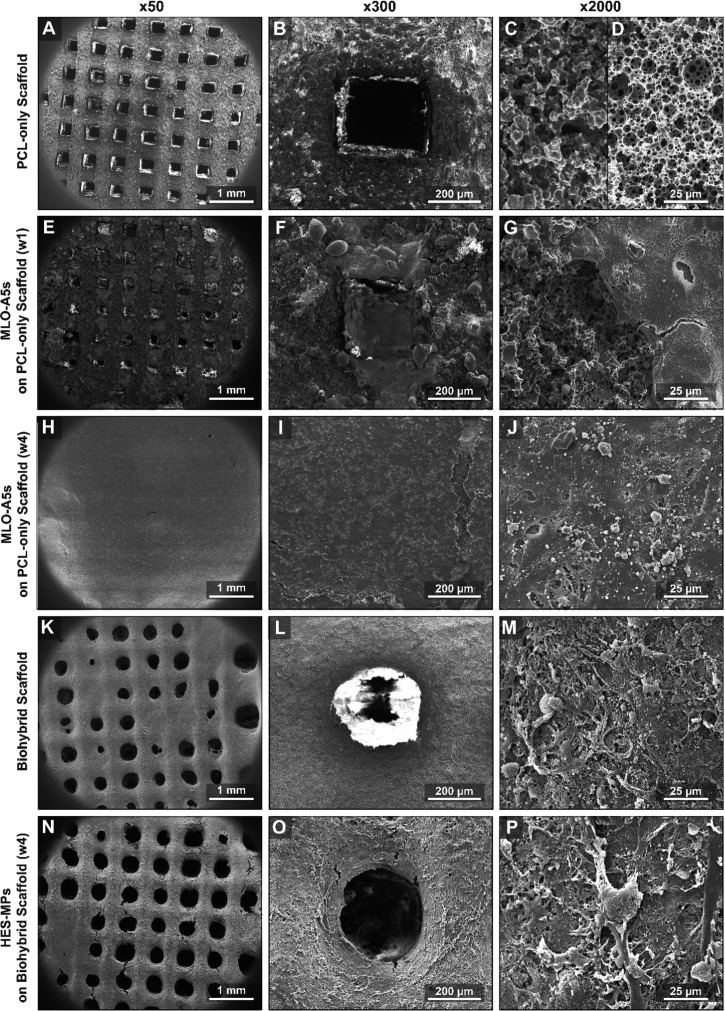
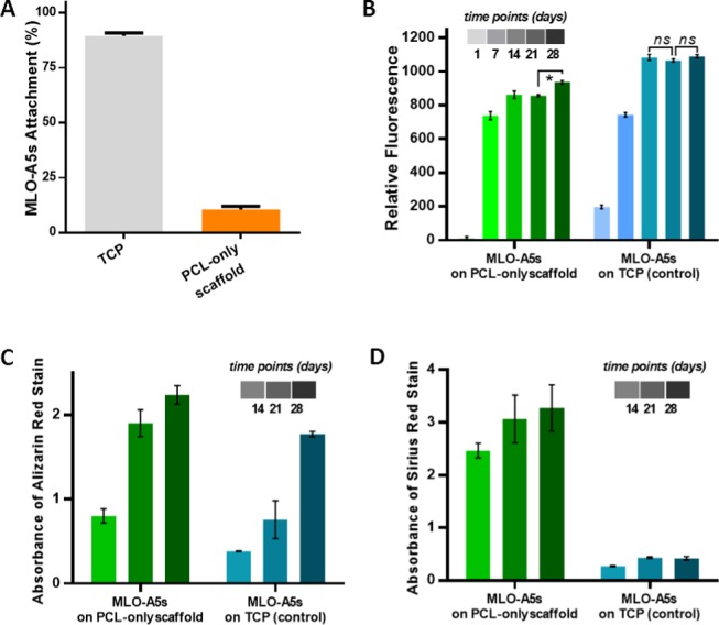
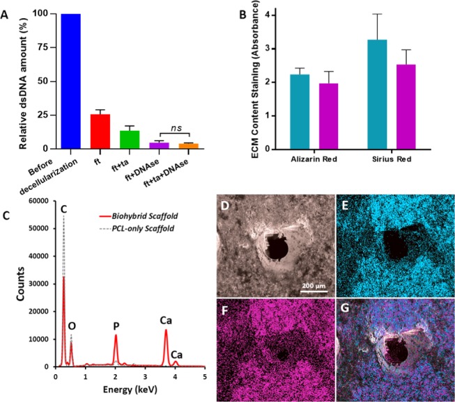
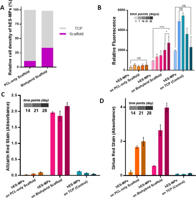
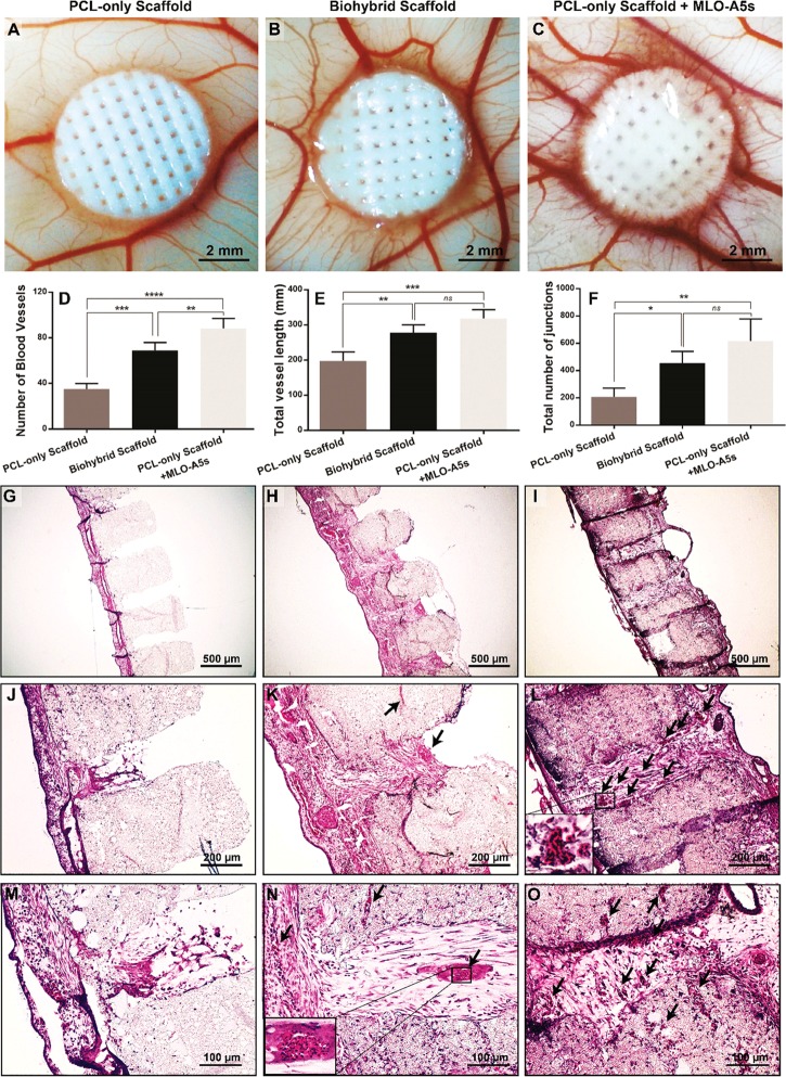
Tissue engineering (TE)-based bone grafts are favorable alternatives to autografts and allografts. Both biochemical properties and the architectural features of TE scaffolds are crucial in their design process. Synthetic polymers are attractive biomaterials to be used in the manufacturing of TE scaffolds, due to various advantages, such as being relatively inexpensive, enabling precise reproducibility, possessing tunable mechanical/chemical properties, and ease of processing. However, such scaffolds need modifications to improve their limited interaction with biological tissues. Structurally, multiscale porosity is advantageous over single-scale porosity; therefore, in this study, we have considered two key points in the design of a bone repair material; (i) manufacture of multiscale porous scaffolds made of photocurable polycaprolactone (PCL) by a combination of emulsion templating and three-dimensional (3D) printing and (ii) decoration of these scaffolds with the in vitro generated bone-like extracellular matrix (ECM) to create biohybrid scaffolds that have improved biological performance compared to PCL-only scaffolds. Multiscale porous scaffolds were fabricated, bone cells were cultured on them, and then they were decellularized. The biological performance of these constructs was tested in vitro and in vivo. Mesenchymal progenitors were seeded on PCL-only and biohybrid scaffolds. Cells not only showed improved attachment on biohybrid scaffolds but also exhibited a significantly higher rate of cell growth and osteogenic activity. The chick chorioallantoic membrane (CAM) assay was used to explore the angiogenic potential of the biohybrid scaffolds. The CAM assay indicated that the presence of the in vitro generated ECM on polymeric scaffolds resulted in higher angiogenic potential and a high degree of tissue infiltration. This study demonstrated that multiscale porous biohybrid scaffolds present a promising approach to improve bioactivity, encourage precursors to differentiate into mature bones, and to induce angiogenesis.

Keywords: 3D printing; angiogenesis; biohybrid; decellularization; emulsion templating; polyHIPE; tissue engineering.


Conflict of interest statement

The authors declare no competing financial interest.

Figures

Figure 1
Figure 1
Manufacturing routes of the multiscale porous photocurable polycaprolactone (PCL) scaffolds (steps 1, 2) and multiscale porous biohybrid scaffolds (steps 1–3). (1) Preparation of the emulsion made of photocurable PCL and water, (2) the transfer of the PCL-based high internal phase emulsion (HIPE) into the syringe, pressure-assisted 3D printing, and simultaneous cross-linking, and (3) the culture of bone cells on the PCL-only scaffold to be decellularized and generation of the biohybrid scaffolds.
Figure 2
Figure 2
Steps of the morphometric quantification of angiogenesis: (A) macroimage as captured, (B) improved image using Photoshop (PS), (C) drawn discernable blood vessels, (D) exported blood vessel layer from PS, (E) binary and inverted images in ImageJ, (F) analyzed image using Angiotool.
Figure 3
Figure 3
Synthesis scheme of four-arm photocurable polycaprolactone: (A, B) monomer and the initiator were used for the synthesis of hydroxyl-terminated four-arm polycaprolactone (4PCL). (B,C) 4PCL was methacrylated (4PCLMA). (D) Schematic demonstration of the photocured (UV-cross-linked) network showing a building block made of 4PCLMA. (E) 1H NMR spectrum of 4PCL, 4PCLMA, and relative assignments. Dark gray region: peaks of the hydroxyl group, light yellow regions: peaks of the methacrylate group, which only showed up after methacrylation reaction while they are absent in 4PCL.
Figure 4
Figure 4
(A) Viscosity of the polycaprolactone (PCL)-based high internal phase emulsion (HIPE) prepared to be used in the printing process. (B) Three-dimensional (3D) printing and simultaneous cross-linking of PCL HIPE. (C) Morphological characterization (nmacropore = 20, nstrut = 20, and nmicropore = 50) and (D) micropore size distribution of the scaffolds in terms of the diameter frequency and the volume frequency.
Figure 5
Figure 5
SEM micrographs of (A-D) multiscale porous PCL-only scaffolds immediately after manufacture, (E–G) after 1 week of MLO-A5 culture, (H–J) after 4 weeks of MLO-A5 culture, (K–M) after the decellularization process (biohybrid scaffold), (N–P) after 4 weeks of the culture of hES-MPs on the biohybrid scaffolds. First column macroview of the scaffold, the second column shows the single pore, and the third column shows the microsurface of the scaffold at different stages of the experiment.
Figure 6
Figure 6
(A) Cell seeding efficiency of MLO-A5s on multiscale porous PCL-only scaffolds (n = 5). (B) Metabolic activity (n = 5), (C) mineral, and (D) collagen deposition of MLO-A5s on multiscale porous PCL-only scaffolds and TCP as control over 28 days (n = 3, *: p < 0.05, ns: not significant, p > 0.05).
Figure 7
Figure 7
(A) Comparison of the various decellularization techniques in terms of remaining DNA content (n = 3), (B) Calcium and collagen content of the scaffolds cultured with MLOs for 4 weeks (blue) and scaffolds that are decellularized (purple) (n = 3, ns: not significant, p > 0.05), (C) EDX spectrum of the decellularized scaffold showing the peaks of carbon (C), phosphorus (P), calcium (Ca), and oxygen (O), (D) SEM image of the decellularized scaffold, (E–G) EDX elemental mapping of Ca (blue), P (pink), and merged mapping (Ca and P), respectively.
Figure 8
Figure 8
(A) Seeding efficiencies of human embryonic stem cell-derived mesenchymal progenitor cells (hES-MPs) on polycaprolactone (PCL)-only and biohybrid scaffolds (n = 6), (B) the metabolic activity (n = 6), (C) mineral (n = 3), and (D) collagen deposition of hES-MPs on PCL-only, biohybrid scaffolds, and on the tissue culture plate (TCP) as a control in 28 days culture (n = 3, *: p < 0.05, ****: p < 0.001, ns: not significant, p > 0.05).
Figure 9
Figure 9
Evaluation of the angiogenic potential of polycaprolactone (PCL)-only, PCL-only populated with murine long bone osteocyte cells (MLO-A5s), and biohybrid scaffolds using chick chorioallantoic membrane (CAM) assay. (A–C) Macroimages were taken on embryonic development day 14, (D–F) quantification of the number of blood vessels, total vessel length, and the total number of junctions. (n = 4, *: p < 0.05, **: p < 0.01, ***: p < 0.005, ****: p < 0.001, ns: not significant, p > 0.05.) (G–O) Histological evaluation of the scaffolds isolated from CAM. (Black arrows indicate the blood vessels).

Similar articles

Cited by

References

    1. Campana V.; Milano G.; Pagano E.; Barba M.; Cicione C.; Salonna G.; Lattanzi W.; Logroscino G. Bone Substitutes in Orthopaedic Surgery: From Basic Science to Clinical Practice. J. Mater. Sci. Mater. Med. 2014, 25, 2445–2461. 10.1007/s10856-014-5240-2. - DOI - PMC - PubMed
    1. Pham Q. P.; Kasper F. K.; Mistry A. S.; Sharma U.; Yasko A. W.; Jansen J. A.; Mikos A. G. Analysis of the Osteoinductive Capacity and Angiogenicity of an in Vitro Generated Extracellular Matrix. J. Biomed. Mater. Res., Part A 2009, 88A, 295–303. 10.1002/jbm.a.31875. - DOI - PubMed
    1. Lichte P.; Pape H. C.; Pufe T.; Kobbe P.; Fischer H. Scaffolds for Bone Healing: Concepts, Materials and Evidence. Injury 2011, 42, 569–573. 10.1016/j.injury.2011.03.033. - DOI - PubMed
    1. Khan W. S.; Rayan F.; Dhinsa B. S.; Marsh D. An Osteoconductive, Osteoinductive, and Osteogenic Tissue-Engineered Product for Trauma and Orthopaedic Surgery: How Far Are We?. Stem Cells Int. 2012, 2012, 23623110.1155/2012/236231. - DOI - PMC - PubMed
    1. Van Der Stok J.; Hartholt K. A.; Schoenmakers D. A. L.; Arts J. J. C. The Available Evidence on Demineralised Bone Matrix in Trauma and Orthopaedic Surgery. Bone Jt. Res. 2017, 6, 423–432. 10.1302/2046-3758.67.BJR-2017-0027.R1. - DOI - PMC - PubMed

MeSH terms