. 2020 May;101(5):467-472.
doi: 10.1099/jgv.0.001399. Epub 2020 Feb 25.

Parainfluenza virus 5 fusion protein maintains pre-fusion stability but not fusogenic activity following mutation of a transmembrane leucine/isoleucine domain

Affiliations

Parainfluenza virus 5 fusion protein maintains pre-fusion stability but not fusogenic activity following mutation of a transmembrane leucine/isoleucine domain

Jean Mawuena Branttie et al. J Gen Virol. 2020 May.

Abstract

The paramyxoviruses Hendra virus (HeV) and parainfluenza virus 5 (PIV5) require the fusion (F) protein to efficiently infect cells. For fusion to occur, F undergoes dramatic, essentially irreversible conformational changes to merge the viral and cell membranes into a continuous bilayer. Recently, a transmembrane (TM) domain leucine/isoleucine (L/I) zipper was shown to be critical in maintaining the expression, stability and pre-fusion conformation of HeV F, allowing for fine-tuned timing of membrane fusion. To analyse the effect of the TM domain L/I zipper in another paramyxovirus, we created alanine mutations to the TM domain of PIV5 F, a paramyxovirus model system. Our data show that while the PIV5 F TM L/I zipper does not significantly affect total expression and only modestly affects surface expression and pre-fusion stability, it is critical for fusogenic activity. These results suggest that the roles of TM L/I zipper motifs differ among members of the family Paramyxoviridae.

Keywords: fusogenic activity; leucine/isoleucine zipper; pre-fusion stability; transmembrane domain.


Conflict of interest statement

The authors declare that there are no conflicts of interest.

Figures
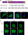
Fig. 1.
Fig. 1.
Mutations to the L/I zipper of HeV F and PIV5 F have variable effects. (a). Schematic of the paramyxovirus fusion protein highlighting the TM domain L/I zipper of HeV F and PIV5 F, and the mutant constructs. FP, fusion peptide; HRA, heptad repeat A; HRB, heptad repeat B; TMD, transmembrane domain; CT, cytoplasmic tail); S–S, disulfide bond. (b). Immunofluorescence to visualize localization of HeV and PIV5 F proteins. Vero cells were seeded in eight-well chamber plates and transfected with 0.75 µg PIV5 F WT or LIZ mutant (left), and HeV F WT or LIZ mutant (right). Localization of HeV F was analysed with anti-F 5G7 antibodies, and PIV5 F analysed with mAb F1a (green). Images were taken with a Nikon 1A confocal microscope. Images are representative. Scale bars represent 10 µm. (c). Z-stack images from (b) were collected in 0.3 µm sections, and images corresponding to top, bottom and middle slices are shown. Images are representative of two independent experiments carried out in triplicate. Scale bars represent 10 µm.
Fig. 2.
Fig. 2.
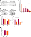
Expression and stability of PIV5 F WT and LIZ are comparable. (a). Assessment of stability of PIV5 F WT or LIZ over time course. A pulse-chase experiment was carried out 18 h after cells were transfected with 2.5 µg of indicated DNA for Vero cells in six-well plates. Following a 30 minute S35 metabolic radiolabel, samples were chased for indicated times. (b). Quantitation of PIV5 F and LIZ expression shown in (a). Expression levels of total F protein (F0+F1) were determined by band densitometry normalized to WT levels. (c). Percentage of cleaved F compared to total F (F1/F0+F1) normalized to WT. The averages represent three independent experiments, each carried out in duplicate. (d). Surface and total expression of PIV5 F protein. Quantitation of expression levels (e) and percentage cleavage (f) of surface and total PIV5 F protein. (g). Flow cytometry to quantify expression of pre-fusion PIV5 F only present at the surface of cells. The averages represent three independent experiments, each carried out in duplicate. The LIZ mutant was compared to WT using Student’s t-test. *, P<0.05; **, P<0.005; ****P<0.0001
Fig. 3.
Fig. 3.
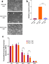
Mutations to the L/I zipper of PIV5 reduce F-mediated fusion activity. (a). Syncytia assay. BHK cells plated in six-well plates were transfected with 2.5 µg of total DNA with the PIV5 HN attachment protein alone, PIV5 WT F and HN or PIV5 LIZ F and HN. Syncytia formation was analysed 24 h post-transfection. Images were taken with a Nikon TS100 microscope. White arrows indicate syncytia. Images are representative of two independent experiments, each carried out in triplicate. (b). Luciferase reporter gene assay to quantify F fusogenic activity. Vero cells in 24-well plates were transfected with 1.0 µg total DNA with a T7 promoter plasmid and PIV5 F WT+HN or pIV5 F LIZ+HN. The following day, Vero cells were overlaid with BSR cells and incubated for 3 h to allow for luciferase production. Luciferase activity was measured using a luciferase assay system. The average represents three independent experiments, each performed in duplicate. (c). Thermal triggering assay to observe PIV5 F WT and LIZ pre-fusion thermostability. Cells expressing surface PIV5 F or WT were exposed to 4, 37, 55, 60 or 65 °C for 15 min. Cells were immediately placed on ice for 15 min and prepared for flow cytometry using PIV5 mAb F1a. The average represents two independent experiments, each performed in triplicate. The LIZ mutant was compared to WT using a using Student’s t-test. ****P<0.0001

Similar articles

Cited by

References

    1. Rima B, Collins P, Easton A, Fouchier R, Kurath G, et al. Problems of classification in the family Paramyxoviridae. Arch Virol. 2018;163:1395–1404. doi: 10.1007/s00705-018-3720-2. - DOI - PMC - PubMed
    1. Cox RM, Plemper RK. Structure and organization of paramyxovirus particles. Curr Opin Virol. 2017;24:105–114. doi: 10.1016/j.coviro.2017.05.004. - DOI - PMC - PubMed
    1. Harrison MS, Sakaguchi T, Schmitt AP. Paramyxovirus assembly and budding: building particles that transmit infections. Int J Biochem Cell Biol. 2010;42:1416–1429. doi: 10.1016/j.biocel.2010.04.005. - DOI - PMC - PubMed
    1. Smith EC, Smith SE, Carter JR, Webb SR, Gibson KM, et al. Trimeric transmembrane domain interactions in paramyxovirus fusion proteins: roles in protein folding, stability, and function. J Biol Chem. 2013;288:35726–35735. doi: 10.1074/jbc.M113.514554. - DOI - PMC - PubMed
    1. Aguilar HC, Henderson BA, Zamora JL, Johnston GP. Paramyxovirus glycoproteins and the membrane fusion process. Curr Clin Microbiol Rep. 2016;3:142–154. doi: 10.1007/s40588-016-0040-8. - DOI - PMC - PubMed

Publication types

LinkOut - more resources