Case Reports
. 2020 Apr;8(4):e1178.
doi: 10.1002/mgg3.1178. Epub 2020 Feb 26.

Expanding the spectrum of SMAD3-related phenotypes to agnathia-otocephaly

Affiliations
Case Reports

Expanding the spectrum of SMAD3-related phenotypes to agnathia-otocephaly

Nicole Meier et al. Mol Genet Genomic Med. 2020 Apr.

Abstract

Background: Agnathia-otocephaly is a rare and lethal anomaly affecting craniofacial structures derived from the first pharyngeal arch. It is characterized by agnathia, microstomia, aglossia, and abnormally positioned auricles with or without associated anomalies. Variants affecting function of OTX2 and PRRX1, which together regulate the neural crest cells and the patterning of the first pharyngeal arch as well as skeletal and limb development, were identified to be causal for the anomaly in a few patients.

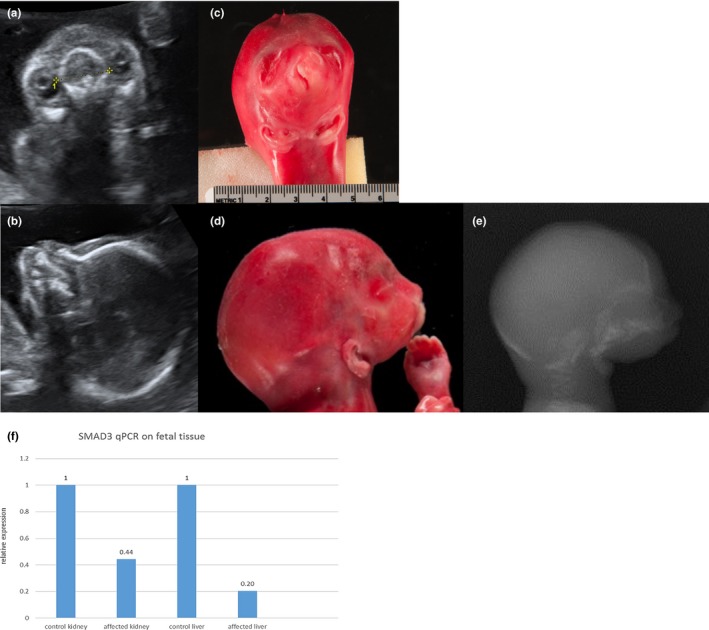
Methods: Family-based exome sequencing (ES) on a fetus with severe agnathia-otocephaly, cheilognathopalatoschisis, laryngeal hypoplasia, fused lung lobes and other organ abnormalities and mRNA expression analysis were performed.

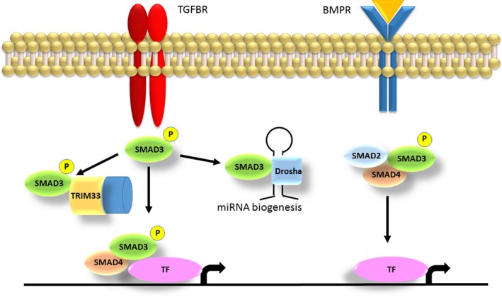
Results: Exome sequencing detected a de novo SMAD3 missense variant in exon 6 (c.860G>A) associated with decreased mRNA expression. Variants in SMAD3 cause Loeys-Dietz syndrome 3 presenting with craniofacial anomalies such as mandibular hypoplasia, micro- or retro-gnathia, bifid uvula and cleft palate as well as skeletal anomalies and arterial tortuosity. The SMAD3 protein acts as a transcriptional regulator in the transforming growth factor β (TGFB) and bone morphogenetic (BMP) signaling pathways, which play a key role in the development of craniofacial structures originating from the pharyngeal arches.

Conclusion: Agnathia-otocephaly with or without associated anomalies may represent the severe end of a phenotypic spectrum related to variants in genes in the interacting SMAD/TGFB/BMP/SHH/FGF developmental pathways.

Keywords: SMAD3; agnathia-otocephaly; exome sequencing; prenatal.


Conflict of interest statement

The authors declare no conflict of interest.

Figures

Figure 1
Figure 1
(a–e) The affected fetus at 19 + 2 weeks of gestation in prenatal ultrasound (a and b) and post mortem (c–e) showing cheilognathopalatoschisis, microstomia, absent mandible and ventral median positioned auricles. (f) Relative SMAD3 mRNA expression in kidney and liver tissue of an age‐matched control fetus and the affected fetus (f). RNA was extracted from FFPE tissue, the expression of SMAD3 was normalized to GUSB expression. FFPE, formalin fixed paraffin embedded
Figure 2
Figure 2
Roles of SMAD3 in the TGF‐β and the BMP pathway. The SMAD3 protein gets activated through TGF‐β signaling and can act in different downstream pathways. If SMAD3 binds to TRIM33, a chromatin reading and remodeling protein, the complex opens the histone to allow other TFs to enter the DNA. Complexes of SMAD3 and SMAD4 get recruited to different genes, and the transcription of a specific gene is determined by varying partner TFs. In those effector pathways SMAD3 is involved in chondrocyte and osteoblast maturation (Massagué, 2012). If SMAD3 binds to DROSHA it regulates the processing of different miRNA precursors. SMAD3 forming a complex with other SMAD proteins driven by BMP signaling also activates transcription via other TFs, regulating various bone formation processes (Rahman et al., 2015). TGF‐β, transforming growth factor‐β; TFs, transcription factors

Similar articles

  • Re-focusing on Agnathia-Otocephaly complex.
    Dubucs C, Chassaing N, Sergi C, Aubert-Mucca M, Attié-Bitach T, Lacombe D, Thauvin-Robinet C, Arpin S, Perez MJ, Cabrol C, Chen CP, Aziza J, Colin E, Martinovic J, Calvas P, Plaisancié J. Dubucs C, et al. Clin Oral Investig. 2021 Mar;25(3):1353-1362. doi: 10.1007/s00784-020-03443-w. Epub 2020 Jul 9. Clin Oral Investig. 2021. PMID: 32643087 Review.
  • Prenatal Diagnosis of Otocephaly: A Rare Facial Anomaly.
    Goenka S, Sahithi K, Ratha C. Goenka S, et al. J Obstet Gynaecol India. 2022 Aug;72(4):364-368. doi: 10.1007/s13224-021-01494-x. Epub 2021 Jul 7. J Obstet Gynaecol India. 2022. PMID: 35923505 Free PMC article.
  • A de novo variant in OTX2 in a lamb with otocephaly.
    Paris JM, Letko A, Häfliger IM, Švara T, Gombač M, Klinc P, Škibin A, Pogorevc E, Drögemüller C. Paris JM, et al. Acta Vet Scand. 2020 Jan 22;62(1):5. doi: 10.1186/s13028-020-0503-z. Acta Vet Scand. 2020. PMID: 31969185 Free PMC article.
  • Agnathia-Otocephaly Complex Due to a De Novo Deletion in the OTX2 Gene.
    Fabiani M, Libotte F, Margiotti K, Tannous DKI, Sparacino D, D'Aleo MP, Monaco F, Dello Russo C, Mesoraca A, Giorlandino C. Fabiani M, et al. Genes (Basel). 2022 Dec 2;13(12):2269. doi: 10.3390/genes13122269. Genes (Basel). 2022. PMID: 36553536 Free PMC article.
  • Current perspectives on the etiology of agnathia-otocephaly.
    Gekas J, Li B, Kamnasaran D. Gekas J, et al. Eur J Med Genet. 2010 Nov-Dec;53(6):358-66. doi: 10.1016/j.ejmg.2010.09.002. Epub 2010 Sep 16. Eur J Med Genet. 2010. PMID: 20849990 Review.

References

    1. Aubart, M. , Gobert, D. , Aubart‐Cohen, F. , Detaint, D. , Hanna, N. , d’Indya, H. , … Jondeau, G. (2014). Early‐onset osteoarthritis, Charcot‐Marie‐Tooth like neuropathy, autoimmune features, multiple arterial aneurysms and dissections: An unrecognized and life threatening condition. PLoS ONE, 9(5), e96387 10.1371/journal.pone.0096387 - DOI - PMC - PubMed
    1. Bakrania, P. , Efthymiou, M. , Klein, J. C. , Salt, A. , Bunyan, D. J. , Wyatt, A. , … Ragge, N. K. (2008). Mutations in BMP4 cause eye, brain, and digit developmental anomalies: Overlap between the BMP4 and hedgehog signaling pathways. The American Journal of Human Genetics, 82(2), 304–319. 10.1016/J.AJHG.2007.09.023 - DOI - PMC - PubMed
    1. Casari, A. , Schiavone, M. , Facchinello, N. , Vettori, A. , Meyer, D. , Tiso, N. , … Argenton, F. (2014). A Smad3 transgenic reporter reveals TGF‐beta control of zebrafish spinal cord development. Developmental Biology, 396(1), 81–93. 10.1016/j.ydbio.2014.09.025 - DOI - PubMed
    1. Çelik, T. , Simsek, P. O. , Sozen, T. , Ozyuncu, O. , Utine, G. E. , Talim, B. , … Kamnasaran, D. (2012). PRRX1 is mutated in an otocephalic newborn infant conceived by consanguineous parents. Clinical Genetics, 81(3), 294–297. 10.1111/j.1399-0004.2011.01730.x - DOI - PubMed
    1. Dudas, M. , Sridurongrit, S. , Nagy, A. , Okazaki, K. , & Kaartinen, V. (2004). Craniofacial defects in mice lacking BMP type I receptor Alk2 in neural crest cells. Mechanisms of Development, 121(2), 173–182. 10.1016/j.mod.2003.12.003 - DOI - PubMed

Publication types

Supplementary concepts