. 2020 Feb 26;18(1):19.
doi: 10.1186/s12915-020-0749-y.

Ca2+ mobilization-dependent reduction of the endoplasmic reticulum lumen is due to influx of cytosolic glutathione

Affiliations

Ca2+ mobilization-dependent reduction of the endoplasmic reticulum lumen is due to influx of cytosolic glutathione

Beáta Lizák et al. BMC Biol. .

Abstract

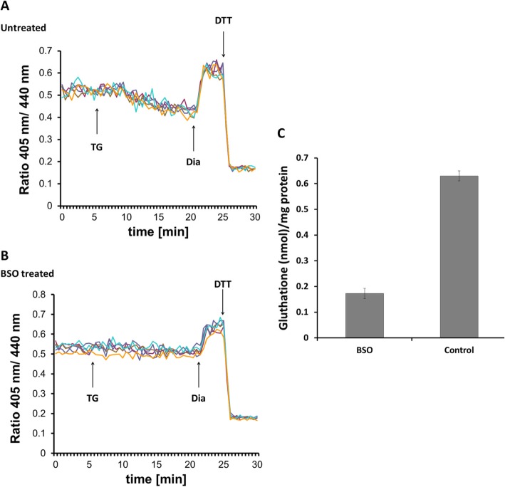
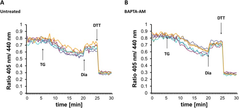
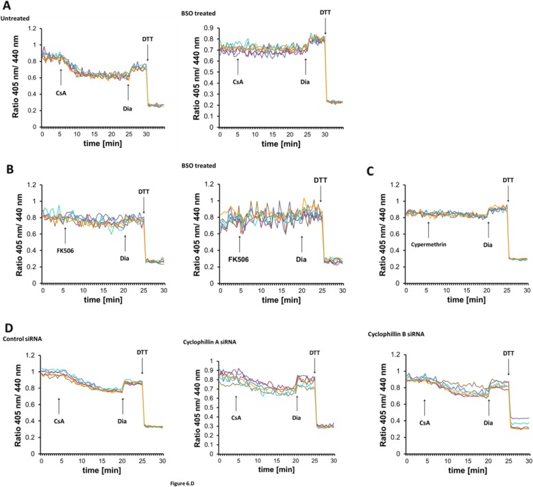
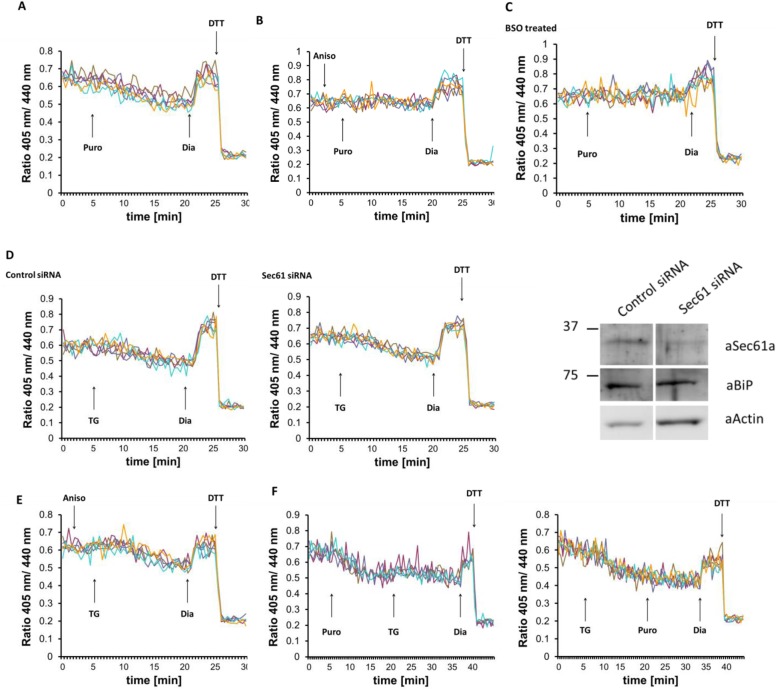
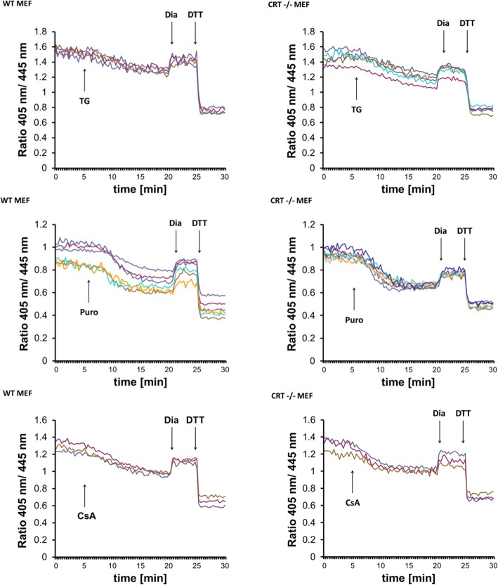
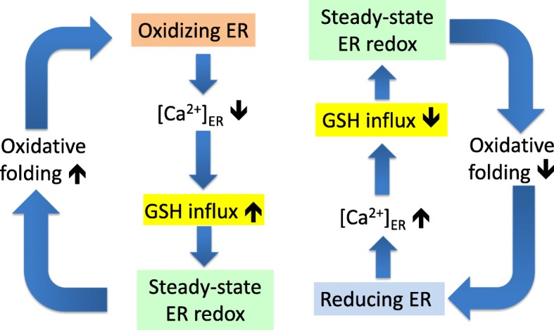
Background: The lumen of the endoplasmic reticulum (ER) acts as a cellular Ca2+ store and a site for oxidative protein folding, which is controlled by the reduced glutathione (GSH) and glutathione-disulfide (GSSG) redox pair. Although depletion of luminal Ca2+ from the ER provokes a rapid and reversible shift towards a more reducing poise in the ER, the underlying molecular basis remains unclear.

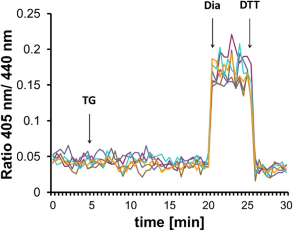
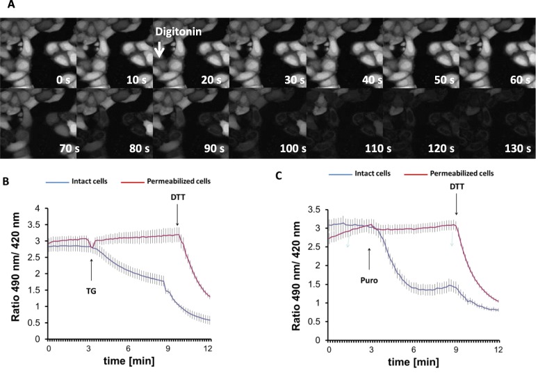
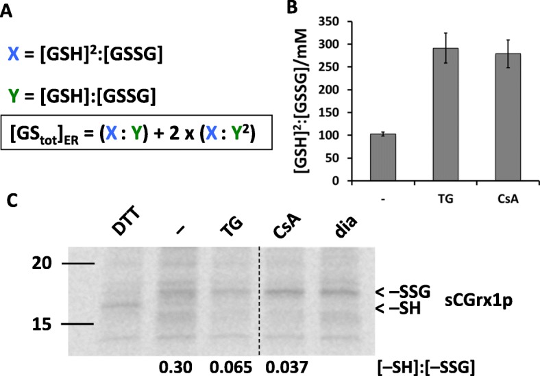
Results: We found that Ca2+ mobilization-dependent ER luminal reduction was sensitive to inhibition of GSH synthesis or dilution of cytosolic GSH by selective permeabilization of the plasma membrane. A glutathione-centered mechanism was further indicated by increased ER luminal glutathione levels in response to Ca2+ efflux. Inducible reduction of the ER lumen by GSH flux was independent of the Ca2+-binding chaperone calreticulin, which has previously been implicated in this process. However, opening the translocon channel by puromycin or addition of cyclosporine A mimicked the GSH-related effect of Ca2+ mobilization. While the action of puromycin was ascribable to Ca2+ leakage from the ER, the mechanism of cyclosporine A-induced GSH flux was independent of calcineurin and cyclophilins A and B and remained unclear.

Conclusions: Our data strongly suggest that ER influx of cytosolic GSH, rather than inhibition of local oxidoreductases, is responsible for the reductive shift upon Ca2+ mobilization. We postulate the existence of a Ca2+- and cyclosporine A-sensitive GSH transporter in the ER membrane. These findings have important implications for ER redox homeostasis under normal physiology and ER stress.

Keywords: Calcium; Calreticulin; Cyclophilins; Cyclosporine A; Endoplasmic reticulum; Endoplasmic reticulum stress; Glutathione; Membrane transport proteins; Redox homeostasis; Sec61 translocon.


Conflict of interest statement

The authors declare that they have no competing interests.

Figures

Fig. 1
Fig. 1
Ca2+ depletion-triggered ER reduction is sensitive to glutathione depletion by BSO. HEK293 cells were stably transfected with Grx1-roGFP1-iEER constructs and subjected to ratiometric laser scanning microscopy on a temperature-controlled stage with CO2 control. Fluorescence ratio changes were monitored over time. Each trace corresponds to the data recorded from one cell; traces were obtained from two independent experiments. One micromolar TG were applied to untreated (a) or BSO-treated (b) cells as indicated by the arrow. At the end of each experiment, 500 μM diamide (Dia) and 20 mM DTT were added to ensure the functionality of the probe. c Determination of total glutathione concentration by glutathione reductase assay as described in the “Materials and methods” section. One millimolar BSO treatment was performed overnight prior to experiment
Fig. 2
Fig. 2
Cytosolic redox sensor Grx1-roGFP2 is not detectably disturbed upon thapsigargin-induced Ca2+ release. Fluorescence ratio changes of cytosolic Grx1-roGFP2 transiently expressed in HEK293. Traces correspond to data recorded from one cell; traces were obtained from two independent experiments. Cells were pretreated for 3 h before imaging with 100 μM of the GR inhibitor carmustine (BCNU) to prevent GSSG re-reduction. One micromolar TG were applied to cells as indicated by the arrow. At the end of the experiment, 500 μM diamide (Dia) and 20 mM DTT were added to ensure the functionality of the probe
Fig. 3
Fig. 3
Permeabilization of the plasma membrane prevents thapsigargin-induced ER lumen reduction. a Sequential images of digitonin (25 μg/ml)-treated HeLa cells loaded with BCECF-AM fluorescent dye. b, c Fluorescence ratio changes of HyPer-ER sensor 24 h after transfection in digitonin-permeabilized (red line) or intact (blue line) HeLa cells. Cells were pretreated with digitonin for 3 min and washed with intracellular medium as described in the “Materials and methods” section prior to the experiment. TG (200 nM, b) or puromycin (100 μM, c) were applied at 3 min of imaging as indicated by the arrow. Experiments were terminated by addition of 0.5 mM DTT. Traces represent average intensity ratios acquired from 14 to 34 cells of 4 independent experiments
Fig. 4
Fig. 4
Thapsigargin and cyclosporine A increase glutathione levels in the ER lumen. a Formula for the calculation of [GStot]ER from [GSH]2:[GSSG] and [GSH]:[GSSG] in the ER. b [GSH]2:[GSSG] was quantified in the ER of Grx1-roGFP1-iEER-expressing HEK293 cells that were left untreated (−) or treated with TG or CsA for 15 min by measuring the ratiometric emission intensity values of Grx1-roGFP1-iEER at steady state, fully oxidized, and fully reduced conditions. c sCGrx1pER-transfected HEK293 cells were left untreated (−) or treated with TG or CsA for 15 min. Glutationylation state ([−SH]:[−SSG]) of sCGrx1p was determined by immunoprecipitation and TMMPEG modification of the radiolabelled protein. [−SH]:[−SSG] was quantified by SDS-PAGE, phosphor imaging, and densitometric analysis. Samples obtained from cells that were treated with 10 mM DTT or 5 mM diamide (dia) served as mobility markers for −SH and −SSG, respectively. The vertical dashed line indicates where an intervening lane has been removed. Note that [GSH]:[GSSG] is directly proportional to [−SH]:[−SSG]. One of three representative experiments is shown
Fig. 5
Fig. 5
Chelation of cytosolic Ca2+ does not inhibit glutathione transport. Effect of 1 μM TG on the fluorescence ratio changes of Grx1-roGFP1-iEER in HEK293 cells left untreated (a) or pretreated with the Ca2+ chelator BAPTA-AM (b). Each trace corresponds to the data recorded from one cell
Fig. 6
Fig. 6
GSH transport can be triggered by cyclosporine A. a Real-time fluorescence ratio changes of Grx1-roGFP1-iEER in response to 10 micromolar CsA in HEK293 cells stably expressing the sensor. Each trace corresponds to the data recorded from one cell; traces were obtained from two independent experiments. At the end of each experiment, 500 μM diamide (Dia) and 20 mM DTT were added to ensure the functionality of the probe. Cells were left untreated or treated overnight with 1 mM BSO prior to the experiment. b, c Experiment performed as in a, but 50 μM FK506 (b) or 10 μM cyphermethrin (c) was applied as marked instead of CsA. d HEK293 cells stably expressing Grx1-roGFP1-iEER were transfected with control, cyclophillin A or B siRNA for 48 h before imaging; 10 μM CsA were applied as indicated by the arrow. Knockdown efficiency was verified by qPCR
Fig. 7
Fig. 7
The Sec61 translocon polypeptide channel does not participate in glutathione transport. Effects of manipulating the translocon on fluorescence ratio changes of Grx1-roGFP1-iEER in HEK293 cells stably expressing the sensor. Each trace corresponds to the data recorded from one cell. At the end of each experiment, 500 μM diamide (Dia) and 20 mM DTT were added to ensure the functionality of the probe. a One hundred micromolar puromycin, b 200 μM anisomycin followed by 100 μM puromycin, e 200 μM anisomycin followed by 1 μM TG, and f 100 μM puromycin followed by 1 μM TG were applied as indicated by the arrow. c Cells were treated overnight with 1 mM BSO prior to experiment, and 100 μM puromycin were applied as marked. d HEK293 cells stably expressing Grx1-roGFP1-iEER were transfected with control or Sec61 siRNA for 48 h before imaging as above; 1 μM TG were applied as indicated by the arrow. Knockdown efficiency was verified by Western blot (aSec61a, anti-Sec61α antibody; aBiP, anti-BiP antibody; aActin, anti-actin antibody)
Fig. 8
Fig. 8
Calreticulin is dispensable for ER reduction induced by Ca2+ depletion or Cyclosporin A. Wild-type and CRT −/− mouse embryonic fibroblasts were transfected with Grx1-roGFP1-iEER, and real-time fluorescent ratio changes were monitored. Reductive shift-provoking agents were applied as indicated. Each trace represents the data recorded from one cell; traces shown are representative of three independent experiments
Fig. 9
Fig. 9
Schematic representation of feedback loops that connect ER Ca2+ loading, GSH influx, and oxidative protein folding. Hyper-oxidizing conditions in the ER (orange box) due to peak oxidative protein folding leads to Ca2+ depletion via opening of IP3R calcium channels and inhibition of SERCA pumps. Ca2+ depletion can in turn activate a GSH transporter (yellow box), which will restore the proper steady-state ER redox environment (green box). Conversely, hyper-reducing conditions in the ER (blue box) lower GSH influx via increased [Ca2+]ER, thereby rescuing steady-state ER redox and commensurate oxidative protein folding. These feedback mechanisms regulate the pace of oxidative protein folding and contribute to the robustness of ER luminal redox balance

Similar articles

Cited by

References

    1. Braakman I, Bulleid NJ. Protein folding and modification in the mammalian endoplasmic reticulum. Annu Rev Biochem. 2011;80:71–99. doi: 10.1146/annurev-biochem-062209-093836. - DOI - PubMed
    1. Csala M, Kereszturi E, Mandl J, Banhegyi G. The endoplasmic reticulum as the extracellular space inside the cell: role in protein folding and glycosylation. Antioxid Redox Signal. 2012;16(10):1100–1108. doi: 10.1089/ars.2011.4227. - DOI - PubMed
    1. Margittai E, Sitia R. Oxidative protein folding in the secretory pathway and redox signaling across compartments and cells. Traffic. 2011;12(1):1–8. doi: 10.1111/j.1600-0854.2010.01108.x. - DOI - PubMed
    1. Hetz C, Papa FR. The unfolded protein response and cell fate control. Mol Cell. 2018;69(2):169–181. doi: 10.1016/j.molcel.2017.06.017. - DOI - PubMed
    1. Camello C, Lomax R, Petersen OH, Tepikin AV. Calcium leak from intracellular stores--the enigma of calcium signalling. Cell Calcium. 2002;32(5–6):355–361. doi: 10.1016/S0143416002001926. - DOI - PubMed

Publication types