

Emerging roles of the αC-β4 loop in protein kinase structure, function, evolution, and disease
- PMID: 32101380
- PMCID: PMC8375298
- DOI: 10.1002/iub.2253
Emerging roles of the αC-β4 loop in protein kinase structure, function, evolution, and disease
Abstract
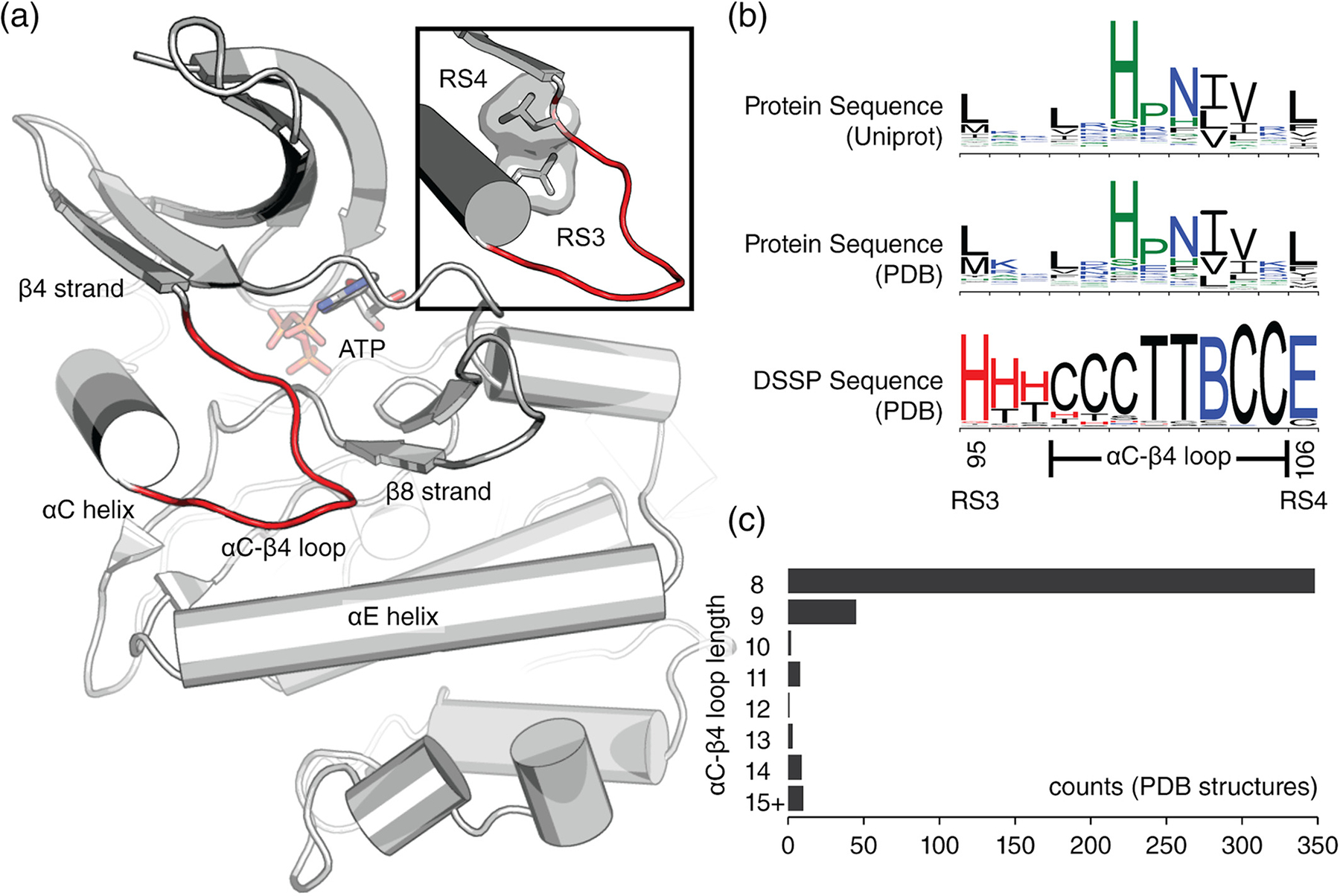
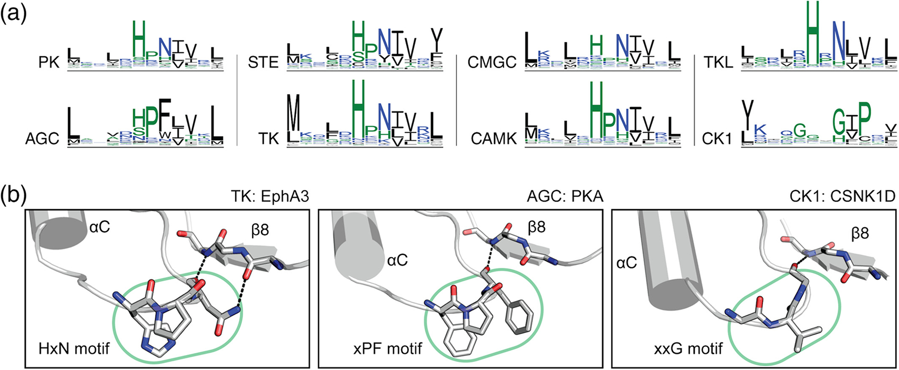
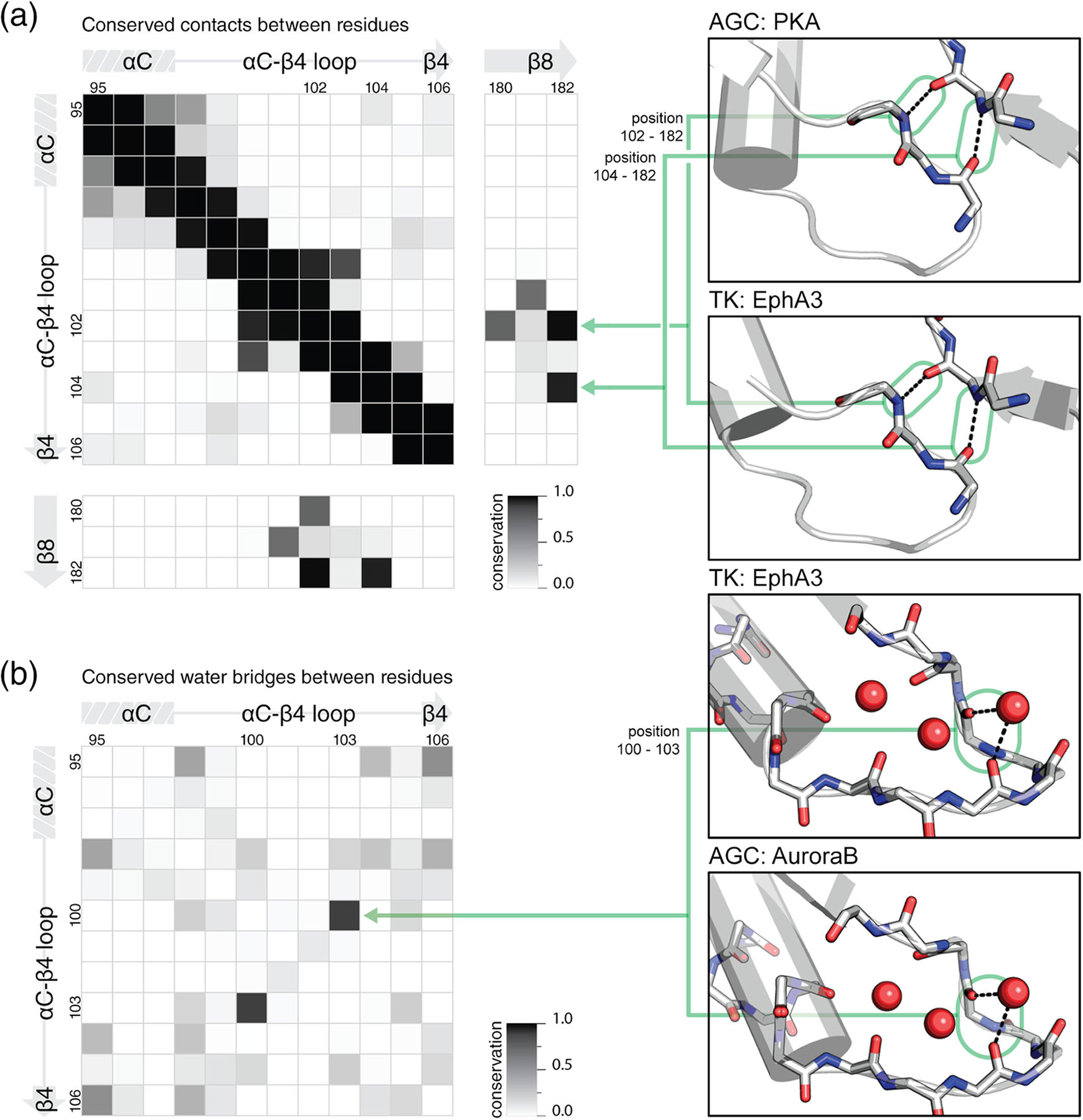
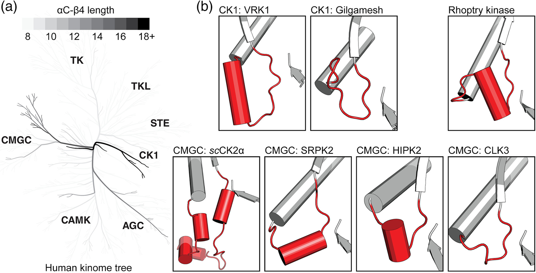
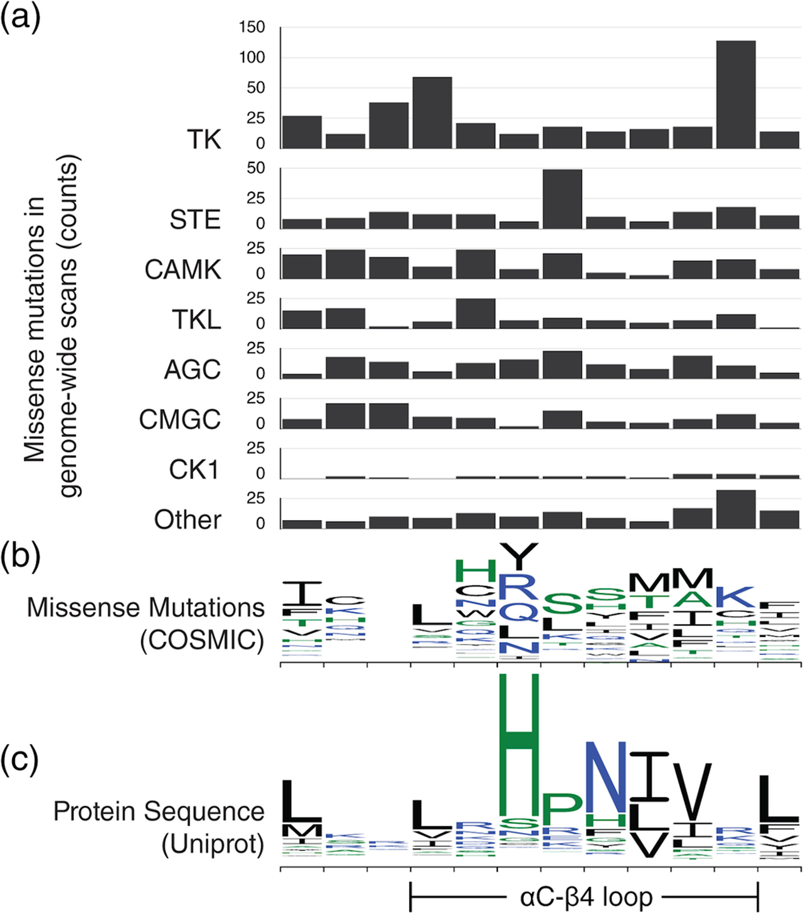
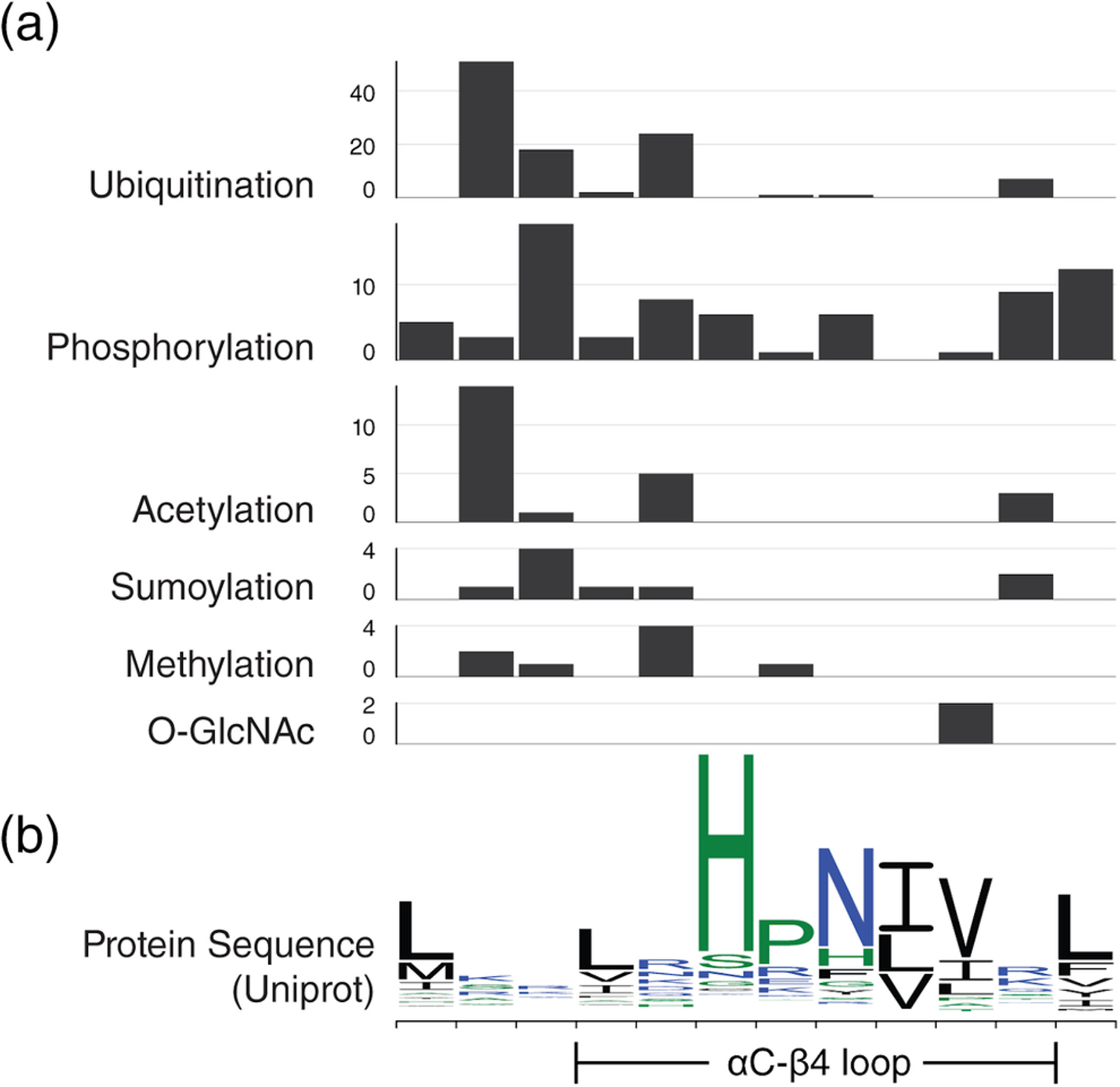
The faithful propagation of cellular signals in most organisms relies on the coordinated functions of a large family of protein kinases that share a conserved catalytic domain. The catalytic domain is a dynamic scaffold that undergoes large conformational changes upon activation. Most of these conformational changes, such as movement of the regulatory αC-helix from an "out" to "in" conformation, hinge on a conserved, but understudied, loop termed the αC-β4 loop, which mediates conserved interactions to tether flexible structural elements to the kinase core. We previously showed that the αC-β4 loop is a unique feature of eukaryotic protein kinases. Here, we review the emerging roles of this loop in kinase structure, function, regulation, and diseases. Through a kinome-wide analysis, we define the boundaries of the loop for the first time and show that sequence and structural variation in the loop correlate with conformational and regulatory variation. Many recurrent disease mutations map to the αC-β4 loop and contribute to drug resistance and abnormal kinase activation by relieving key auto-inhibitory interactions associated with αC-helix and inter-lobe movement. The αC-β4 loop is a hotspot for post-translational modifications, protein-protein interaction, and Hsp90 mediated folding. Our kinome-wide analysis provides insights for hypothesis-driven characterization of understudied kinases and the development of allosteric protein kinase inhibitors.
Keywords: Hsp-90; cancer mutation; conformational regulation; disease mutation; drug resistance; molecular brake; post-translational modifications; protein kinase.
© 2020 International Union of Biochemistry and Molecular Biology.
Figures
Similar articles
-
Analogous regulatory sites within the alphaC-beta4 loop regions of ZAP-70 tyrosine kinase and AGC kinases.Biochim Biophys Acta. 2008 Jan;1784(1):27-32. doi: 10.1016/j.bbapap.2007.09.007. Epub 2007 Sep 29. Biochim Biophys Acta. 2008. PMID: 17977811 Free PMC article. Review.
-
Integration of signaling in the kinome: Architecture and regulation of the αC Helix.Biochim Biophys Acta. 2015 Oct;1854(10 Pt B):1567-74. doi: 10.1016/j.bbapap.2015.04.007. Epub 2015 Apr 17. Biochim Biophys Acta. 2015. PMID: 25891902 Free PMC article.
-
Role of the αC-β4 loop in protein kinase structure and dynamics.Elife. 2024 Dec 4;12:RP91980. doi: 10.7554/eLife.91980. Elife. 2024. PMID: 39630082 Free PMC article.
-
The αC-β4 loop controls the allosteric cooperativity between nucleotide and substrate in the catalytic subunit of protein kinase A.Elife. 2024 Jun 24;12:RP91506. doi: 10.7554/eLife.91506. Elife. 2024. PMID: 38913408 Free PMC article.
-
The ABC of protein kinase conformations.Biochim Biophys Acta. 2015 Oct;1854(10 Pt B):1555-66. doi: 10.1016/j.bbapap.2015.03.009. Epub 2015 Apr 1. Biochim Biophys Acta. 2015. PMID: 25839999 Review.
Cited by
-
Saturation mutagenesis of a predicted ancestral Syk-family kinase.Protein Sci. 2022 Oct;31(10):e4411. doi: 10.1002/pro.4411. Protein Sci. 2022. PMID: 36173161 Free PMC article.
-
The Role of Hsp90 in Retinal Proteostasis and Disease.Biomolecules. 2022 Jul 12;12(7):978. doi: 10.3390/biom12070978. Biomolecules. 2022. PMID: 35883534 Free PMC article. Review.
-
Survivin Mediates Mitotic Onset in HeLa Cells Through Activation of the Cdk1-Cdc25B Axis.Res Sq [Preprint]. 2024 Feb 28:rs.3.rs-3949429. doi: 10.21203/rs.3.rs-3949429/v1. Res Sq. 2024. PMID: 38464014 Free PMC article. Preprint.
-
An update on evolutionary, structural, and functional studies of receptor-like kinases in plants.Front Plant Sci. 2024 Jan 31;15:1305599. doi: 10.3389/fpls.2024.1305599. eCollection 2024. Front Plant Sci. 2024. PMID: 38362444 Free PMC article. Review.
-
Conformation and dynamics of the kinase domain drive subcellular location and activation of LRRK2.Proc Natl Acad Sci U S A. 2021 Jun 8;118(23):e2100844118. doi: 10.1073/pnas.2100844118. Proc Natl Acad Sci U S A. 2021. PMID: 34088839 Free PMC article.
References
-
- Fountas A, Diamantopoulos L-N, Tsatsoulis A. Tyrosine kinase inhibitors and diabetes: A novel treatment paradigm? Trends Endocrinol. Metabolism. 2015;26:643–656. - PubMed
-
- Kumar R, Singh VP, Baker KM. Kinase inhibitors for cardiovascular disease. J Mol Cell Cardiol. 2007;42:1–11. - PubMed
-
- Ubersax JA, Ferrell JE. Mechanisms of specificity in protein phosphorylation. Nat Rev Mol Cell Biol. 2007;8:530–541. - PubMed
Publication types
MeSH terms
Substances
Grants and funding
LinkOut - more resources
Full Text Sources
