. 2020 Apr 1;318(4):R772-R780.
doi: 10.1152/ajpregu.00338.2019. Epub 2020 Feb 26.

Enhancement by TNF-α of TTX-resistant NaV current in muscle sensory neurons after femoral artery occlusion

Affiliations

Enhancement by TNF-α of TTX-resistant NaV current in muscle sensory neurons after femoral artery occlusion

Qin Li et al. Am J Physiol Regul Integr Comp Physiol. .

Abstract

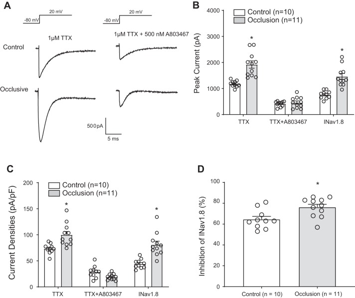
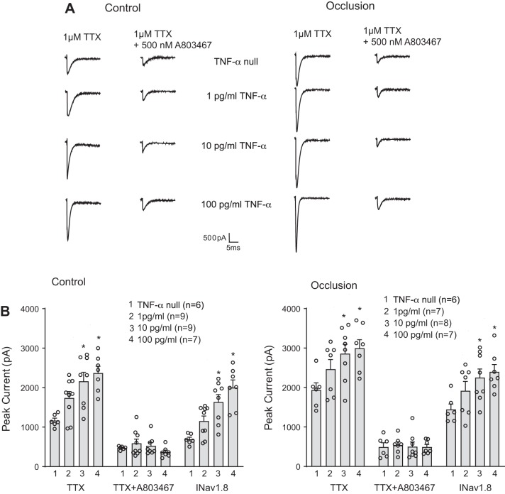
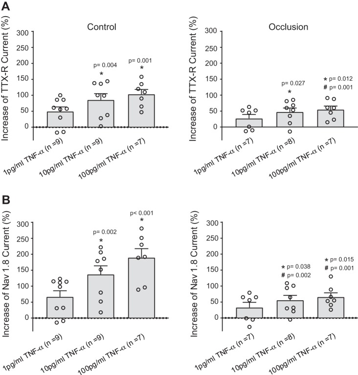
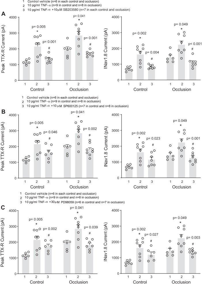
Femoral artery occlusion in rats has been used to study human peripheral artery disease (PAD). Using this animal model, a recent study suggests that increases in levels of tumor necrosis factor-α (TNF-α) and its receptor lead to exaggerated responses of sympathetic nervous activity and arterial blood pressure as metabolically sensitive muscle afferents are activated. Note that voltage-dependent Na+ subtype NaV1.8 channels (NaV1.8) are predominately present in chemically sensitive thin fiber sensory nerves. The purpose of this study was to examine the role played by TNF-α in regulating activity of NaV1.8 currents in muscle dorsal root ganglion (DRG) neurons of rats with PAD induced by femoral artery occlusion. DRG neurons from control and occluded limbs of rats were labeled by injecting the fluorescent tracer DiI into the hindlimb muscles 5 days before the experiments. A voltage patch-clamp mode was used to examine TTX-resistant (TTX-R) NaV currents. Results were as follows: 72 h of femoral artery occlusion increased peak amplitude of TTX-R [1,922 ± 139 pA in occlusion (n = 11 DRG neurons) vs. 1,178 ± 39 pA in control (n = 10), means ± SE; P < 0.001 between the 2 groups] and NaV1.8 currents [1,461 ± 116 pA in occlusion (n = 11) and 766 ± 48 pA in control (n = 10); P < 0.001 between groups] in muscle DRG neurons. TNF-α exposure amplified TTX-R and NaV1.8 currents in DRG neurons of occluded muscles in a dose-dependent manner. Notably, the amplification of TTX-R and NaV1.8 currents induced by TNF-α was attenuated in DRG neurons with preincubation with respective inhibitors of the intracellular signaling pathways p38-MAPK, JNK, and ERK. In conclusion, our data suggest that NaV1.8 is engaged in the role of TNF-α in amplifying muscle afferent inputs as the hindlimb muscles are ischemic; p38-MAPK, JNK, and ERK pathways are likely necessary to mediate the effects of TNF-α.

Keywords: NaV1.8 channels; TNF-α; dorsal root ganglion neuron; muscle afferent; peripheral artery disease.


Conflict of interest statement

No conflicts of interest, financial or otherwise, are declared by the authors.

Figures

Fig. 1.
Fig. 1.
Effects of femoral artery occlusion on TTX-resistant (TTX-R) and voltage-dependent Na+ channel subtype NaV1.8 (NaV1.8) currents in rat dorsal root ganglion (DRG) neurons. All rat DRG neurons were dissociated 72 h after femoral occlusion and recorded in voltage patch mode with −80 mV holding followed by a pulse of 20 mV. One micromolar TTX was used to block TTX-sensitive currents, and 500 nM of A803467 was used to block NaV1.8 currents. A: representative current traces after application of TTX and TTX plus A803467 in DRG neurons of control limb and occluded limb. A greater amplitude of currents was observed in DRG neurons of the occluded limb. B: averaged data showing that femoral artery occlusion amplified current amplitude after TTX application. The data also show that the currents were largely decreased after application of TTX and A803467. NaV1.8 currents were assessed as INaV1.8 (peak current after TTX − peak currents after both TTX and A803467). INaV1.8 was also amplified by femoral artery occlusion. *P < 0.001 between control and occlusion for TTX-R and NaV1.8 current in DRG neurons. C: femoral artery occlusion also increased current densities in DRG neurons compared with the control group. *P = 0.002 between control and occlusion after TTX; *P < 0.001 between control and occlusion for INaV1.8. D: %inhibition of INaV1.8 by A803467 was greater in DRG neurons of occluded limbs than that in DRG neurons of control limbs. *P = 0.010 between control and occlusion. Individual data points are also shown; n = 10 DRG neurons in the control group, and n = 11 in the occlusion group.
Fig. 2.
Fig. 2.
Effects of TNF-α on TTX-resistant (TTX-R) and voltage-dependent Na+ channel subtype NaV1.8 (NaV1.8) currents in rat dorsal root ganglion (DRG) neurons of control and occlusion groups. TTX-R currents were examined in DRG neurons after 24 h of exposure to TNF-α. A: representative current traces showing that TNF-α amplified TTX-R current in a dose-related manner in DRG neurons of both control limbs and occluded limbs. B: averaged data demonstrate that TNF-α amplified TTX-R currents and NaV1.8 currents in DRG neurons of control limbs and occluded limbs. ○, Individual data points; n is the number of DRG neurons. INaV1.8, peak current after TTX − peak currents after both TTX and A803467. In the control group, *P = 0.005 and P = 0.001 compared with TNF-α null after TTX and *P = 0.002 and P < 0.001 for INaV1.8 current. In the occlusion group, *P = 0.041 and P = 0.020 compared with TNF-α null after TTX and *P = 0.047 and P = 0.021 for INaV1.8 current.
Fig. 3.
Fig. 3.
Percent effects of TNF-α on TTX-resistant (TTX-R; A) and voltage-dependent Na+ channel subtype NaV1.8 (NaV1.8) currents (B) of dorsal root ganglion (DRG) neurons of control limbs and occluded limbs. TNF-α amplified TTX-R and NaV1.8 currents of DRG neurons in a dose-dependent manner, and the effects of TNF-α appeared to a lesser degree in the occlusion group. ○Individual data points. All the P values are shown in the figure.
Fig. 4.
Fig. 4.
Involvement of intracellular signaling pathways p38-MAPK, JNK, and ERK in the effects of TNF-α on voltage-dependent Na+ channel (NaV) currents of dorsal root ganglion (DRG) neurons. To inhibit these signaling pathways, before application of 10 pg/mL of TNF-α, SB203580 (10 μM; A), SP600125 (10 μM; B), and PD98059 (30 μM; C) were applied, respectively. TNF-α amplified TTX-resistant (TTX-R) and NaV1.8 currents in rat DRG neurons of the control and occlusion groups compared with the neurons without TNF-α application. Note that the effects of TNF-α were significantly attenuated by respective inhibitors of p38-MAPK, JNK, and ERK. The numbers of DRG neurons (n) are indicated in the figure, and individual data points are also shown. *P < 0.05 compared with the group without application of TNF-α; #P < 0.05 group with TNF-α vs. group with TNF-α plus respective inhibitors SB203580, SP600125, and PD98059.

Similar articles

Cited by

References

    1. Antonopoulos AS, Papanikolaou E, Vogiatzi G, Oikonomou E, Tousoulis D. Anti-inflammatory agents in peripheral arterial disease. Curr Opin Pharmacol 39: 1–8, 2018. doi:10.1016/j.coph.2017.11.001. - DOI - PubMed
    1. Baccelli G, Reggiani P, Mattioli A, Corbellini E, Garducci S, Catalano M. The exercise pressor reflex and changes in radial arterial pressure and heart rate during walking in patients with arteriosclerosis obliterans. Angiology 50: 361–374, 1999. doi:10.1177/000331979905000502. - DOI - PubMed
    1. Bakke EF, Hisdal J, Kroese AJ, Jørgensen JJ, Stranden E. Blood pressure response to isometric exercise in patients with peripheral atherosclerotic disease. Clin Physiol Funct Imaging 27: 109–115, 2007. doi:10.1111/j.1475-097X.2007.00720.x. - DOI - PubMed
    1. Catterall WA, Goldin AL, Waxman SG. International Union of Pharmacology. XLVII. Nomenclature and structure-function relationships of voltage-gated sodium channels. Pharmacol Rev 57: 397–409, 2005. doi:10.1124/pr.57.4.4. - DOI - PubMed
    1. Chen W, Sheng J, Guo J, Gao F, Zhao X, Dai J, Wang G, Li K. Tumor necrosis factor-α enhances voltage-gated Na+ currents in primary culture of mouse cortical neurons. J Neuroinflammation 12: 126, 2015. doi:10.1186/s12974-015-0349-x. - DOI - PMC - PubMed

Publication types

MeSH terms