. 2020 Apr 15;31(9):917-929.
doi: 10.1091/mbc.E20-01-0026. Epub 2020 Feb 26.

DYRK kinase Pom1 drives F-BAR protein Cdc15 from the membrane to promote medial division

Affiliations

DYRK kinase Pom1 drives F-BAR protein Cdc15 from the membrane to promote medial division

Rahul Bhattacharjee et al. Mol Biol Cell. .

Abstract

In many organisms, positive and negative signals cooperate to position the division site for cytokinesis. In the rod-shaped fission yeast Schizosaccharomyces pombe, symmetric division is achieved through anillin/Mid1-dependent positive cues released from the central nucleus and negative signals from the DYRK-family polarity kinase Pom1 at cell tips. Here we establish that Pom1's kinase activity prevents septation at cell tips even if Mid1 is absent or mislocalized. We also find that Pom1 phosphorylation of F-BAR protein Cdc15, a major scaffold of the division apparatus, disrupts Cdc15's ability to bind membranes and paxillin, Pxl1, thereby inhibiting Cdc15's function in cytokinesis. A Cdc15 mutant carrying phosphomimetic versions of Pom1 sites or deletion of Cdc15 binding partners suppresses division at cell tips in cells lacking both Mid1 and Pom1 signals. Thus, inhibition of Cdc15-scaffolded septum formation at cell poles is a key Pom1 mechanism that ensures medial division.


Figures
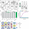
FIGURE 1:
FIGURE 1:
Pom1 kinase activity is required to inhibit tip septa. (A) Representative single z-section images of the indicated strains treated for 2 h with vehicle (DMSO) or ATP analog (3MB-PP1). Cells were fixed and stained with calcofluor. (B) Quantification of tip septa from images acquired as in A from three biological replicates, n > 300 septated cells. Graph shows mean and SEM. (C) Schematics and example images of the three types of tip septa scored. (D) Time course of tip septa appearance. 3MB-PP1 was added at time 0. Samples were prepared as in A. Measurements are from three biological replicates, n > 300 septated cells. (E) Model of the effects of Pom1 kinase activity and Mid1 in division site placement in the context of previous work (Huang et al., 2007; Rincon and Paoletti, 2016).
FIGURE 2:
FIGURE 2:
Pom1 kinase inhibits division site placement even if the positive Mid1 cue is proximal to the cell tip. (A) Representative images of pom1as1nmt81::myo52N-RFP-nup146 cells grown for 24 h in medium lacking thiamine to induce expression of Myo52-Nup146 and treated for 4 h with vehicle (MeOH) or 1 μM 3MB-PP1, and vehicle (DMSO) or 25 μg/ml MBC to displace the nucleus, and stained with calcofluor. (B) Quantification of tip septa as in A from four biological replicates, n > 490 septated cells. Graphs show mean and SEM. (C) Representative images of Myo52N-GFP-Nup146 in pom1+ and pom1Δ cells (left) and cells stained with calcofluor (right). (D) Quantification of tip septa as in C from three biological replicates, n > 326 septated cells. Graphs show mean and SEM. Scale bar, 5 µm.
FIGURE 3:
FIGURE 3:
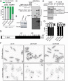
Pom1 can phosphorylate Cdc15 on 22 sites. (A) Quantification of tip septa as in Figure 1, A and B. (B) Denatured protein lysates separated by SDS–PAGE and immunoblotted for the indicated proteins. (C) In vitro kinase reactions were separated by SDS–PAGE. Coomassie-stained gel of inputs and autoradiographs detecting 32P incorporation are shown. (D) Schematic of Cdc15 to scale with Pom1 phosphorylation sites indicated by black lines. (E) Denatured lysates and IP-phosphatase assays were separated by SDS–PAGE and immunoblotted for the indicated proteins. (F) Representative images of DAPI and methyl blue (top panels) or calcofluor (bottom panels) stained cells of the indicated cdc15 genotype. Single z-sections of 0.5 µm are shown in the top panels and maximum projections are shown in the bottom panels. Scale bar, 5 μm. (G) Quantification of nuclei and septation indices from images acquired as in E for the DAPI/methyl blue-stained cells. Graph shows mean and SEM from three biological replicates, n ≥ 940 cells.
FIGURE 4:
FIGURE 4:
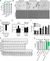
A Cdc15 phosphomimetic suppresses tip septa formation. (A) Quantification of tip septa as described in Figure 1, A–C. (B) Representative live-cell images of the indicated strains. Fluorescent images were deconvolved and maximum projected. (C) Average arrival of Cdc15 at the division site relative to SPB separation (time 0). Measurements are from three biological replicates, n ≥ 12 cells. (D) Quantification of the fluorescence intensity of Cdc15 in the CR (left) or whole cell (right) of the indicated strains from three biological replicates, n ≥ 33 cells. All images used for quantification were not deconvolved and were sum projected. (E) Representative montages from live-cell, time-lapse imaging of indicated strains. Images were acquired every 2 min and every 4 min are shown. Numbers indicate minutes from SPB separation. (F) Length of cytokinesis stage based on imaging as in E. Measurements are from three biological replicates, n ≥ 18 cells. (G) Quantification of tip septa phenotype as described in Figure 1, A–C, except n > 250 cells. Graphs show mean and SEM; wt, wildtype. *p < 0.05, one-way ANOVA with Tukey’s post-hoc analysis. ****p < 0.0001, Brown–Forsythe and Welch ANOVA tests with Dunnett T3 correction for multiple comparisons to wt. Scale bar, 5 µm.
FIGURE 5:
FIGURE 5:
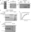
Pom1 inhibits Cdc15 binding to membranes and Pxl1. (A) Coomassie-stained SDS–PAGE of proteins purified from bacteria expressing Cdc15-E30K E152K in the presence or absence of Pom1. (B) Cdc15-E30K E152K coexpressed with Pom1 was purified from bacteria and then treated with lambda phosphatase (PPase) or control before adding to Folch fraction liposomes (lipos). Coomassie staining of supernatant (S) and pellet (P) after pelleting and SDS–PAGE. (C) GST-Cdc15C was phosphorylated in vitro with GST-Pom1 and incubated with MBP or MBP-tagged Pxl1 bound to amylose resin. After pelleting, proteins bound to the amylose resin were separated by SDS–PAGE and stained with Coomassie. PD, pull down. (D) Coomassie-stained SDS–PAGE of binding assays between the indicated MBP-tagged and GST-tagged proteins. (E) Binding curves with varying concentrations of MBP-Pxl1 on MBP nanobody resin and GST-Cdc15 C-terminal variants in solution. Quantification was done from three biological replicates as detailed in the methods. Error bars show SEM. (F) Coomassie-stained SDS–PAGE of binding assays between the indicated MBP-tagged and GST-tagged proteins.
FIGURE 6:
FIGURE 6:
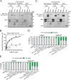
Pxl1 interacts with the central region and SH3 domain of Cdc15. (A, C) Coomassie-stained SDS–PAGE of binding assays between the indicated MBP-tagged and GST-tagged proteins. Amylose resin was used to pull down (PD) proteins. (B) Binding curves with varying concentrations of MBP-Pxl1 on MBP nanobody resin and GST-Cdc15C and GST-Cdc15C (W903S) in solution. Quantification was done from three biological replicates. Error bars show SEM. (D, E) Quantification of tip septa as in Figure 1, A–C.
FIGURE 7:
FIGURE 7:
Models of Pom1 inhibition of Cdc15 function. (A) Model of Pom1 inhibition of Cdc15 membrane binding at cell tips in a single interphase cell with the nucleus in the middle. (B) Molecular model of Pom1 inhibition of Cdc15 membrane binding.

Similar articles

Cited by

References

    1. Almonacid M, Moseley JB, Janvore J, Mayeux A, Fraisier V, Nurse P, Paoletti A. (2009). Spatial control of cytokinesis by Cdr2 kinase and Mid1/anillin nuclear export. Curr Biol , 961–966. - PubMed
    1. Arasada R, Pollard TD. (2011). Distinct roles for F-BAR proteins Cdc15p and Bzz1p in actin polymerization at sites of endocytosis in fission yeast. Curr Biol , 1450–1459. - PMC - PubMed
    1. Arasada R, Pollard TD. (2014). Contractile ring stability in S. pombe depends on F-BAR protein Cdc15p and Bgs1p transport from the Golgi complex. Cell Rep , 1533–1544. - PMC - PubMed
    1. Bahler J, Pringle JR. (1998). Pom1p, a fission yeast protein kinase that provides positional information for both polarized growth and cytokinesis. Genes Dev , 1356–1370. - PMC - PubMed
    1. Bahler J, Wu JQ, Longtine MS, Shah NG, McKenzie A, 3rd, Steever AB, Wach A, Philippsen P, Pringle JR. (1998). Heterologous modules for efficient and versatile PCR-based gene targeting in Schizosaccharomyces pombe . Yeast , 943–951. - PubMed

Publication types

MeSH terms

LinkOut - more resources