Review
. 2020 Jul 8:43:337-353.
doi: 10.1146/annurev-neuro-091619-022657. Epub 2020 Feb 26.

Neuropod Cells: The Emerging Biology of Gut-Brain Sensory Transduction

Affiliations
Review

Neuropod Cells: The Emerging Biology of Gut-Brain Sensory Transduction

Melanie Maya Kaelberer et al. Annu Rev Neurosci. .

Abstract

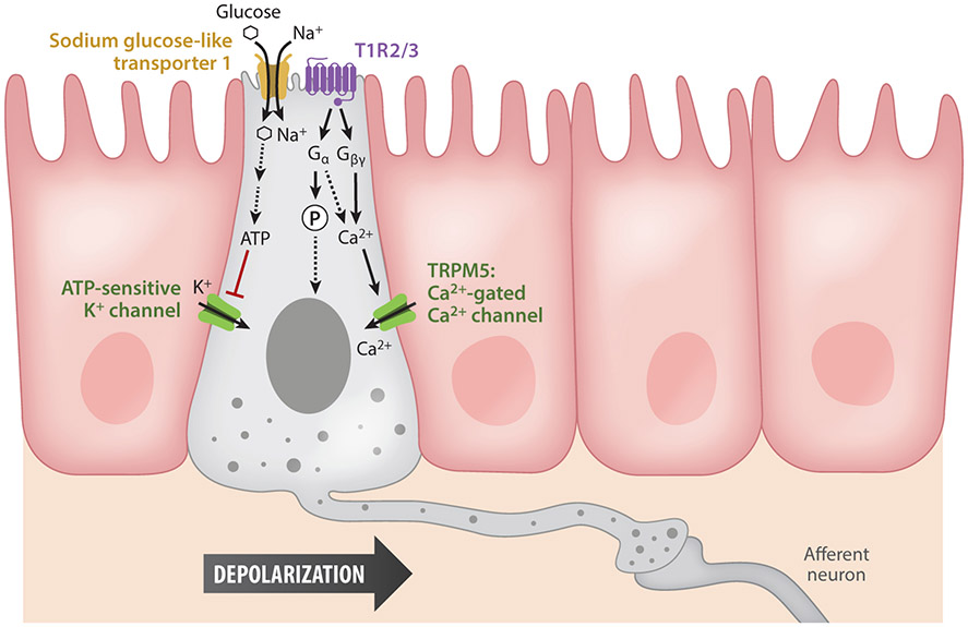
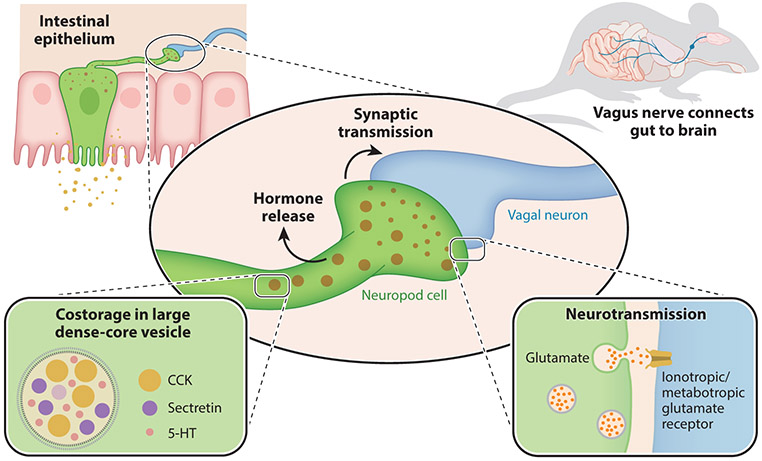
Guided by sight, scent, texture, and taste, animals ingest food. Once ingested, it is up to the gut to make sense of the food's nutritional value. Classic sensory systems rely on neuroepithelial circuits to convert stimuli into signals that guide behavior. However, sensation of the gut milieu was thought to be mediated only by the passive release of hormones until the discovery of synapses in enteroendocrine cells. These are gut sensory epithelial cells, and those that form synapses are referred to as neuropod cells. Neuropod cells provide the foundation for the gut to transduce sensory signals from the intestinal milieu to the brain through fast neurotransmission onto neurons, including those of the vagus nerve. These findings have sparked a new field of exploration in sensory neurobiology-that of gut-brain sensory transduction.

Keywords: enteroendocrine cells; glutamatergic transmission; gut-brain biology; neuropod cells; sensory transduction; vagus nerve.


Figures

Figure 1
Figure 1
Molecular pathways of activation in neuropod cells. A nutrient such as glucose is sensed by neuropod cells in two ways. First, through substrate Na+ transporters, specifically Na+ glucose-like transporter 1, the entry of Na+ depolarizes the cells, leading to vesicle release and the activation of synaptically connected afferent neurons. Glucose is also metabolized, producing ATP that closes on ATP-sensitive K+ channels and further depolarizes the cell. Second, neuropod cells also express the sweet taste receptor T1R2/3, a G protein–coupled receptor. Activated G proteins either phosphorylate transcription factors or release intracellular Ca2+, which activates transient receptor potential channel M5 (TRPM5), the Ca2+-activated Ca2+ channel. The intracellular Ca2+ cascade induces vesicle fusion and the further activation of afferent neurons.
Figure 2
Figure 2
Glutamatergic synaptic transmission of neuropod cells. Neuropod cells in the intestinal epithelium contain both large dense-core neuropeptide vesicles and small neurotransmitter vesicles. The large vesicles contain multiple neuropeptides with endocrine functions such as cholecystokinin (CCK), secretin, and serotonin (5-HT) and are co-released with neurotransmitters. Activation of neuropod cells stimulates synaptic vesicle release, including the neurotransmitter glutamate. When this fast neurotransmission acts on afferent vagal neurons, it serves to transduce signals from nutrients directly to the brain in milliseconds. Mouse image adapted from Kaelberer et al. (2018).

Similar articles

Cited by

References

    1. Alcaino C, Knutson KR, Treichel AJ, Yildiz G, Strege PR, et al. 2018. A population of gut epithelial enterochromaffin cells is mechanosensitive and requires Piezo2 to convert force into serotonin release. PNAS 115:E7632–41 - PMC - PubMed
    1. Altschuler SM, Ferenci DA, Lynn RB, Miselis RR. 1991. Representation of the cecum in the lateral dorsal motor nucleus of the vagus nerve and commissural subnucleus of the nucleus tractus solitarii in rat. J. Comp. Neurol 304:261–74 - PubMed
    1. Ball GG. 1974. Vagotomy: effect on electrically elicited eating and self-stimulation in the lateral hypothalamus. Science 184:484–85 - PubMed
    1. Bayliss WM, Starling EH. 1902. The mechanism of pancreatic secretion. J. Physiol 28:325–53 - PMC - PubMed
    1. Bellono NW, Bayrer JR, Leitch DB, Castro J, Zhang C, et al. 2017. Enterochromaffin cells are gut chemosensors that couple to sensory neural pathways. Cell 170:185–98.e16 - PMC - PubMed

Publication types

LinkOut - more resources