

Experimental validation of the prestretch-strain relationship as a non-invasive index of left ventricular myocardial contractility
- PMID: 32101554
- PMCID: PMC7043779
- DOI: 10.1371/journal.pone.0228027
Experimental validation of the prestretch-strain relationship as a non-invasive index of left ventricular myocardial contractility
Abstract
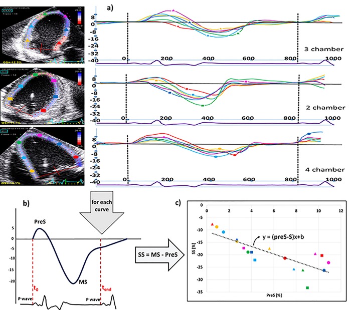
Background: The slope of the relationship between segmental PreS and total systolic shortening (S) has been proposed as a non-invasive index of left ventricular contractility. The aim of this study was to correlate this novel parameter to invasive gold standard measurements of contractility and to investigate how it is influenced by afterload.
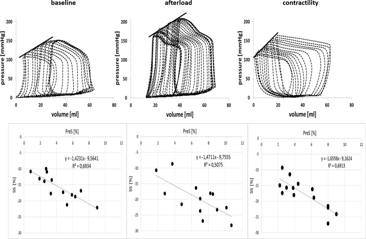
Methods: In domestic pigs, afterload was increased by either balloon inflation in the aorta or by administration of phenylephrine while contractility was increased by dobutamine infusion. During all interventions, left ventricular pressure-volume measurements and trans-diaphragmatic two-dimensional echocardiographic images were acquired. The PreS-S slope was constructed from 18 segmental strain curves obtained by speckle tracking analysis and compared to the slope of the end systolic PV relationship (Emax) and the pre-load recruitable stroke work (PRSW).
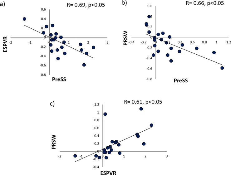
Results: Sixteen datasets of increased contractility and afterload were analyzed. During dobutamine infusion, the LV volumes decreased (p<0.05) while ejection fraction increased (p<0.05). Emax, PRSW and the slope of the intra-ventricular PreS-S relation increased significantly during dobutamine infusion. Afterload increase led to increase in systolic blood pressure (105±16mmHg vs. 138±25mmHg; p<0.01) and decrease of LV stroke volume and ejection fraction (p<0.01). The PreS-S slope was not influenced by loading conditions in concordance with the PRSW findings. The absolute values of the PreS-S slope did not correlate with Emax or PRSW. However, the change of the PreS-S slope in relation with different interventions demonstrated good correlation with changes in PRSW or Emax, (r = 0.66, p<0.05 and r = 0.69, p<0.05).
Conclusions: The slope of the PreS-S relationship is sensitive to changes in inotropy and is less load-dependent than conventional non-invasive parameters of left ventricular function. The magnitude of the change of this slope correlates well with changes in invasive contractility measurements making it an attractive parameter to assess contractile reserve or contractile changes during longitudinal follow-up of patients.
Conflict of interest statement
The authors have declared that no competing interests exist.
Figures
References
Publication types
MeSH terms
LinkOut - more resources
Full Text Sources
Other Literature Sources
