. 2020 Feb 26;15(2):e0229570.
doi: 10.1371/journal.pone.0229570. eCollection 2020.

Herpes simplex virus type 1 inflammasome activation in proinflammatory human macrophages is dependent on NLRP3, ASC, and caspase-1

Affiliations

Herpes simplex virus type 1 inflammasome activation in proinflammatory human macrophages is dependent on NLRP3, ASC, and caspase-1

Andrew H Karaba et al. PLoS One. .

Abstract

The proinflammatory cytokines interleukin (IL)-1β and IL-18 are products of activation of the inflammasome, an innate sensing system, and important in the pathogenesis of herpes simplex virus type 1 (HSV-1). The release of IL-18 and IL-1β from monocytes/macrophages is critical for protection from HSV-1 based on animal models of encephalitis and genital infection, yet if and how HSV-1 activates inflammasomes in human macrophages is unknown. To investigate this, we utilized both primary human monocyte derived macrophages and human monocytic cell lines (THP-1 cells) with various inflammasome components knocked-out. We found that HSV-1 activates inflammasome signaling in proinflammatory primary human macrophages, but not in resting macrophages. Additionally, HSV-1 inflammasome activation in THP-1 cells is dependent on nucleotide-binding domain and leucine-rich repeat-containing receptor 3 (NLRP3), apoptosis-associated speck-like molecule containing a caspase recruitment domain (ASC), and caspase-1, but not on absent in melanoma 2 (AIM2), or gamma interferon-inducible protein 16 (IFI16). In contrast, HSV-1 activates non-canonical inflammasome signaling in proinflammatory macrophages that results in IL-1β, but not IL-18, release that is independent of NLRP3, ASC, and caspase-1. Ultraviolet irradiation of HSV-1 enhanced inflammasome activation, demonstrating that viral replication suppresses inflammasome activation. These results confirm that HSV-1 is capable of activating the inflammasome in human macrophages through an NLRP3 dependent process and that the virus has evolved an NLRP3 specific mechanism to inhibit inflammasome activation in macrophages.


Conflict of interest statement

The authors have declared that no competing interests exist.

Figures
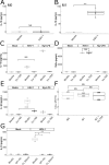
Fig 1
Fig 1. HSV-1 activates inflammasomes in primary human macrophages.
A and B. Primary human MDMs cultured without (M0 A) or with IFNγ (M1 B) were incubated with HSV-1 or media for 24 hours. Cell culture supernatants were collected and assayed for IL-18 (B represents the combination of two experiments). C, D, and E. Primary human MDMs stimulated with IFNγ were cultured in media alone or in media containing 100 μg/mL of VX-765 (Invivogen, San Diego, California) and then incubated with HSV-1, nigericin and LPS (Ng+LPS), or media for 24 hours as outlined in Materials and Methods. Cell culture supernatants were collected and assayed for (C) IL-18, (D) TNFα, and (E) IL-1β. F. MDMs cultured without IFNγ (M0), with IFNγ (M1), or with IFNγ and VX-765 (M1+Vx-765) were infected with HSV-1 for 1 hour followed by citrate wash to inactivate any extracellular virus. Supernatants were collected 24 hours later and plaque forming units (PFU) were determined via standard plaque assay on Vero cells. Data shown are combined from two independent experiments. G. MDMs stimulated with IFNγ (M1) were incubated with HSV-1 or media as well as a neutralizing antibody (WT Ab) and a neutralizing antibody unable to bind to Fc receptors (Fc Silent) for 24 hours. Cell culture supernatants were collected and assayed for IL-18. Differences between groups indicated by brackets were determined by a Student’s t-test. NS, *,**,*** indicate p-values >0.05, <0.05, <0.01, <0.001, respectively. The lower and upper borders of the boxplots represent the 1st quartile and 3rd quartile respectively. The median is represented by a horizontal line in the box. The lower and upper whiskers represent 1.5x the interquartile range (IQR) beyond the quartile lines. Each dot represents an individual sample.
Fig 2
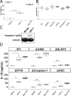
Fig 2. HSV-1 inflammasome activation in THP-1 cells is dependent on NLRP3, ASC, and caspase-1.
A. THP-1 cells were stimulated overnight with PMA (100 ng/mL) and then incubated with HSV-1, nigericin and LPS (Ng+LPS), or media, as outlined in Materials and Methods, for 24 hours. Cell culture supernatants were collected and IL-18 was measured via ELISA. B. THP-1 cells with the indicated gene disrupted via CRISPR-cas9 (Δ) were stimulated overnight with PMA, infected with HSV-1 for 1 hour followed by citrate wash to inactivate any extracellular virus. Supernatants were collected 24 hours later and PFU were determined via standard plaque assay on Vero cells. Data shown are combined from two independent experiments. C. Cell lysates from THP-1 cells infected with HSV-1 or mock infected were probed for caspase-1 or β-actin via western blot. D. THP-1 cells with the indicated gene disrupted via CRISPR-cas9 (Δ) were stimulated overnight with PMA and then incubated with HSV-1, nigericin and LPS (Ng+LPS), or media for 24 hours before IL-18 was measured in cell supernatants. ΔHUMCYC cells are labeled as “WT.” Differences between groups indicated by brackets were determined by a Student’s t-test. NS, *,**,*** indicate p-values >0.05, <0.05, <0.01, <0.001, respectively.
Fig 3
Fig 3. IL-18 produced by MDMs after infection with UV-irradiated HSV-1.
A and B. Primary human MDMs cultured without (M0 A) or with IFNγ (M1 B) were incubated with HSV-1, UV irradiated HSV-1 (HSV-1/UV), or media for 24 hours. Cell culture supernatants were collected and assayed for IL-18. C. Primary human MDMs cultured with IFNγ were incubated with HSV-1, UV irradiated HSV-1 (HSV-1/UV), or media for 4 hours and 8 hours. Cell culture supernatants were collected and assayed for IL-18. Differences between indicated conditions within a cell type were determined by a one-way ANOVA with Tukey HSD post-hoc analysis. NS, *,**,*** indicate p-values >0.05, <0.05, <0.01, <0.001 respectively.
Fig 4
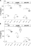
Fig 4. IL-18 produced by THP-1 cells after infection with UV-irradiated HSV-1.
A. THP-1 cells were stimulated overnight with PMA (5 ng/mL) and then incubated with HSV-1, UV irradiated HSV-1 (HSV-1/UV), or media for 24 hours. Cell culture supernatants were collected and IL-18 was measured via ELISA. B. THP-1 cells were stimulated with PMA (5 ng/mL) and then with IFN0γ (25 ng/mL) the following day for 24 hours prior to incubation with HSV-1, UV irradiated HSV-1 (HSV-1/UV), or media alone for 24 hours. Cell culture supernatants were collected and IL-18 was measured via ELISA. ΔHUMCYC cells are labeled as “WT.” Differences between indicated conditions within a cell type were determined by a one-way ANOVA with Tukey HSD post-hoc analysis. NS, *,**,*** indicate p-values >0.05, <0.05, <0.01, <0.001 respectively.
Fig 5
Fig 5. IL-1β produced by THP-1 cells after infection with UV-irradiated HSV-1.
A and B. THP-1 cells were stimulated with PMA (5 ng/mL) and then with IFNγ (25 ng/mL) the following day for 24 hours prior to incubation with HSV-1, UV irradiated HSV-1 (HSV-1/UV), or media alone for 24 hours. Cell culture supernatants were collected and IL-1β was measured. ΔHUMCYC cells are labeled as “WT.” Differences between indicated conditions within a cell type were determined by a one-way ANOVA with Tukey HSD post-hoc analysis. NS, *,**,*** indicate p-values >0.05, <0.05, <0.01, <0.001 respectively.

Similar articles

Cited by

References

    1. Brubaker SW, Bonham KS, Zanoni I, Kagan JC. Innate immune pattern recognition: a cell biological perspective. 2015;33: 257–290. 10.1146/annurev-immunol-032414-112240 - DOI - PMC - PubMed
    1. Frank D, Vince JE. Pyroptosis versus necroptosis: similarities, differences, and crosstalk. Cell Death Differ. 2019;26: 99–114. 10.1038/s41418-018-0212-6 - DOI - PMC - PubMed
    1. Mantovani A, Dinarello CA, Molgora M, Garlanda C. Interleukin-1 and Related cytokines in the regulation of inflammation and immunity. Immunity. 2019;50: 778–795. 10.1016/j.immuni.2019.03.012 - DOI - PMC - PubMed
    1. Costa LS, Outlioua A, Anginot A, Akarid K, Arnoult D. RNA viruses promote activation of the NLRP3 inflammasome through cytopathogenic effect-induced potassium efflux. Cell Death Dis. 2019; 1–15. 10.1038/s41419-019-1579-0 - DOI - PMC - PubMed
    1. Chen W, Xu Y, Li H, Tao W, Xiang Y, Huang B, et al. HCV genomic RNA activates the NLRP3 inflammasome in human myeloid cells. PloS One. 2014;9: e84953–10. 10.1371/journal.pone.0084953 - DOI - PMC - PubMed

Publication types

MeSH terms