. 2020 Feb 25;30(8):2512-2525.e9.
doi: 10.1016/j.celrep.2020.01.099.

Type I Interferons Suppress Anti-parasitic Immunity and Can Be Targeted to Improve Treatment of Visceral Leishmaniasis

Affiliations

Type I Interferons Suppress Anti-parasitic Immunity and Can Be Targeted to Improve Treatment of Visceral Leishmaniasis

Rajiv Kumar et al. Cell Rep. .

Abstract

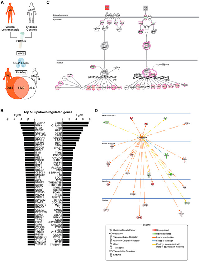
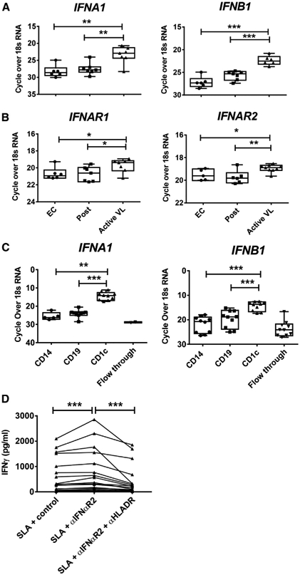
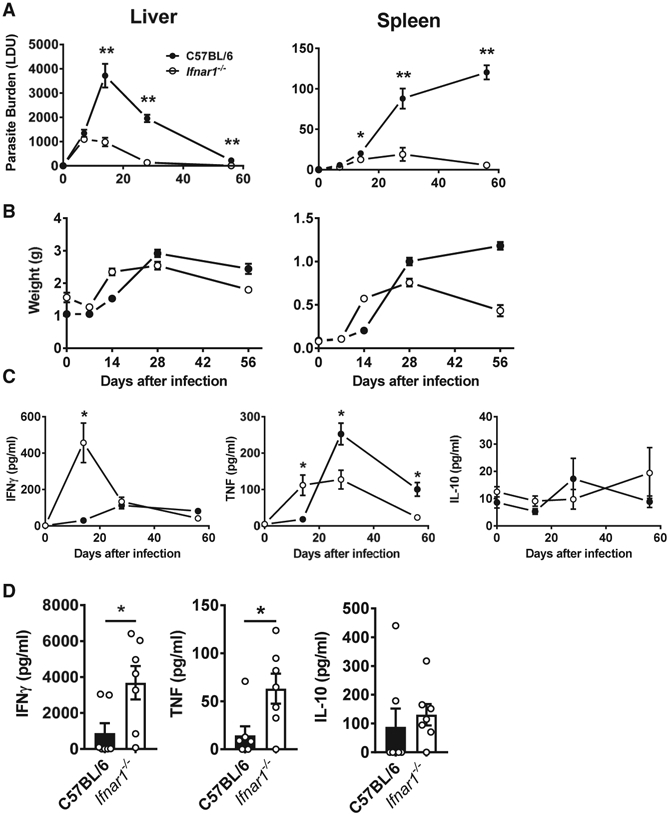
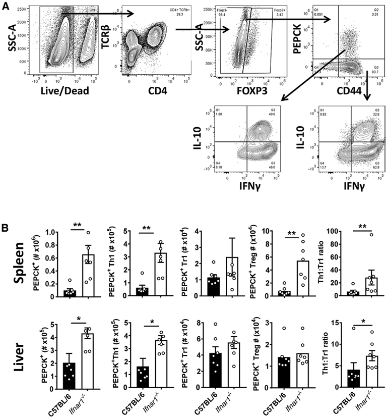
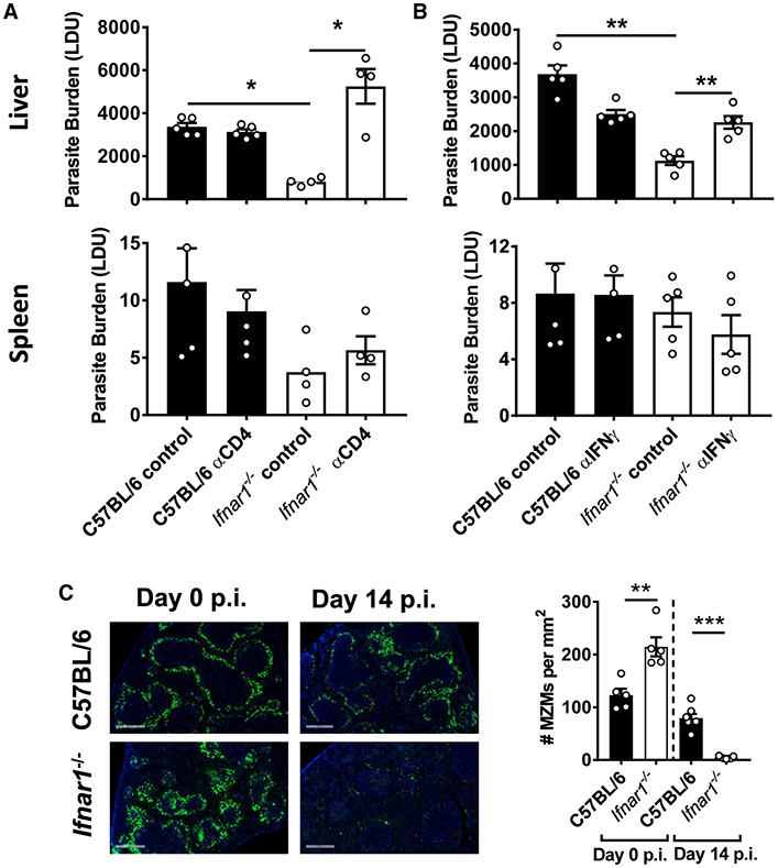
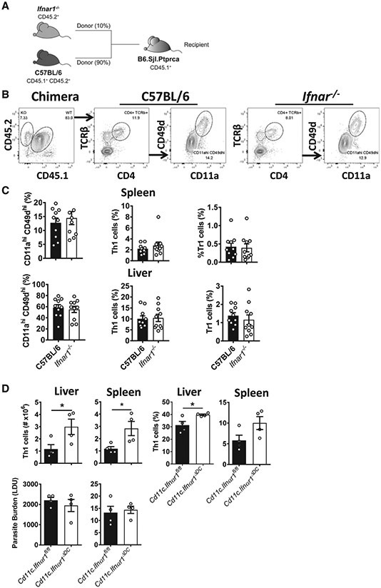
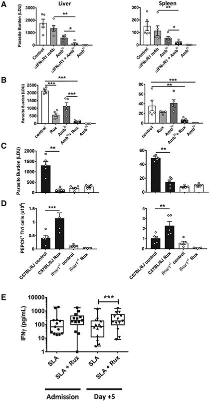
Type I interferons (IFNs) play critical roles in anti-viral and anti-tumor immunity. However, they also suppress protective immune responses in some infectious diseases. Here, we identify type I IFNs as major upstream regulators of CD4+ T cells from visceral leishmaniasis (VL) patients. Furthermore, we report that mice deficient in type I IFN signaling have significantly improved control of Leishmania donovani, a causative agent of human VL, associated with enhanced IFNγ but reduced IL-10 production by parasite-specific CD4+ T cells. Importantly, we identify a small-molecule inhibitor that can be used to block type I IFN signaling during established infection and acts synergistically with conventional anti-parasitic drugs to improve parasite clearance and enhance anti-parasitic CD4+ T cell responses in mice and humans. Thus, manipulation of type I IFN signaling is a promising strategy for improving disease outcome in VL patients.

Keywords: CD4(+) T cells; type I interferons; visceral leishmaniasis.


Conflict of interest statement

Declaration of Interests S.W.L. has served on an advisory board for Novartis, the owner of Jakavi (ruxolitinib), a drug used in this study.

Figures

Figure 1.
Figure 1.. Type I IFNs Are Major Upstream Regulators of VL Patient CD4+ T Cell Responses
(A) Schematic showing a brief outline of the workflow for isolating peripheral blood CD4+ T cells from visceral leishmaniasis (VL) patients (n = 12) and endemic controls (ECs; n = 12) for RNA-seq analysis and subsequent identification of differentially expressed genes indicated in the Venn diagram. (B) The top 50 up- and downregulated genes identified in VL patient CD4+ T cells relative to EC CD4+ T cells are listed. (C) The type I interferon (IFN) signaling pathway was identified as a canonical pathway (Z score of 2.828, p = 1.38 × 10⁻⁴) in CD4+ T cells from VL patients by Ingenuity Pathways Analysis. The pathway visualization shows upregulated (red) and downregulated (green) genes within the dataset, relative to CD4+ T cells from ECs. IFNα and IFNβ were found to be major upstream regulators (predicted activation state, activated; activation Z score of 2.490) of VL patient CD4+ T cells in this analysis. (D) The network shows all upregulated (red) and downregulated (green) genes within the dataset that were predicted to be regulated by IFNα and IFNβ and illustrates the predicted relationship between IFNα and IFNβ and these genes.
Figure 2.
Figure 2.. PBMCs from VL Patients Have a Type I IFN Gene Signature
(A and B) RNA isolated from peripheral blood mononuclear cells (PBMCs) from VL patients upon admission to the clinic (active VL, n = 7–9), 3–6 months post-treatment (post, n = 6–7), and from ECs (n = 6) was subjected to qPCR to measure IFNA1 and IFNB1 (A) and IFNAR1 and IFNAR2 (B) mRNA transcripts. (C) IFNA1 and IFNB1 mRNA levels were also measured by qPCR in CD14+, CD19+, and CD1c+ cells isolated from VL patient PBMCs (n = 10) by magnetic activated cell sorting (MACS) as well as the flow through cells. (D) Antigen-specific IFNγ production was measured in whole-blood cells from VL patients upon admission to the clinic (n = 17), cultured for 24 h with soluble leishmania antigen (SLA) with control, anti-IFNαR1, or anti-HLA-DR antibodies as indicated. Median + minimum and maximum; *p < 0.05, **p < 0.01, and ***p < 0.001; significance assessed by Wilcoxon matched-pairs signed-rank test or Mann-Whitney test, as appropriate.
Figure 3.
Figure 3.. Ifnar1-Deficient Mice Have Improved Parasite Clearance and an Improved Inflammatory Response
(A and B) Ifnar1−/− (open circles and columns) and control B6 WT (closed circles and columns) mice were infected with L. donovani, and liver and spleen parasite burdens (A) and organ weights (B) were measured on days 7, 14, 28, and 56 p.i., as indicated. (C and D) Serum cytokine (IFNγ, TNF, and IL-10) levels were also measured (C), as well as cytokine production by splenic mononuclear cells isolated from infected mice on day 14 p.i., after 72 h in culture with fixed L. donovani amastigotes (D). n = 4–7 mice per group. Each experiment was conducted 3–5 times. Mean ± SEM; *p < 0.05 and **p < 0.01; significance assessed by one-way ANOVA or Mann-Whitney test, as appropriate.
Figure 4.
Figure 4.. Ifnar1-Deficient Mice Have Improved Parasite-Specific CD4+ T Cell Responses
(A) A Leishmania-specific MHCII-specific tetramer presenting the PEPCK peptide was used to measure parasite-specific CD4+ T cell responses using the gating strategy for liver mononuclear cells. (B) Ifnar1−/− (open columns) and control B6 WT (closed columns) mice were infected with L. donovani for 14 days prior to measuring PEPCK+ CD4+ T cells and Th1 cells, Tr1 cells, and Treg cells in the spleen and liver, as indicated. n = 7 mice per group. Experiments were conducted 3 times. Mean ± SEM; *p < 0.05 and **p < 0.01; significance assessed by Mann-Whitney test.
Figure 5.
Figure 5.. Improved Control of Parasite Growth in the Absence of Type I IFNs Required CD4+ T Cells and IFNγ
(A and B) Ifnar1−/− (open columns) and control B6 WT (closed columns) mice were infected with L. donovani and treated with a depleting anti-CD4 mAb (A) or a blocking anti-IFNγ mAb (B). Parasite burdens were measured in the liver and spleen, as indicated, on day 14 post-infection (p.i.) and compared with mice treated with control antibodies. (C) The number of MZ macrophages (MZMs) per square millimeter of spleen tissue was determined on day 14 p.i., as indicated. Representative images show nucleated cells (blue, DAPI) and MZMs (indicated by uptake of fluorescein isothiocyanate (FITC)-dextran, green) (objective, 20×; scale bars, 500 μm). n = 4–7 mice per group. Experiments were conducted 2–3 times. Mean ± SEM; *p < 0.05, **p < 0.01, and ***p < 0.001; significance assessed by one-way ANOVA or Mann-Whitney test, as appropriate.
Figure 6.
Figure 6.. Type I IFNs Act on DCs to Suppress Development of Antigen-Specific CD4+ T Cells
(A) A 90:10 mix of B6.CD45.1/CD45.2 and Ifnar1−/− (CD45.2) BM cells was engrafted into lethally irradiated CD45.1 recipients. (B) Following 10–12 weeks of engraftment, chimeric mice were infected with L. donovani, and 14 days later, spleen and liver mononuclear cells were isolated to assess the activation status of control B6 (CD45.1/CD45.2) and Ifnar1−/− (CD45.2) CD4+ T cells from the same mouse using the gating strategy shown. (C) The frequency of splenic and hepatic CD4+ T cells recently activated by parasite antigen (CD11ahi/CD49dhi), Th1 cells, and Tr1 cells (gated as shown in Figure S2) was measured by flow cytometry, as indicated. (D) Ifnar1ΔDC mice generated by crossing Cd11c-Cre mice with Ifnar1-floxed animals were infected with L. donovani, and 14 days later, Th1 cell number and frequency as well as parasite burden in the spleen and liver were measured, as indicated. n = 4–10 mice per group. Experiments were conducted 3 times. Mean ± SEM (C and D), *p < 0.05; significance assessed by Mann-Whitney test.
Figure 7.
Figure 7.. Type I IFN Signaling Blockade Acts Synergistically with Ambisome to Improve Anti-parasitic Immunity
(A–D) B6 WT mice were infected with L. donovani and, 14 days later, treated with either vehicle control, a single suboptimal dose of Ambisome (Amblo, 1 mg/kg) with or without 0.1 mg anti-IFNαR1 mAb every 3 days for 14 days (A), or with or without 60 mg/kg ruxolitinib (Rux) twice a day for 5 days (B). Parasite burdens in the liver and spleen were measured on day 28 p.i., as indicated, and a full dose of Ambisome (Ambhi, 10 mg/kg) was used as a positive control for drug efficacy. Ifnar1−/− (open columns) and control B6 WT (closed columns) mice were infected with L. donovani for 14 days and then treated with 60 mg/kg Rux twice a day for 5 days. Parasite burden (C) and number of parasite-specific CD4+ T cells (gated as in Figure 4A) (D) were measured on day 28 p.i., as indicated. n = 4–7 mice per group. Experiments were conducted twice. Mean ± SEM; *p < 0.05, **p < 0.01, and ***p < 0.001; significance assessed by one-way ANOVA or Mann-Whitney test, as appropriate. (E) Antigen-specific IFN-γ production was measured in whole-blood cells from VL patients upon admission to the clinic and, 3 days after Ambisome treatment (n = 13), cultured for 24 h with SLA with vehicle control or Rux (1 μM), as indicated. Median + minimum and maximum; *p < 0.05; significance assessed by Wilcoxon matched-pairs signed-rank test.

References

    1. Abram CL, Roberge GL, Hu Y, and Lowell CA (2014). Comparative analysis of the efficiency and specificity of myeloid-Cre deleting strains using ROSA-EYFP reporter mice. J. Immunol. Methods 408, 89–100. - PMC - PubMed
    1. Alvar J, Vélez ID, Bern C, Herrero M, Desjeux P, Cano J, Jannin J, and den Boer M; WHO Leishmaniasis Control Team (2012). Leishmaniasis world-wide and global estimates of its incidence. PLoS ONE 7, e35671. - PMC - PubMed
    1. Ansari NA, Kumar R, Gautam S, Nylén S, Singh OP, Sundar S, and Sacks D (2011). IL-27 and IL-21 are associated with T cell IL-10 responses in human visceral leishmaniasis. J. Immunol 186, 3977–3985. - PMC - PubMed
    1. Bhagwat N, Koppikar P, Keller M, Marubayashi S, Shank K, Rampal R, Qi J, Kleppe M, Patel HJ, Shah SK, et al. (2014). Improved targeting of JAK2 leads to increased therapeutic efficacy in myeloproliferative neoplasms. Blood 123, 2075–2083. - PMC - PubMed
    1. Boring L, Gosling J, Chensue SW, Kunkel SL, Farese RV Jr., Broxmeyer HE, and Charo IF (1997). Impaired monocyte migration and reduced type 1 (Th1) cytokine responses in C-C chemokine receptor 2 knockout mice. J. Clin. Invest 100, 2552–2561. - PMC - PubMed

Publication types

MeSH terms