Review
. 2020 Apr 9;82(4):399-407.
doi: 10.1292/jvms.19-0690. Epub 2020 Feb 26.

Ultrasonography of the liver in healthy and diseased camels (Camelus dromedaries)

Affiliations
Review

Ultrasonography of the liver in healthy and diseased camels (Camelus dromedaries)

Mohamed Tharwat. J Vet Med Sci. .

Abstract

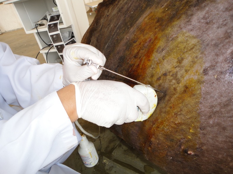
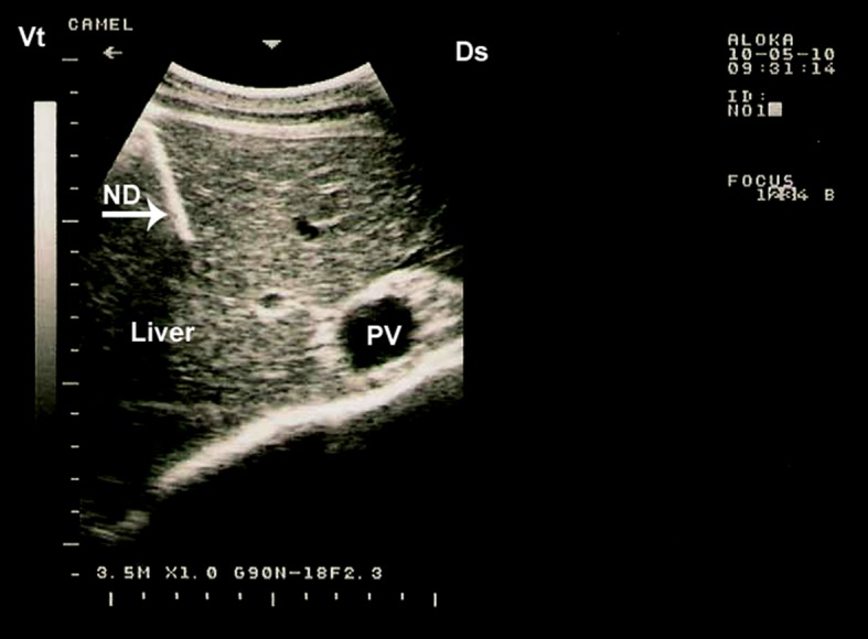
In camels, hepatic diseases are relatively common and most of them are misdiagnosed as a cause of illness because signs may be subtle. In addition, diagnostic laboratory methods are insufficient as hepatic enzymes can also be elevated in camels with cardiac or skeletal muscle damage. Examples of liver diseases in camels are hepatic lipidosis, hepatitis, cirrhosis, hepatic necrosis, choleostasis, hyperplasia of biliary epithelium, hydatid cysts, glycogen deposition, cholangitis, cholangiohepatitis, calcified hydatid cyst and hepatic abscesses. When the liver is examined by ultrasonography, the clinician gets sufficient information about the size, position, echopatterns of the hepatic parenchyma, bile ducts and outlines of the hepatic blood vessels. Ultrasonography has been used previously in camels only for reproductive purposes. However, during the past decade, it has been used for scanning of the healthy organs as well as evaluation and determining the diagnosis and prognosis of non-reproductive disorders. Examples of diseases evaluated by ultrasonography in camels are paratuberculosis, trypanosomiasis, abdominal and urinary disorders, thoracic diseases, renal tumors, pyelonephritis, renal abscessation, gastrointestinal tumors, chronic peritonitis and splenic abscessation. Ultrasound-guidance in biopsy of hepatic lesions and in portocentesis has also been reported in camels. This mini review article is written to shed light on ultrasonography of the liver and its blood vessels in healthy camels as well as finding in camels with hepatic disorders such as fatty infiltration of the liver, hepatic abscesses and calcification of the bile ducts.

Keywords: camel; dromedary; imaging; liver; ultrasonography.


Conflict of interest statement

The author declares no conflict of interest related to this review article.

Figures

Fig. 1.
Fig. 1.
Anatomical position of the liver in a camel, right side view. The liver is positioned in a camel carcass preserved in 10% formalin solution (a) and at postmortem examination without preservation (b).
Fig. 2.
Fig. 2.
Lobulation of the liver in an adult camel (a, parietal surface; b, visceral surface).
Fig. 3.
Fig. 3.
Ultrasonogram of the hepatic parenchyma and portal vein (PV) in a healthy camel. The vein is positively differentiated from the hepatic vein in the area of the portal fissure because the portal veins in this region are characterized by stellate ramifications. This image is taken at the right 10th intercostal space. Ds, dorsal; Vt, ventral.
Fig. 4.
Fig. 4.
Ultrasonogram of the caudal vena cava (CVC) in a healthy camel. In this animal, the imaged vein was triangular (arrow). The image was taken from right 11th intercostal space. Ds, dorsal; Vt, ventral.
Fig. 5.
Fig. 5.
Ultrasonogram of the hepatic vein (HV) in a healthy camel. The vein wall appeared to be anechoic. The image was taken from right 9th intercostal space. 1 =hepatic parenchyma. Ds, dorsal; Vt, ventral.
Fig. 6.
Fig. 6.
A free-hand, ultrasound-guided advancement of the biopsy needle towards the hepatic parenchyma.
Fig. 7.
Fig. 7.
Hepatic biopsy in a camel. The needle is clearly visible within the hepatic parenchyma. PV, portal vein; ND, needle; Ds, dorsal; Vt, ventral.
Fig. 8.
Fig. 8.
Liver biopsy in camels. A 14G × 150 mm spinal biopsy needle can be used for liver biopsy in camels (a). Image (b) shows a hepatic biopsy specimen from a camel.
Fig. 9.
Fig. 9.
Portocentesis in a healthy camel. The needle (ND) appears on the screen as a sharp bright line. The image was taken in the right 10th intercostal space using a 3.5 MHz convex transducer. PV, portal vein; Ds, dorsal; Vt, ventral.
Fig. 10.
Fig. 10.
Profound depression in a female camel with fatty infiltration of the liver (a). Yellowish discoloration of the oral mucosa (b) and sclera (c) are shown. Diagnosis was confirmed histologically.
Fig. 11.
Fig. 11.
Ultrasonograms of fatty infiltration of the liver in a camel (b) compared to healthy one (a). The liver appears hyperechogenic on ultrasonograms compared to normal imaging picture. PV, portal vein; HV, hepatic vein.
Fig. 12.
Fig. 12.
Ultrasonogram of severe fatty degeneration in 2 camels. The transducer was placed in the 11th intercostal space using a 7.5 MHz linear transducer (a) and 3.5 MHz sector transduced (b). The liver is hyperechoic near the abdominal wall and cannot be visualized very far from the abdominal wall.
Fig. 13.
Fig. 13.
Hepatic ultrasonogram of a hepatic abscess in a camel (arrow, a) which was detected at postmortem examination (b).
Fig. 14.
Fig. 14.
Hepatic ultrasonogram of multiple hepatic abscesses of different sized in a camel. Differential diagnosis includes hepatic cirrhosis, calcified hydatid cysts, calcification of the bile ducts and hepatic tumors.
Fig. 15.
Fig. 15.
Hydatid cyst in a camel liver. Image a shows ultrasonographic finding in a water bath for the distal part of the affected liver (b).
Fig. 16.
Fig. 16.
Calcified bile ducts and hepatic abscess detected by ultrasonography in 2 apparent healthy camels. Intense echoes are imaged accompanied by a distal acoustic shadow (a, b). AS, acoustic shadowing; AB, abscess; PV, portal vein. Arrow in image (b) points to the calcified bile duct.
Fig. 17.
Fig. 17.
Ultrasonogram of the caudal vena cava in a camel with systemic venous congestion due to cardiac insufficiency. Image was triangular from the 10th intercostal space. Ds, dorsal; Vt, ventral.

References

    1. Acorda J. A., Yamada H., Ghamsari S. M.1994. Ultrasonographic features of diffuse hepatocellular disorders in dairy cattle. Vet. Radiol. Ultrasound 35: 196–200. doi: 10.1111/j.1740-8261.1994.tb01592.x - DOI
    1. Al-Ani F. K.2004. The urinary system. pp. 263–267. In: Camel Management and Diseases (Al-Ani, F. K. ed.), Al-Sharq printing press, Amman.
    1. Aljameel M. A., Halima M. O., ElTigani-Asil A. E., Abdalla A. S., Abdellatif M. M.2014. Liver abscesses in dromedary camels: Pathological characteristics and aerobic bacterial aetiology. Open Vet. J. 4: 118–123. - PMC - PubMed
    1. Bobe G., Amin V. R., Hippen A. R., She P., Young J. W., Beitz D. C.2008. Non-invasive detection of fatty liver in dairy cows by digital analyses of hepatic ultrasonograms. J. Dairy Res. 75: 84–89. doi: 10.1017/S002202990700297X - DOI - PubMed
    1. Braun U.2009. Ultrasonography of the liver in cattle. Vet. Clin. North Am. Food Anim. Pract. 25: 591–609. doi: 10.1016/j.cvfa.2009.07.003 - DOI - PubMed

MeSH terms