. 2020 May;132(5):1080-1090.
doi: 10.1097/ALN.0000000000003179.

Desflurane Anesthesia Alters Cortical Layer-specific Hierarchical Interactions in Rat Cerebral Cortex

Affiliations

Desflurane Anesthesia Alters Cortical Layer-specific Hierarchical Interactions in Rat Cerebral Cortex

Anthony G Hudetz et al. Anesthesiology. 2020 May.

Abstract

Background: Neurocognitive investigations suggest that conscious sensory perception depends on recurrent neuronal interactions among sensory, parietal, and frontal cortical regions, which are suppressed by general anesthetics. The purpose of this work was to investigate if local interactions in sensory cortex are also altered by anesthetics. The authors hypothesized that desflurane would reduce recurrent neuronal interactions in cortical layer-specific manner consistent with the anatomical disposition of feedforward and feedback pathways.

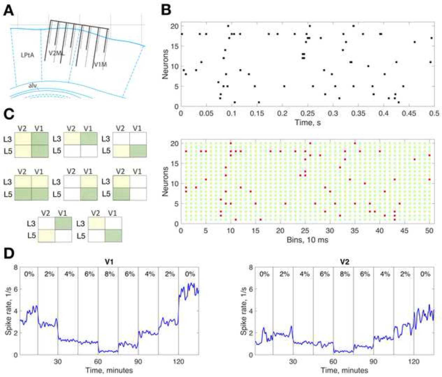
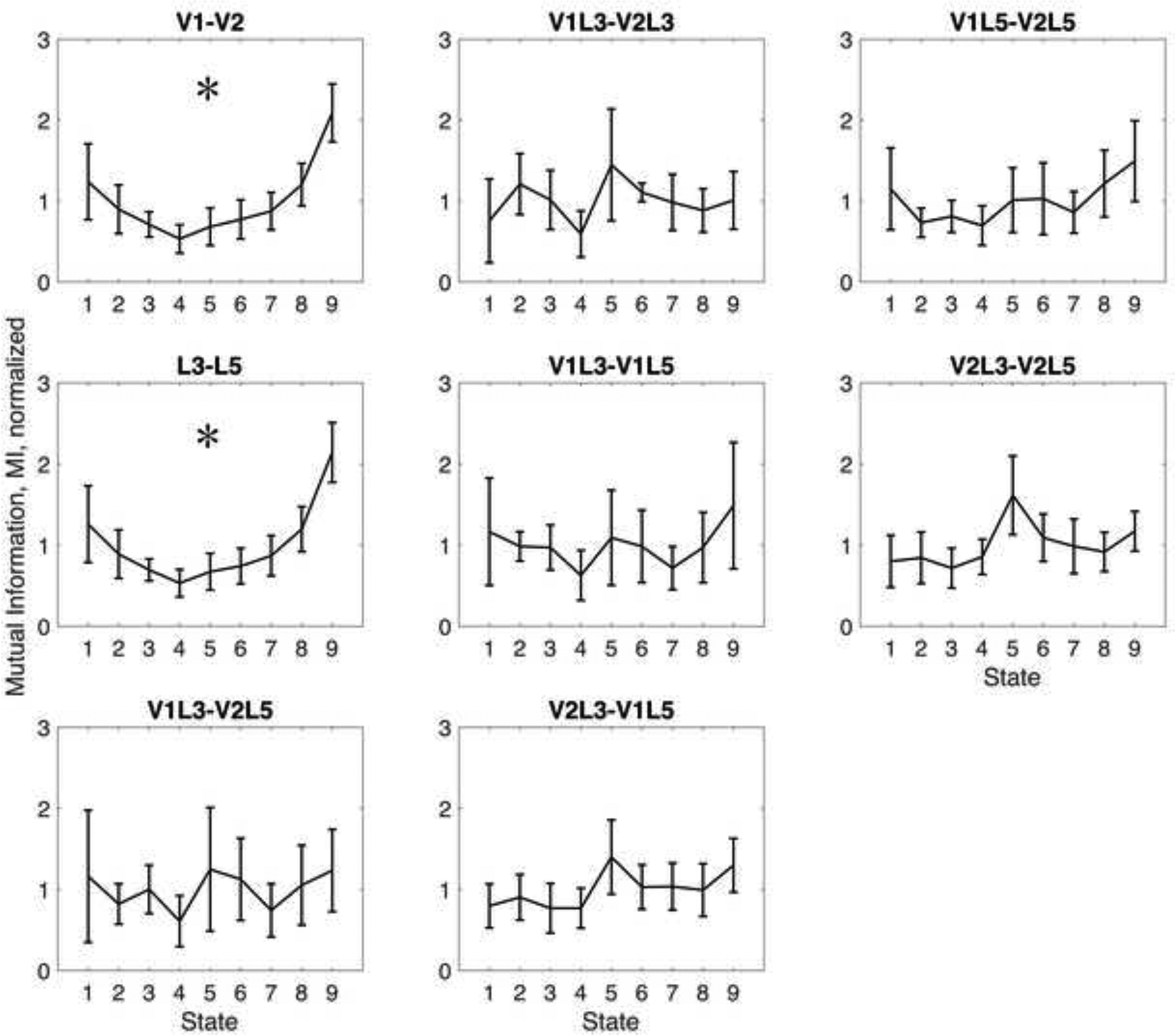
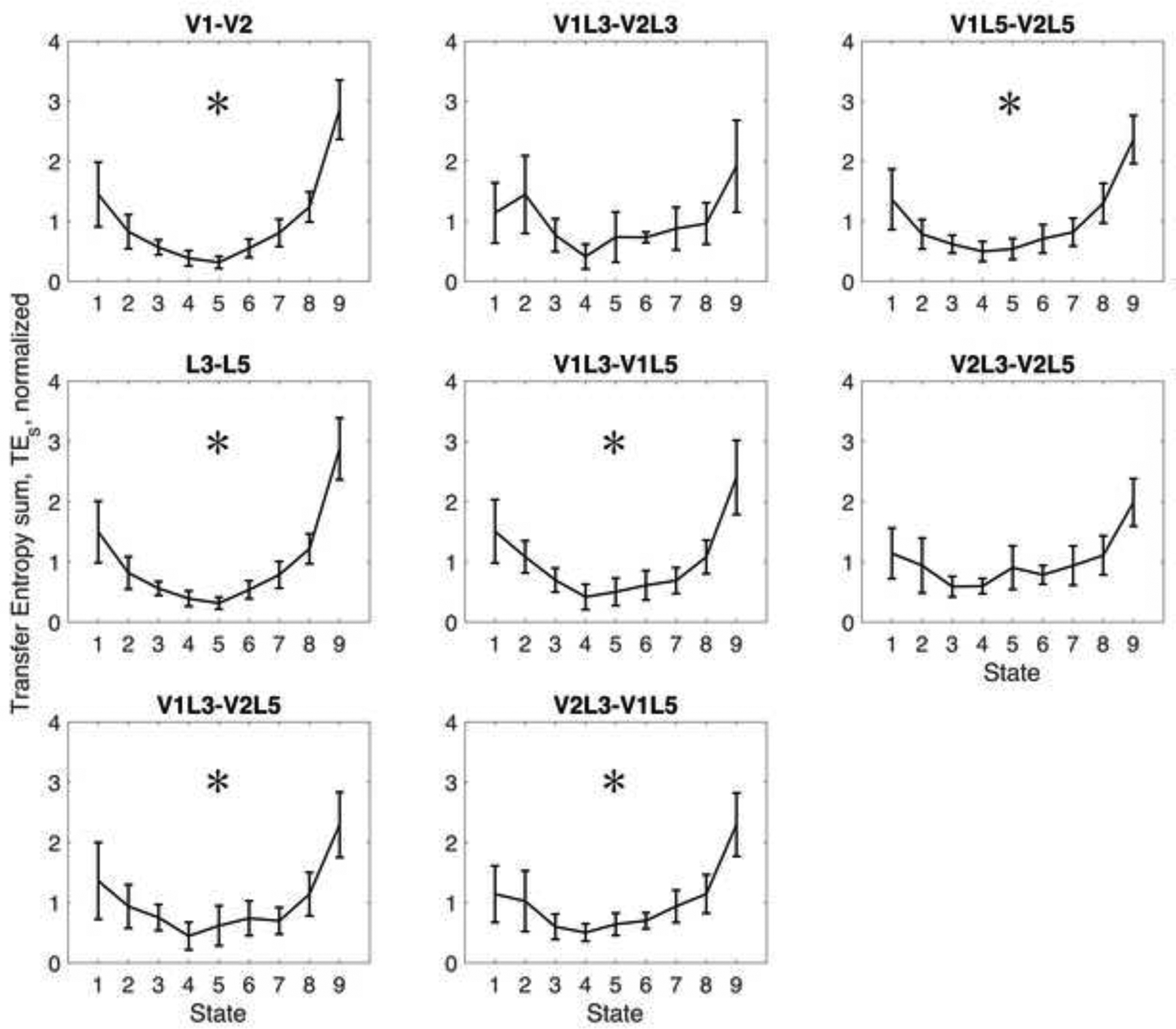
Methods: Single-unit neuronal activity was measured in freely moving adult male rats (268 units; 10 animals) using microelectrode arrays chronically implanted in primary and secondary visual cortex. Layer-specific directional interactions were estimated by mutual information and transfer entropy of multineuron spike patterns within and between cortical layers three and five. The effect of incrementally increasing and decreasing steady-state concentrations of desflurane (0 to 8% to 0%) was tested for statistically significant quadratic trend across the successive anesthetic states.

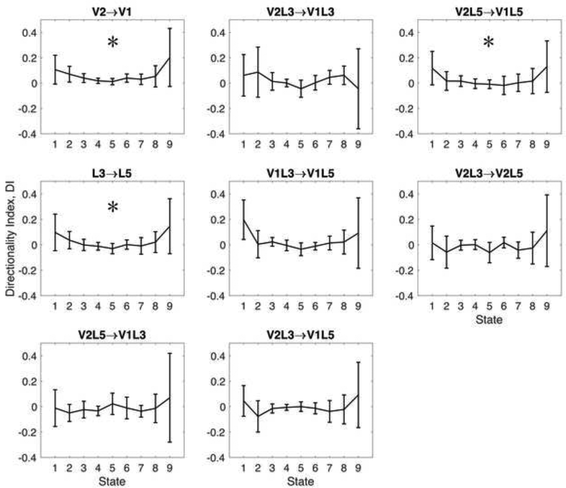
Results: Desflurane produced robust, state-dependent reduction (P = 0.001) of neuronal interactions between primary and secondary visual areas and between layers three and five, as indicated by mutual information (37 and 41% decrease at 8% desflurane from wakeful baseline at [mean ± SD] 0.52 ± 0.51 and 0.53 ± 0.51 a.u., respectively) and transfer entropy (77 and 78% decrease at 8% desflurane from wakeful baseline at 1.86 ± 1.56 a.u. and 1.87 ± 1.67 a.u., respectively). In addition, a preferential suppression of feedback between secondary and primary visual cortex was suggested by the reduction of directional index of transfer entropy overall (P = 0.001; 89% decrease at 8% desflurane from 0.11 ± 0.18 a.u. at baseline) and specifically, in layer five (P = 0.001; 108% decrease at 8% desflurane from 0.12 ± 0.19 a.u. at baseline).

Conclusions: Desflurane anesthesia reduces neuronal interactions in visual cortex with a preferential effect on feedback. The findings suggest that neuronal disconnection occurs locally, among hierarchical sensory regions, which may contribute to global functional disconnection underlying anesthetic-induced unconsciousness.


Conflict of interest statement

Conflicts of Interest: None

Figures

Figure 1.
Figure 1.
Microwire array and data analysis. (A) Location of the microwire array in rat visual cortex. Eight wires target primary visual cortex monocular area (V1M) and 8 wires target secondary visual cortex mediolateral area (V2ML). The wires have alternating lengths with wire tips residing approximately in cortical layer 3 and layer 5. Only two rows of wires are illustrated; the second row is shown with dotted lines. Scale above is the distance in mm from Bregma at 3.4 mm lateral position. LPtA: lateral parietal association area. Drawing is based on Paxinos and Watson atlas. (B) Spike raster plot for 20 recorded neurons in one experiments (top) and binned binary representation of spiking (bottom). Red dots indicate at least one spike present in each 10ms bin, green open circles indicate no spike. Each configuration of coincident red dots across neurons represents a population activity pattern. (C) Pairwise cortical regions (yellow and green areas) for which mutual information and transfer entropy were calculated. (D) Time course of average spike rate from all units within V1 and V2 in one experiment. Spike rate was calculated for 10-second time bins and 10-point Gaussian smoothing was applied. Data from consecutive anesthetic states were concatenated, thus the time axis shows cumulative recording time, not real time. Vertical gray lines demarcate anesthetic conditions with percent inhaled concentration of desflurane indicated.
Figure 2.
Figure 2.
Effect of desflurane on mutual information between neurons recorded in primary and secondary visual cortex (V1, V2), cortical layers 3 and 5 (L3, L5), and their subregions. Anesthetic states 1 to 9 correspond to consecutive desflurane concentrations of 0, 2, 4, 6, 8, 6, 4, 2, 0 percent, respectively. Mutual information is in relative units, plotted as mean and 95% confidence interval. *: statistically significant quadratic trend with consecutive anesthetic states.
Figure 3.
Figure 3.
Robust concentration-dependent effect of desflurane on the sum of transfer entropies calculated between primary and secondary visual cortex (V1-V2), between cortical layers 3 and 5 (L3, L5), and between various subregions. Anesthetic states 1 to 9 correspond to consecutive desflurane concentrations of 0, 2, 4, 6, 8, 6, 4, 2, 0 percent, respectively. Transfer entropy sum is in relative units, plotted as mean and 95% confidence interval. *: statistically significant quadratic trend with consecutive anesthetic states.
Figure 4.
Figure 4.
Effect of desflurane on the directionality index. Statistically significant change occurred in layer 5 secondary visual to layer 5 primary visual cortex (V2L5-V1L5) implying a reduction of feedback connectivity at higher desflurane concentration. Anesthetic states 1 to 9 correspond to consecutive desflurane concentrations of 0, 2, 4, 6, 8, 6, 4, 2, 0 percent, respectively. Directionality index is in relative units, plotted as mean and 95% confidence interval. *: statistically significant quadratic trend with consecutive anesthetic states.

References

    1. John ER, Prichep LS, Kox W, Valdes-Sosa P, Bosch-Bayard J, Aubert E, Tom M, diMichele F, Gugino LD: Invariant reversible QEEG effects of anesthetics. Conscious Cogn 2001; 10: 165–83. - PubMed
    1. Alkire MT, Hudetz AG, Tononi G: Consciousness and anesthesia. Science 2008; 322: 876–80 - PMC - PubMed
    1. Bonhomme V, Boveroux P, Brichant JF, Laureys S, Boly M: Neural correlates of consciousness during general anesthesia using functional magnetic resonance imaging (fMRI). Arch Ital Biol 2012; 150: 155–63 - PubMed
    1. Kelz MB, Garcia PS, Mashour GA, Solt K: Escape From Oblivion: Neural Mechanisms of Emergence From General Anesthesia. Anesth Analg 2019; 128: 726–736 - PMC - PubMed
    1. Voss LJ, Garcia PS, Hentschke H, Banks MI: Understanding the Effects of General Anesthetics on Cortical Network Activity Using Ex Vivo Preparations. Anesthesiology 2019; 130: 1049–1063 - PMC - PubMed

Publication types