. 2020 Jul;161(7):1636-1649.
doi: 10.1097/j.pain.0000000000001846.

Distribution of functional opioid receptors in human dorsal root ganglion neurons

Affiliations

Distribution of functional opioid receptors in human dorsal root ganglion neurons

Jamie K Moy et al. Pain. 2020 Jul.

Abstract

Preclinical evidence has highlighted the importance of the μ-opioid peptide (MOP) receptor on primary afferents for both the analgesic actions of MOP receptor agonists, as well as the development of tolerance, if not opioid-induced hyperalgesia. There is also growing interest in targeting other opioid peptide receptor subtypes (δ-opioid peptide [DOP], κ-opioid peptide [KOP], and nociceptin/orphanin-FQ opioid peptide [NOP]) on primary afferents, as alternatives to MOP receptors, which may not be associated with as many deleterious side effects. Nevertheless, results from several recent studies of human sensory neurons indicate that although there are many similarities between rodent and human sensory neurons, there may also be important differences. Thus, the purpose of this study was to assess the distribution of opioid receptor subtypes among human sensory neurons. A combination of pharmacology, patch-clamp electrophysiology, Ca imaging, and single-cell semiquantitative polymerase chain reaction was used. Our results suggest that functional MOP-like receptors are present in approximately 50% of human dorsal root ganglion neurons. δ-opioid peptide-like receptors were detected in a subpopulation largely overlapping that with MOP-like receptors. Furthermore, KOP-like and NOP-like receptors are detected in a large proportion (44% and 40%, respectively) of human dorsal root ganglion neurons with KOP receptors also overlapping with MOP receptors at a high rate (83%). Our data confirm that all 4 opioid receptor subtypes are present and functional in human sensory neurons, where the overlap of DOP, KOP, and NOP receptors with MOP receptors suggests that activation of these other opioid receptor subtypes may also have analgesic efficacy.


Figures
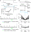
Figure 1.
Figure 1.. Voltage-gated Ca2+ currents (VGCCs) in human DRG neurons as a target for opioid receptor activation, and as a mechanism for initiating depolarization-induced increases in intracellular Ca2+.
A. VGCC were evoked from a holding potential of −60 mV, to a test potential of 0 mV, before and after the application of DAMGO (1 μM). Inset: A diary plot of the peak inward current before, during and after DAMGO as indicated. B. A pre-pulse potentiation protocol involving an 80 ms pre-pulse to either −70 mV (thin lines) or +80 mV (heavier lines) prior to a test pulse to 0 mV, was employed before (top traces) and after (bottom traces) the application of DAMGO. Current suppressed by DAMGO could be recovered by a pre-pulse to +80 mV. C. Typical Ca2+ imaging protocol used to assess the impact of opioid receptor agonists on Ca2+ transients evoked with a depolarization associated with the application (800 ms) of 50 mM KCl. The evoked transient was reversibly eliminated by the application of a Ca2+ free bath solution. The neuron illustrated was responsive to capsaicin (500 nM). D. Pooled data from 23 neurons from three donors confirm the dependence of the depolarization evoked transient on Ca2+ influx (F (1.3, 35.4) = 107.9, df=2, n=29, **p<0.01, RM ANOVA). E. The depolarization evoked Ca2+ transient was also reversibly attenuated by the non-specific VGCC blockers Cd2+ (100μM) and Ni2+ (100μM). F. Pooled data from 9 neurons from 3 donors confirmed that the attenuation of the evoked Ca2+ transient was significant (F (1.5, 11.7) = 37.3, df=2, n=9, **p=0.01, RM ANOVA).
Figure 2.
Figure 2.. Characterization of functional MOP receptors in human DRG neurons.
A. DAMGO responsive neuron that was also responsive to capsaicin, but that appeared un-responsive to nociceptin. B. Pooled data from DAMGO responsive neurons challenged with DAMGO at concentrations between 30 nM and 1 μM. Data were fitted with a Hill equation yielding an EC50 of 56 nM and a maximal inhibition of 40%. Inset: Percent of DAMGO responsive neurons (Responders) plotted as function of DAMGO concentration. C. The MOP receptor antagonists naloxone (top trace) and CTOP (bottom trace) attenuated the magnitude of DAMGO-mediated inhibition and/or percentage of DAMGO responsive neurons (Naloxone; p=0.19, chi-squared test. CTOP; *p=0.045, fisher’s exact test). Interestingly, however, CTOP itself, significantly attenuated the evoked Ca2+ transient. D. Pooled data from neurons challenged with DAMGO alone, or DAMGO in the presence of either naloxone or CTOP (P<0.01, F(2, 78)=17.03, ANOVA, post-hoc Dunnett’s test for multiple comparisons, **p=0.01).
Figure 3.
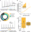
Figure 3.. Characterization of DOP receptor agonist, SNC80, in human DRG neurons.
A. Example of a neuron pre-treated (for 30 min) with the PTEN inhibitor, bpV(Phen), that was responsive to SNC80 (1 μM), DAMGO (1 μM), and capsaicin (500 nM). B. Without pretreatment with bpV(Phen), very few neurons were responsive to SNC80 at a concentration of 1 μM. However, the fractional inhibition and percentage of responsive neurons (inset) increased as the concentration of SNC80 was increased to 3 and 10 μM. C. Pooled data of neurons pre-treated with bpV(Phen) or vehicle that were subsequently challenged with 1 μM SCN80 (p=0.026, chi-square). D. Distribution of vehicle and bpV(Phen) treated neurons that were responsive to SNC80, DAMGO, and/or capsaicin. In this and subsequent figures, each spoke in the wheel represents a single neuron. Of note, the vehicle wheel includes the 2/15 neurons responsive to SNC80 when challenged with the agonist alone, as well as the 2/2 neurons responsive to SNC80 when challenged with the agonist following application of naltrindole. E. Example of a neuron challenged with SCN80 following application of the DOP receptor antagonist naltrindole. The neuron was subsequently challenged with DAMGO and capsaicin. Note the inhibition associated with naltrindole alone. F. Pooled data of bpV(Phen) treated neurons that were subsequently challenged with SCN80 alone or following the application of naltrindole. While there was no significant influence of naltrindole on the percentage of neurons responsive to SNC80 (p=0.24, chi-square), the attenuation of the magnitude of the suppression of the evoked transient was significant (t=2.97, df=17, **p=0.01, unpaired t-test).
Figure 4.
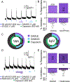
Figure 4.. Further characterization functional DOP receptors with peptide agonist, DADLE, in human DRG neurons.
A. Example of Ca2+ signaling in neuron responsive to the DOP receptor agonist DADLE. This neuron was also responsive to DAMGO and capsaicin. B. In contrast to the influence of bpV(Phen) on the proportion of neurons responsive to SNC80, there was no detectable influence of PTEN inhibition on either the proportion of neurons responsive to DADLE (p=0.40, chi-squared), or the magnitude of the response (t=0.16, df=25, p > 0.05, t-test). C. Distribution of vehicle and bpV(Phen) treated neurons that were responsive to DADLE, DAMGO, and/or capsaicin. D. Example of a neuron in which naltrindole attenuated the response to DADLE, that was also responsive to DAMGO and capsaicin. E. Pooled data from neurons challenged with DADLE alone or DADLE following the application of naltrindole. While there was no detectable influence of naltrindole on the proportion of neurons responsive to DADLE (p=0.86, chi-square), there was a significant attenuation of the magnitude of the DADLE effect (**p=0.0037, t=3.183, df=27, t-test).
Figure 5.
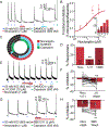
Figure 5.. Characterization of functional KOP receptors in human DRG neurons.
A. Example of Ca2+ signaling in a neuron responsive to dynorphin and DAMGO, but not capsaicin. B. Both the magnitude of dynorphin-induced suppression of the evoked Ca2+ transient, and the proportion of neurons responsive to this KOP receptor agonist (inset), were concentration dependent. C. Distribution of neurons responsive to dynorphin, DAMGO, and capsaicin. D. Example of Ca2+ signaling in a neuron in which pre-application of the KOP receptor antagonist, nor-BNI was associated with the absence of a response to dynorphin. This neuron was responsive to capsaicin, however. E. Pooled data for neurons challenged with dynorphin alone or following application of nor-BNI. The proportion of neurons responsive to dynorphin (**p=0.0025, chi-square) and the magnitude of the suppression of the evoked Ca2+ transient (*p=0.027, Mann Whitney t-test) were significantly reduced by nor-BNI.
Figure 6.
Figure 6.. Characterization of functional NOP receptors in human DRG neurons.
A. Example of Ca2+ signaling in a neuron responsive to nociceptin. This neuron was also responsive to DAMGO and capsaicin. B. Pooled data indicate that both the magnitude of the nociceptin-induced suppression of the evoked Ca2+ transient and the proportion of neurons responsive to nociceptin (inset) was concentration dependent. C. The distribution of neurons responsive to nociceptin, DAMGO, and/or capsaicin. D. The proportion of neurons responsive to nociceptin when applied alone, or following application of the putative NOP receptor antagonists J113397 or SB612111 (J113397; **p=0.01, chi-square). E. Example of Ca2+ signaling in a neuron that was responsive to nociceptin following wash of J113397. F. Pooled magnitude of transient inhibition data from neurons treated with nociceptin in the presence of J113397, and again following wash of the antagonist (t=2.977, df=28, **p=0.01, paired t-test). G. Example of Ca2+ signaling in a neuron in which a nociceptin-induced decrease in the magnitude of the evoked transient was only detected following wash of the NOP receptor antagonist SB612111. H. Pooled data of neurons challenged with nociceptin in the presence of and then following wash of SB612111. The increase in the magnitude of the suppression of the evoked transient was significant (nociceptin [300nM] alone, t=2.977, df=15, **p=0.01, paired t-test).
Figure 7.
Figure 7.. Expression of opioid receptor subtypes among human DRG neurons.
A. The pattern of opioid receptor subtype and TRPV1 expression in neurons pooled from five different organ donors. B. The sex and age of each donor is indicated in the center of each wheel. As with previous figures, each spoke is a single neuron, except that mRNA was assessed rather than functional responses to agonists. One donor was African American (29yo female).

Similar articles

Cited by

References

    1. Abdelhamid EE, Sultana M, Portoghese PS, Takemori AE. Selective blockage of delta opioid receptors prevents the development of morphine tolerance and dependence in mice. J Pharmacol Exp Ther 1991;258(1):299–303. - PubMed
    1. Aley KO, Levine JD. Different mechanisms mediate development and expression of tolerance and dependence for peripheral mu-opioid antinociception in rat. J Neurosci 1997;17(20):8018–8023. - PMC - PubMed
    1. Anand P, Yiangou Y, Anand U, Mukerji G, Sinisi M, Fox M, McQuillan A, Quick T, Korchev YE, Hein P. Nociceptin/orphanin FQ receptor expression in clinical pain disorders and functional effects in cultured neurons. Pain 2016;157(9):1960–1969. - PubMed
    1. Bardoni R, Tawfik VL, Wang D, Francois A, Solorzano C, Shuster SA, Choudhury P, Betelli C, Cassidy C, Smith K, de Nooij JC, Mennicken F, O’Donnell D, Kieffer BL, Woodbury CJ, Basbaum AI, MacDermott AB, Scherrer G. Delta Opioid Receptors Presynaptically Regulate Cutaneous Mechanosensory Neuron Input to the Spinal Cord Dorsal Horn. Neuron 2014;81(6):1443. - PubMed
    1. Biggs JE, Yates JM, Loescher AR, Clayton NM, Boissonade FM, Robinson PP. Changes in vanilloid receptor 1 (TRPV1) expression following lingual nerve injury. Eur J Pain 2007;11(2):192–201. - PubMed