

Fibrin Clot Mistook as a Worm in the Intravenous Line
- PMID: 32102129
- PMCID: PMC7044686
- DOI: 10.3349/ymj.2020.61.3.267
Fibrin Clot Mistook as a Worm in the Intravenous Line
Abstract
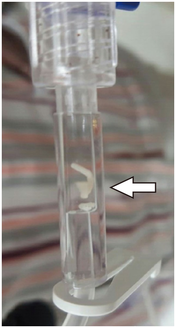
There have been several reports of foreign bodies being discovered in the intravenous set. In this case, the patient complained that he found a worm in his intravenous line. It was later confirmed as a long, white fibrin deposit by pathologic examination. This happened even though there was a non-return valve in the intravenous line. Also, since there were few red blood cells in the deposit, it did not look like a blood clot. In cases like this, we suggest that physicians keep this possibility in mind to reassure their patients.
Keywords: Fibrin clot; foreign body; intravenous line; non-return valve; patient-controlled analgesia.
© Copyright: Yonsei University College of Medicine 2020.
Conflict of interest statement
The authors have no potential conflicts of interest to disclose.
Figures


References
-
- Madsen H, Winding O. Release of foreign bodies (particles) by clinical use of intravenous infusion sets. Biomaterials. 1996;17:663–666. - PubMed
-
- Yorioka K, Oie S, Oomaki M, Imamura A, Kamiya A. Particulate and microbial contamination in in-use admixed intravenous infusions. Biol Pharm Bull. 2006;29:2321–2323. - PubMed
-
- Gschwind CR. The intravenous foreign body: a report of 2 cases. J Hand Surg Am. 2002;27:350–354. - PubMed
Publication types
MeSH terms
Substances
LinkOut - more resources
Full Text Sources
Medical
