Review
. 2020 Feb 23;21(4):1517.
doi: 10.3390/ijms21041517.

Mucopolysaccharidosis IVA: Diagnosis, Treatment, and Management

Affiliations
Review

Mucopolysaccharidosis IVA: Diagnosis, Treatment, and Management

Kazuki Sawamoto et al. Int J Mol Sci. .

Abstract

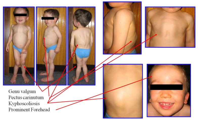
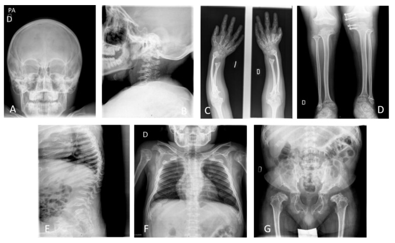
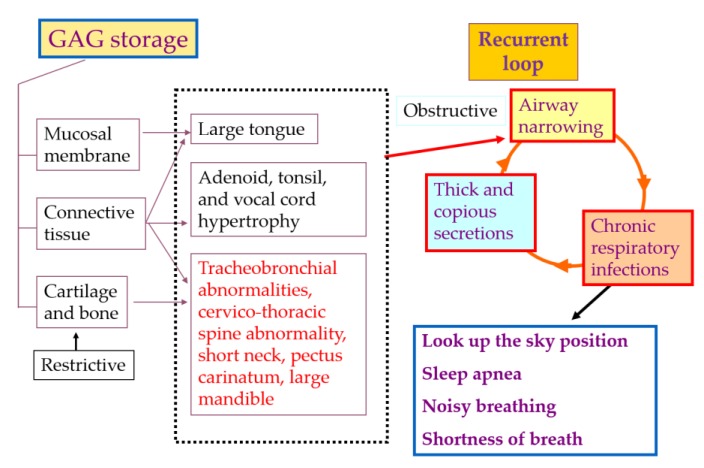
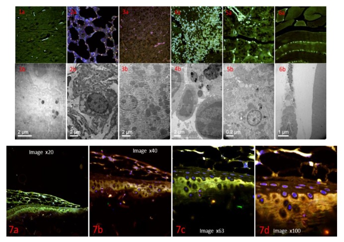
Mucopolysaccharidosis type IVA (MPS IVA, or Morquio syndrome type A) is an inherited metabolic lysosomal disease caused by the deficiency of the N-acetylglucosamine-6-sulfate sulfatase enzyme. The deficiency of this enzyme accumulates the specific glycosaminoglycans (GAG), keratan sulfate, and chondroitin-6-sulfate mainly in bone, cartilage, and its extracellular matrix. GAG accumulation in these lesions leads to unique skeletal dysplasia in MPS IVA patients. Clinical, radiographic, and biochemical tests are needed to complete the diagnosis of MPS IVA since some clinical characteristics in MPS IVA are overlapped with other disorders. Early and accurate diagnosis is vital to optimizing patient management, which provides a better quality of life and prolonged life-time in MPS IVA patients. Currently, enzyme replacement therapy (ERT) and hematopoietic stem cell transplantation (HSCT) are available for patients with MPS IVA. However, ERT and HSCT do not have enough impact on bone and cartilage lesions in patients with MPS IVA. Penetrating the deficient enzyme into an avascular lesion remains an unmet challenge, and several innovative therapies are under development in a preclinical study. In this review article, we comprehensively describe the current diagnosis, treatment, and management for MPS IVA. We also illustrate developing future therapies focused on the improvement of skeletal dysplasia in MPS IVA.

Keywords: GALNS; LC-MS/MS; MPS IVA; bone-targeting; keratan sulfate; skeletal dysplasia; tracheal reconstructive surgery.


Conflict of interest statement

The authors declare no conflict of interest.

Figures

Figure 1
Figure 1
Clinical features of an MPS IVA patient. The patients in this figure had severe form at three years of age and had bone abnormalities of short stature, genu valgum, pectus carinatum, kyphoscoliosis, and prominent forehead. His height is 90 cm with the 50th percentile of male MPS IVA growth chart (adapted from Educational CD for Morquio and permitted by Carol Ann Foundation and Morquio Conference; https://morquioconference.wixsite.com/morquio).
Figure 2
Figure 2
X-ray images of a 17-year-old male patient with MPS IVA. Images show skeletal dysplasia (dysostosis multiplex); (A) incomplete ossification and prominent forehead, (B) incomplete ossification in odontoid process and subluxation of the atlas secondary to odontoid hypoplasia with platyspondyly of cervical vertebrae, (C) cortical thinning and mild widening of the diaphysis of the humerus and tilting of the radial epiphysis towards the ulna producing curvature, (D) genu valgum with cortical thinning of tibia and fibula, (E) the accentuated dorsal thoracolumbar kypholordosis with the advanced platyspondyly, irregularity, and anterior beaking of vertebral bodies characteristic of MPS IVA and flared ribs, (F) abnormal thoracic cage, pectus carinatum and scoliosis with oar shaped ribs: the ribs are wide anteriorly and laterally and overconstricted in their paravertebral portions, (G) spondyloepiphyseal dysplastic femoral heads and oblique acetabular roof with coxa valgus deformity and flared iliac wings.
Figure 3
Figure 3
Pathophysiology of airway compromise in MPS IVA patients (adapted from Educational CD for Morquio and permitted by Carol Ann Foundation and Morquio Conference; https://morquioconference.wixsite.com/morquio).
Figure 4
Figure 4
Distribution of nanostructured lipid carriers containing GALNS in mouse tissues. Images different tissues in wild type mouse, (1a) confocal microscopy brain tissue, (1b) electronic microscopy nanoparticles inside of neuron cell, (2a) confocal microscopy lung tissue, (2b) electronic microscopy nanoparticles inside of pneumocyte cell, (3a) confocal microscopy liver tissue, (3b) electronic microscopy nanoparticles inside of hepatocyte cells, (4a) confocal microscopy spleen tissue, (4b) electronic microscopy nanoparticles inside of macrophages cells, (5a) confocal microscopy kidney tissue, (5b) confocal microscopy renal tubule cells, (6a) confocal microscopy muscle tissue, (6b) confocal microscopy fibroblast cell, (7) images of mouse cartilage of mice at different zoom, showing that the cell inside of the cartilage contains nanoparticles (pink color) (7a) picture of cartilage ×20 zoom, (7b) picture of cartilage ×40 zoom, (7c) picture of cartilage ×63 zoom, (7d) picture of cartilage ×100 zoom.

References

    1. Neufeld E.F., Muenzer J. The mucopolysaccharidoses. In: Scriver C.R., Beaudet A.L., Sly W.S., Valle D., editors. The Metabolic and Molecular Bases of Inherited Disease. 8th ed. McGraw-Hill; New York, NY, USA: 2001. pp. 3421–3452.
    1. Khan S., Alméciga-Díaz C.J., Sawamoto K., Mackenzie W.G., Theroux M.C., Pizarro C., Mason R.W., Orii T., Tomatsu S. Mucopolysaccharidosis IVA and glycosaminoglycans. Mol. Genet. Metab. 2017;120:78–95. doi: 10.1016/j.ymgme.2016.11.007. - DOI - PMC - PubMed
    1. Peracha H., Sawamoto K., Averill L., Kecskemethy H., Theroux M., Thacker M., Nagao K., Pizarro C., Mackenzie W., Kobayashi H., et al. Diagnosis and prognosis of Mucopolysaccharidosis IVA. Mol. Genet. Metab. 2018;125:18–37. doi: 10.1016/j.ymgme.2018.05.004. - DOI - PMC - PubMed
    1. Sawamoto K., Alméciga-Díaz C.J., Mason R.W., Orii T., Tomatsu S. In: Mucopolysaccharidosis type IVA: Clinical features, biochemistry, diagnosis, genetics, and treatment. Mucopolysaccharidoses Update. Tomatsu S., editor. Nova Science Publishers; New York, NY, USA: 2018. pp. 235–272.
    1. Tomatsu S., Averill L.W., Sawamoto K., Mackenzie W.G., Bober M.B., Pizarro C., Goff C.J., Xie L., Orii T., Theroux M. Obstructive airway in Morquio A syndrome, the past, the present and the future. Mol. Genet. Metab. 2016;117:150–156. doi: 10.1016/j.ymgme.2015.09.007. - DOI - PMC - PubMed

MeSH terms

LinkOut - more resources