Review
. 2020 Feb 23;8(2):308.
doi: 10.3390/microorganisms8020308.

The Human Oral Microbiome in Health and Disease: From Sequences to Ecosystems

Affiliations
Review

The Human Oral Microbiome in Health and Disease: From Sequences to Ecosystems

Jesse R Willis et al. Microorganisms. .

Abstract

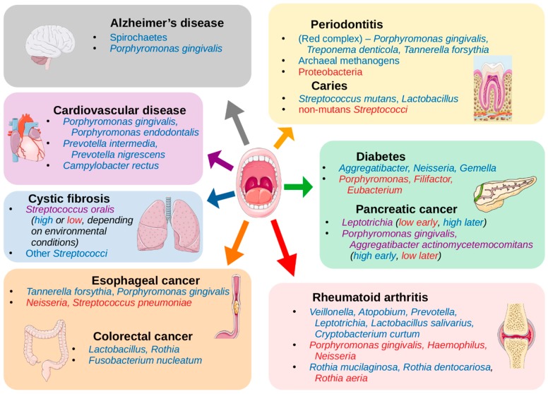
Abstract: The human oral cavity is home to an abundant and diverse microbial community (i.e., the oral microbiome), whose composition and roles in health and disease have been the focus of intense research in recent years. Thanks to developments in sequencing-based approaches, such as 16S ribosomal RNA metabarcoding, whole metagenome shotgun sequencing, or meta-transcriptomics, we now can efficiently explore the diversity and roles of oral microbes, even if unculturable. Recent sequencing-based studies have charted oral ecosystems and how they change due to lifestyle or disease conditions. As studies progress, there is increasing evidence of an important role of the oral microbiome in diverse health conditions, which are not limited to diseases of the oral cavity. This, in turn, opens new avenues for microbiome-based diagnostics and therapeutics that benefit from the easy accessibility of the oral cavity for microbiome monitoring and manipulation. Yet, many challenges remain ahead. In this review, we survey the main sequencing-based methodologies that are currently used to explore the oral microbiome and highlight major findings enabled by these approaches. Finally, we discuss future prospects in the field.

Keywords: Next generation sequencing; Oral microbiome; microbiome perturbations; oral diseases; stomatotypes; systemic diseases.


Conflict of interest statement

The authors declare no conflict of interest.

Figures

Figure 1
Figure 1
Schematics of standard techniques used in microbiome studies. (A) Marker gene sequencing techniques can use primers to target certain conserved regions of a genome to capture intermittent variable regions, which can then be used to identify organisms in a sample rapidly and inexpensively. The 16S rRNA gene is the most commonly used marker gene in bacteria and archaea, and in the figure, primers are used to capture the V3 and V4 variable regions together, a common approach for 16S sequencing. The internal transcribed spacer (ITS) region of the nuclear rRNA cistron in fungi is made of two segments, which can be captured with primers targeting the 18S, 5.8S, and 28S rRNA sections that surround them. (BD) Instead of targeting one small segment of the genome, these techniques capture the entirety of the genetic material from an organism. (B) Single virus genomics (SVG) uses a fluorescent stain to isolate individual virus particles in a sample by fluorescence-activated virus sorting (FAVS), wherein they are embedded in an agarose bead before undergoing whole genome amplification and sequencing. (C) Whole metagenome shotgun sequencing (WMS) involves the fragmentation of all DNA in a sample, sequencing of the fragments, and assembly of the sequences, which can then be mapped to reference genomes, or de novo assembly can be performed. (D) Metatranscriptomics also involves a shotgun sequencing approach, but it is performed after mRNA extraction. The outputs then allow for differential gene expression analysis. (E) Metabolomics and metaproteomics allow for quantification of the metabolites and proteins produced by the microbiome in a sample, respectively. Mass spectrometry is a common approach to quantification. Mock metabolite shapes in Figure 1 were generated using the JSME Molecular Editor by Peter Ertl and Bruno Bienfait licensed under CC-BY-NC-SA 3.0. Images of body sites and organs in Figure 1 and Figure 2 were obtained from Servier Medical Art by Servier licensed under CC-BY 3.0.Traditionally, 16S sequences were clustered into groups with at least 97% identity, called operational taxonomic units (OTUs), which have been used as proxies for species-level or, more commonly, genus-level taxonomic identification. A number of software tools are available, which convert reads to sample-by-OTU feature tables, such as QIIME [55] and mothur [56]. However, newer approaches are better able to control for amplicon sequencing errors, and thereby obviate the use of arbitrary identity thresholds, allowing for single-nucleotide resolution with amplicon sequence variants (ASVs) [57]. Software options for ASV methods include DADA2 [58] and Deblur [59].
Figure 2
Figure 2
Oral and systemic diseases associated with the oral microbiome. A representation of the associations found between diseases with increases or decreases of the abundances of organisms in the oral cavity (listed in Table 1 and Table 2). Organisms listed in blue have been shown to be increased in abundance in the oral cavity in individuals presenting with the noted disease, and organisms listed in red have been shown to be decreased. Those in purple may be either increased or decreased depending on the conditions or progression of the disease. Images of body sites and organs in Figure 1 and Figure 2 were obtained from Servier Medical Art by Servier licensed under CC-BY 3.0.
Figure 3
Figure 3
Gradients of abundances of consensus stomatotype-driving genera. Using a random subset of 500 samples from an oral microbiome dataset [10], samples were clustered into two stomatotypes using the weighted Unifrac distance measure. Type 1 samples are represented by circles and type 2 samples by squares. In each box, samples are colored by the total relative abundance of the indicated organisms. Overlaid are arrows indicating the tendency of the abundances of each organism noted in Table 3. In this subset of samples, Neisseria and Haemophilus strongly associate with stomatotype 1 samples, Prevotella strongly associates with stomatotype 2 samples while Veillonella does so weakly. The “variable stomatotype” drivers are indeed variable in their associations in this instance. Streptococcus shows a clear gradient but does not conform to either stomatotype. Gemella and Rothia, which have been shown to co-occur with Streptococcus in stomatotypes in the literature, do the same here, with Rothia more associated with stomatotype 1. However, Porphyromonas, which has been shown to co-occur with Streptococcus, Gemella, or Neisseria previously, associates with none of these here, and instead is strongly associated with stomatotype 2.

References

    1. Ursell L.K., Metcalf J.L., Parfrey L.W., Knight R. Defining the human microbiome. Nutr. Rev. 2012;70:S38–S44. doi: 10.1111/j.1753-4887.2012.00493.x. - DOI - PMC - PubMed
    1. Integrative HMP (iHMP) Research Network Consortium The Integrative Human Microbiome Project: Dynamic analysis of microbiome-host omics profiles during periods of human health and disease. Cell Host Microbe. 2014;16:276–289. doi: 10.1016/j.chom.2014.08.014. - DOI - PMC - PubMed
    1. Human Microbiome Project Consortium Structure, function and diversity of the healthy human microbiome. Nature. 2012;486:207–214. doi: 10.1038/nature11234. - DOI - PMC - PubMed
    1. Li K., Bihan M., Yooseph S., Methé B.A. Analyses of the microbial diversity across the human microbiome. PLoS ONE. 2012;7:e32118. doi: 10.1371/journal.pone.0032118. - DOI - PMC - PubMed
    1. Marsh P.D., Do T., Beighton D., Devine D.A. Influence of saliva on the oral microbiota. Periodontology. 2000;70:80–92. doi: 10.1111/prd.12098. - DOI - PubMed