. 2020 Feb 24;9(2):515.
doi: 10.3390/cells9020515.

Non-Phosphorylatable PEA-15 Sensitises SKOV-3 Ovarian Cancer Cells to Cisplatin

Affiliations

Non-Phosphorylatable PEA-15 Sensitises SKOV-3 Ovarian Cancer Cells to Cisplatin

Shahana Dilruba et al. Cells. .

Abstract

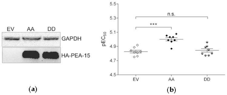
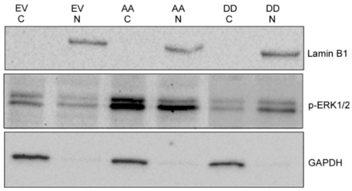
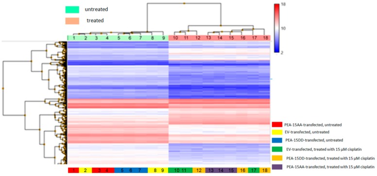
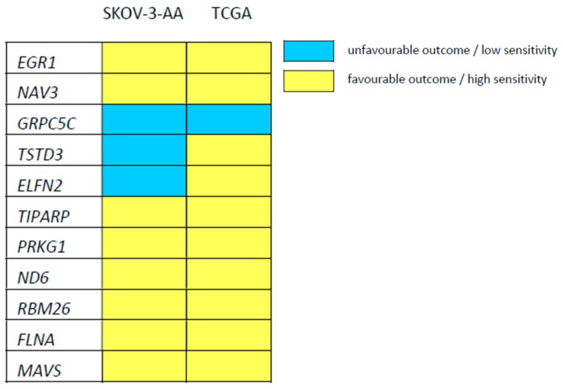
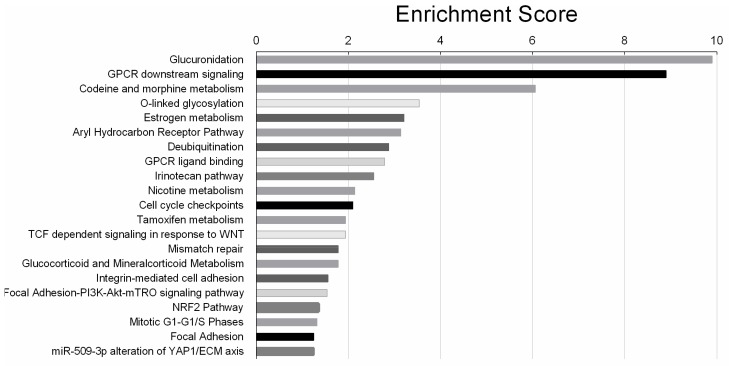
The efficacy of cisplatin-based chemotherapy in ovarian cancer is often limited by the development of drug resistance. In most ovarian cancer cells, cisplatin activates extracellular signal-regulated kinase1/2 (ERK1/2) signalling. Phosphoprotein enriched in astrocytes (PEA-15) is a ubiquitously expressed protein, capable of sequestering ERK1/2 in the cytoplasm and inhibiting cell proliferation. This and other functions of PEA-15 are regulated by its phosphorylation status. In this study, the relevance of PEA-15 phosphorylation state for cisplatin sensitivity of ovarian carcinoma cells was examined. The results of MTT-assays indicated that overexpression of PEA-15AA (a non-phosphorylatable variant) sensitised SKOV-3 cells to cisplatin. Phosphomimetic PEA-15DD did not affect cell sensitivity to the drug. While PEA-15DD facilitates nuclear translocation of activated ERK1/2, PEA-15AA acts to sequester the kinase in the cytoplasm as shown by Western blot. Microarray data indicated deregulation of thirteen genes in PEA-15AA-transfected cells compared to non-transfected or PEA-15DD-transfected variants. Data derived from The Cancer Genome Atlas (TCGA) showed that the expression of seven of these genes including EGR1 (early growth response protein 1) and FLNA (filamin A) significantly correlated with the therapy outcome in cisplatin-treated cancer patients. Further analysis indicated the relevance of nuclear factor erythroid 2related factor 2/antioxidant response element (Nrf2/ARE) signalling for the favourable effect of PEA-15AA on cisplatin sensitivity. The results warrant further evaluation of the PEA-15 phosphorylation status as a potential candidate biomarker of response to cisplatin-based chemotherapy.

Keywords: PEA-15; cisplatin sensitivity; ovarian cancer.


Conflict of interest statement

The authors declare no conflict of interest. The funders had no role in the design of the study; in the collection, analyses, or interpretation of data; in the writing of the manuscript, or in the decision to publish the results.

Figures

Figure 1
Figure 1
(a) Expression of hemagglutinin (HA)-tagged phosphoprotein enriched in astrocytes (PEA-15) in SKOV-3 cells after transfection with the HA-tagged empty vector (EV), PEA-15AA (AA) and PEA-15DD (DD). GAPDH was used as a loading control. (b) Cisplatin sensitivity (pEC50, mean ± SEM, n = 8) of transfected SKOV-3-EV (EV), SKOV-3-AA (AA) and SKOV-3-DD (DD) cells. *** p < 0.001, n.s. = not significant.
Figure 2
Figure 2
A representative Western blot of phosphorylated extracellular signal-regulated kinase1/2 (p-ERK1/2) expression in nuclear and cytosolic fractions of the SKOV-3 cells transfected with empty vector (EV), PEA-15AA (AA) and PEA-15DD (DD). GAPDH and Lamin B1 were used as the markers and loading controls of cytosolic (C) and nuclear fractions (N), respectively.
Figure 3
Figure 3
Heatmap of the transcriptome-wide ClariomTM S array, regulated genes with fold change cut-off at 2.0 for differentially expressed genes and a p-value cut-off at 0.05 are shown.
Figure 4
Figure 4
Heatmap indicating the relationship between low expression of the indicated genes and sensitivity/outcome, favourable (low cisplatin EC50 in SKOV-3-AA cells or prolonged survival of cisplatin-treated patients, indicated in yellow) or unfavourable (high cisplatin EC50 in SKOV-3-AA cells or reduced survival of cisplatin-treated patients, indicated in blue), based on the comparison of gene expression between SKOV-3-AA and EV- or PEA-15DD-transfected variants and TCGA data.
Figure 5
Figure 5
Venn diagram representing the exclusively and commonly regulated genes in different transfected cells upon cisplatin exposure. The diagram shows the total number of genes affected by cisplatin exposure in empty vector—(EV), PEA-15AA—(AA) and PEA-15DD-transfected—(DD) cells.
Figure 6
Figure 6
Twenty-one biological pathways significantly affected by cisplatin treatment in SKOV-3-AA cells, listed according to the significance level (log 2 base) in a descending order.
Figure 7
Figure 7
Representative Western blots and the corresponding densitometric quantification (mean ± SEM, n = 3) of (a) the relative uridine diphosphate-glucuronyl transferase (UGT)1A expression and (b) the relative nuclear factor erythroid 2-related factor 2 (Nrf2) expression in empty vector—(EV), PEA-15AA—(AA) and PEA-15DD-transfected—(DD) cells after treatment with 15 µM cisplatin (+Pt) for 24 h and in untreated transfected SKOV-3 cells. GAPDH was used as a loading control. * p < 0.05, ** p < 0.01.
Figure 8
Figure 8
Representative Western blots and the corresponding densitometric quantification (mean ± SEM, n = 3) of (a) the relative Nrf2 expression and (b) the relative UGT1A expression in the transfected untreated SKOV-3 cells (Ctrl), after exposure to 15 µM cisplatin (Pt), to 20 µM retinoic acid (RA) and after co-incubation with 20 µM retinoic acid and 15 µM cisplatin (Pt + RA) for 24 h are shown. GAPDH was used as a loading control. *p < 0.05, ** p < 0.01, n.s. = not significant.
Figure 9
Figure 9
Sensitivity of SKOV-3 cells (pEC50, mean ± SEM, n = 9–10) of cisplatin alone (Pt), and upon co-incubation with 20 µM retinoic acid (Pt + RA) was determined over 48 h. *** p < 0.001.

References

    1. Dilruba S., Kalayda G.V. Platinum-based drugs: Past, present and future. Cancer Chemother. Pharm. 2016;77:1103–1124. doi: 10.1007/s00280-016-2976-z. - DOI - PubMed
    1. Siddik Z.H. Cisplatin: Mode of cytotoxic action and molecular basis of resistance. Oncogene. 2003;22:7265–7279. doi: 10.1038/sj.onc.1206933. - DOI - PubMed
    1. Chang L., Karin M. Mammalian MAP kinase signalling cascades. Nature. 2001;410:37–40. doi: 10.1038/35065000. - DOI - PubMed
    1. Johnson G.L., Lapadat R. Mitogen-activated protein kinase pathways mediated by ERK, JNK, and p38 protein kinases. Science. 2002;298:1911–1912. doi: 10.1126/science.1072682. - DOI - PubMed
    1. Marshall C.J. Specificity of receptor tyrosine kinase signaling: Transient versus sustained extracellular signal-regulated kinase activation. Cell. 1995;80:179–185. doi: 10.1016/0092-8674(95)90401-8. - DOI - PubMed

Publication types