. 2020 Apr;40(4):929-942.
doi: 10.1161/ATVBAHA.119.313698. Epub 2020 Feb 27.

Platelets Disseminate Extracellular Vesicles in Lymph in Rheumatoid Arthritis

Affiliations

Platelets Disseminate Extracellular Vesicles in Lymph in Rheumatoid Arthritis

Nicolas Tessandier et al. Arterioscler Thromb Vasc Biol. 2020 Apr.

Abstract

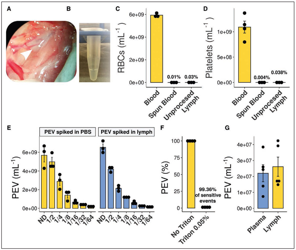
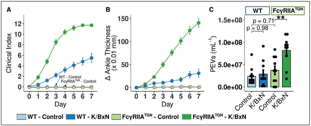
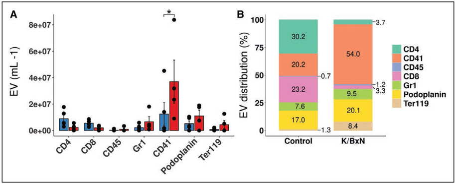
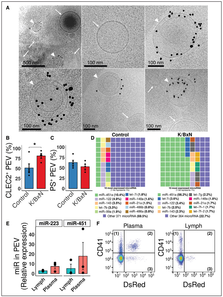
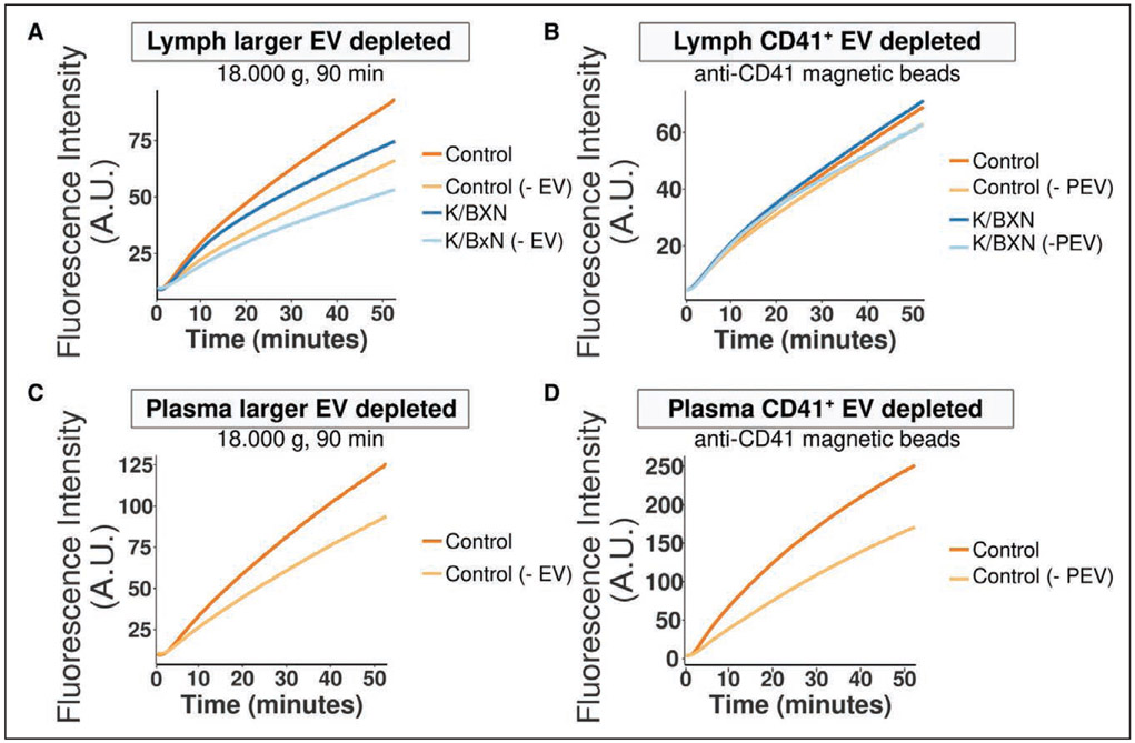
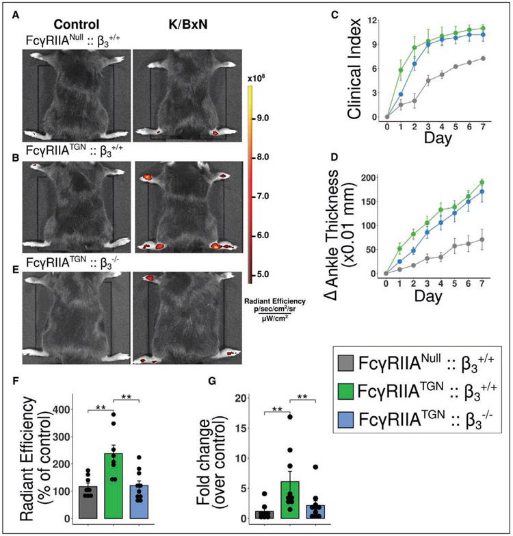
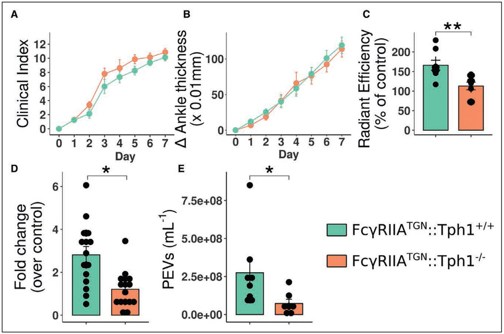
Objective: The lymphatic system is a circulatory system that unidirectionally drains the interstitial tissue fluid back to blood circulation. Although lymph is utilized by leukocytes for immune surveillance, it remains inaccessible to platelets and erythrocytes. Activated cells release submicron extracellular vesicles (EV) that transport molecules from the donor cell. In rheumatoid arthritis, EV accumulate in the joint where they can interact with numerous cellular lineages. However, whether EV can exit the inflamed tissue to recirculate is unknown. Here, we investigated whether vascular leakage that occurs during inflammation could favor EV access to the lymphatic system. Approach and Results: Using an in vivo model of autoimmune inflammatory arthritis, we show that there is an influx of platelet EV, but not EV from erythrocytes or leukocytes, in joint-draining lymph. In contrast to blood platelet EV, lymph platelet EV lacked mitochondrial organelles and failed to promote coagulation. Platelet EV influx in lymph was consistent with joint vascular leakage and implicated the fibrinogen receptor α2bβ3 and platelet-derived serotonin.

Conclusions: These findings show that platelets can disseminate their EV in fluid that is inaccessible to platelets and beyond the joint in this disease.

Keywords: arthritis; extracellular vesicles; inflammation; leukocytes; lymphatic system; platelets.


Figures

Figure 1.
Figure 1.. Lymph collection and lymph platelet extracellular vesicles (PEV) quantification.
A, Lymph is collected at the thoracic duct, between the transverse lumbar artery and the diaphragm in a collection tube coated with EDTA (0.1 M). B, Representative picture of translucent collected lymph. C and D, Red blood cells (RBCs; C) and platelet (D) counts in blood, spun blood (plasma), and unprocessed lymph (n=3). E, Efficient flow cytometry quantification of PEV in lymph was verified by spiking and serial-dilutions of in vitro-generated PEV in either lymph or PBS. Nondiluted (ND), n=3. F, Sensitivity to Triton 0.05% of the PEV quantified by flow cytometry (n=4). G, PEV were quantified in platelet-free plasma and in lymph in C57BL/6 wild-type mice (n=5).
Figure 2.
Figure 2.. Platelet extracellular vesicles (PEV) circulate in lymph in autoimmune inflammatory arthritis.
A and B, Clinical index (A) and delta ankle thickness (B) in wild-type (WT; blue) and FcγRIIATGN (green) mice injected with either 150 μL PBS (respectively light blue and light green) or 150 μL K/BxN serum (respectively dark blue and dark green) on day 0 and day 2 (n=10). C, Flow cytometry quantification of lymph PEV in WT and FcγRIIATGN mice at the peak of arthritis severity, day 7 (n=10). **P≤0.01, using a 1-way ANOVA, followed by post hoc Tukey honestly significant difference for multiple comparisons.
Figure 3.
Figure 3.. Platelet extracellular vesicles (EV) are a predominant EV population in FcγRIIATGN K/BxN lymph.
A, Quantification of EV populations between control and K/BxN lymph, as assessed by flow cytometry. B, Assessment of EV populations proportion by flow cytometry in lymph from control and K/BxN mice. EV distribution are presented as a percentage based on the selected EV markers (CD4, CD41, CD45, CD8, Gr1 [granulocyte marker 1], Podoplanin, Ter119 [terminal 119 antigen of glycophorin A]). EV not presenting those markers were, therefore, not taken into account for the total EV population in this representation (n=4). Same samples were used for creating both A and B. *P≤0.05 using a 2-way ANOVA, followed by post hoc Holm-Šídák for multiple comparisons.
Figure 4.
Figure 4.. Characterization of extracellular vesicles (EV) in lymph.
A, Representative Cryo-electron-microscopy images of vesicles detected in FcγRIIATGN K/BxN lymph. Black dots identify CD41 antibody coupled to gold beads. Top left, the specificity of labeling, as all the black dots can be found in close proximity with EV. White arrowheads identify CD41+ EV, whereas white arrows identify CD41 EV (lymph pooled from 3 FcγRIIATGN K/BxN mice). B, CLEC-2 (C-type lectin domain family 1 member B) exposure and (C) phosphatidylserine (PS) exposure on platelet EV (PEV) in control and K/BxN mice (n=4 and n=5). D, Representations of the proportions of the 10 most expressed extracellular microRNA in Control (left) and K/BxN (right) FcγRIIATGN lymph. Each tile represents 1% (n=1, pooled from 3 mice per condition). E, Proportion of micro RNA (miR)-223 and miR-451 contained in K/BxN immunoprecipitated CD41+ PEV over total extracellular content in lymph vs plasma, as determined by real-time quantitative polymerase chain reaction (n=3). F, Representative gating illustration of plasma and lymph from K/BxN mice (n=2). Samples were labeled with an anti-CD41 (Pacific Blue), whereas mitochondria are endogenously fluorescent (Mitochondria-DsRed [Discosoma Red]). *P≤0.05, using an unpaired t test. let-7 indicates lethal-7.
Figure 5.
Figure 5.. Platelet extracellular vesicles (PEV) do not contribute to lymph coagulation but interact with lymphatic endothelial cells.
A and B, Averaged curves showing fluorescence from cleavage of a fluorogenic substrate of thrombin in control (PBS) or FcγRIIATGN K/BxN lymph, either native or larger EV-depleted (centrifugation at 18,000 g for 90 min; A), or native and CD41+-EV-depleted (B; n=4). C and D, Averaged curves showing fluorescence from cleavage of a fluorogenic substrate of thrombin in control (PBS) or FcγRIIATGN K/BxN plasma, either native or larger EV-depleted (C), or native and CD41+-EV-depleted (D; n=3 and n=2). A.U. indicates arbitrary unit.
Figure 6.
Figure 6.. Role of FcγRIIA and its signaling through β3 in joint vasculature leakage.
A, B and E, FcγRIIANull::β3+/+ (A), FcγRIIATGN:: β3+/+ (B) and FcγRIIATGN:: β3−/− (E) K/BxN mice were injected intravenously with 0.51-μm diameter microspheres and visualized 2 minutes later using a Xenogen IVIS in vivo imaging system. All mice received the same concentration of fluorescent microspheres. C and D, Clinical index (C) and delta ankle thickness measured at the malleoli with the ankle in a fully flexed position (D). Comparison of arthritis severity for FcγRIIANull::β3+/+, FcγRIIATGN:: β3+/+ and FcγRIIATGN:: β3−/−. F, Radiant efficiency quantification of 0.51 μm diameter sky-blue-conjugated microspheres accumulation in arthritic joints 2 min after intravenous injection, in FcγRIIANull:: β3+/+, FcγRIIATGN:: β3+/+ and FcγRIIATGN:: β3−/−. Data are presented as a percentage of nonarthritic control mice injected with the same concentration of microspheres. G, Fold change of sky-blue fluorescence detected in the inguinal lymph nodes, 45 min after microsphere injection in FcγRIIANull:: β3+/+, FcγRIIATGN::β3+/+ vs FcγRIIATGN::β3−/− K/BxN mice relative to nonarthritic control mice injected with the same concentration of microspheres. **P≤0.01 using a one-way ANOVA, followed by post hoc Tukey honestly significant difference for multiple comparisons.
Figure 7.
Figure 7.. Blood vascular permeability induced by platelet-derived serotonin increases platelet extracellular vesicles (PEV) content in the lymphatic system.
A and B, Clinical index (A) and delta ankle thickness measured at the malleoli with the ankle in a fully flexed position (B). Comparison of arthritis severity for FcγRIIATGN:: Tph1+/+ and FcγRIIATGN:: Tph1−/− K/BxN mice. C, Radiant efficiency quantifications of 0.51-μm diameter sky-blue-conjugated microspheres accumulation in arthritic joints 2 min after intravenous injection, in FcγRIIATGN:: Tph1+/+ and FcγRIIATGN:: Tph1−/− K/BxN mice. Data are presented as a percentage of nonarthritic control mice injected with the same concentration of microspheres. D, Fold change of sky-blue fluorescence detected in the inguinal lymph nodes, 45 min after microsphere injection into FcγRIIATGN:: Tph1+/+ and FcγRIIATGN:: Tph1−/− K/BxN mice relative to nonarthritic control mice injected with the same concentration of microspheres. E, PEV quantification in FcγRIIATGN:: Tph1+/+ and FcγRIIATGN:: Tph1−/− thoracic lymph, in arthritic mice.

References

    1. Smolen JS, Aletaha D, Barton A, Burmester GR, Emery P, Firestein GS, Kavanaugh A, McInnes IB, Solomon DH, Strand V, et al. Rheumatoid arthritis. Nat Rev Dis Primers. 2018;4:18001. doi: 10.1038/nrdp.2018.1 - DOI - PubMed
    1. Smolen JS, Aletaha D, McInnes IB. Rheumatoid arthritis. The Lancet. 2016;6736:1–16. - PubMed
    1. Lee DM, Kiener HP, Agarwal SK, Noss EH, Watts GF, Chisaka O, Takeichi M, Brenner MB. Cadherin-11 in synovial lining formation and pathology in arthritis. Science. 2007;315:1006–1010. doi: 10.1126/science.1137306 - DOI - PubMed
    1. Wright HL, Moots RJ, Edwards SW. The multifactorial role of neutrophils in rheumatoid arthritis. Nat Rev Rheumatol. 2014;10:593–601. doi: 10.1038/nrrheum.2014.80 - DOI - PubMed
    1. Ji H, Ohmura K, Mahmood U, Lee DM, Hofhuis FM, Boackle SA, Takahashi K, Holers VM, Walport M, Gerard C, et al. Arthritis critically dependent on innate immune system players. Immunity. 2002;16:157–168. doi: 10.1016/s1074-7613(02)00275-3 - DOI - PubMed

Publication types