. 2020 Apr;40(4):901-913.
doi: 10.1161/ATVBAHA.120.313990. Epub 2020 Feb 27.

Cardiac Myosin Promotes Thrombin Generation and Coagulation In Vitro and In Vivo

Affiliations

Cardiac Myosin Promotes Thrombin Generation and Coagulation In Vitro and In Vivo

Jevgenia Zilberman-Rudenko et al. Arterioscler Thromb Vasc Biol. 2020 Apr.

Abstract

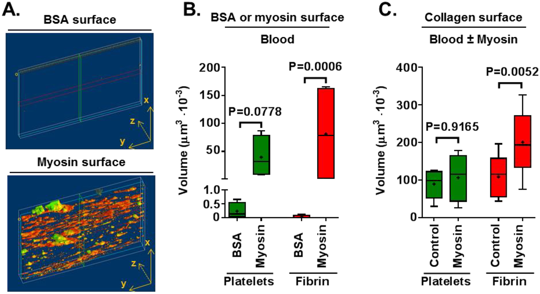
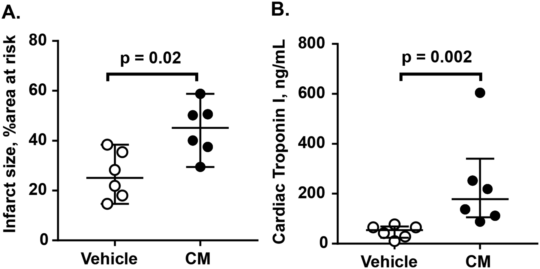
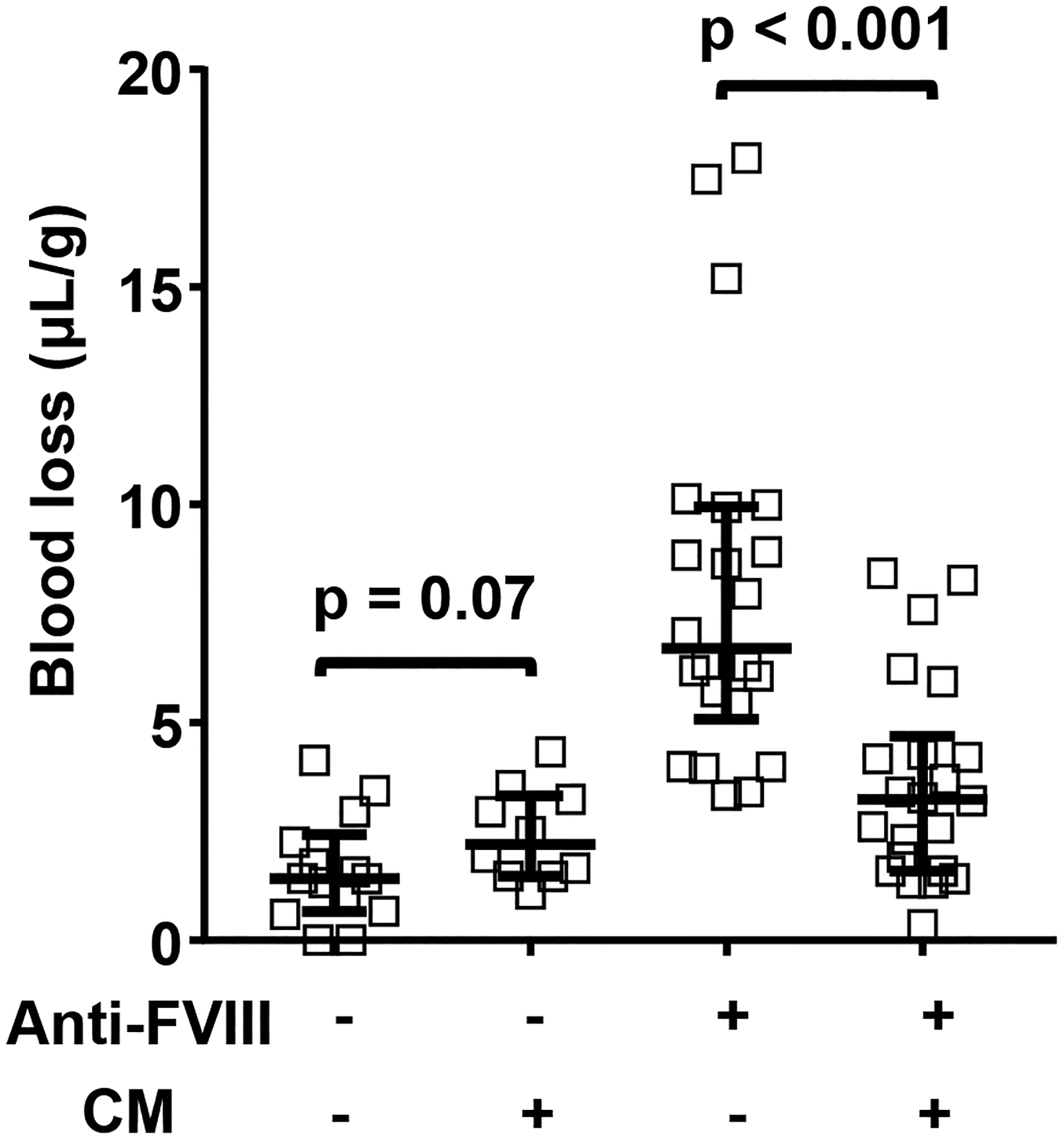
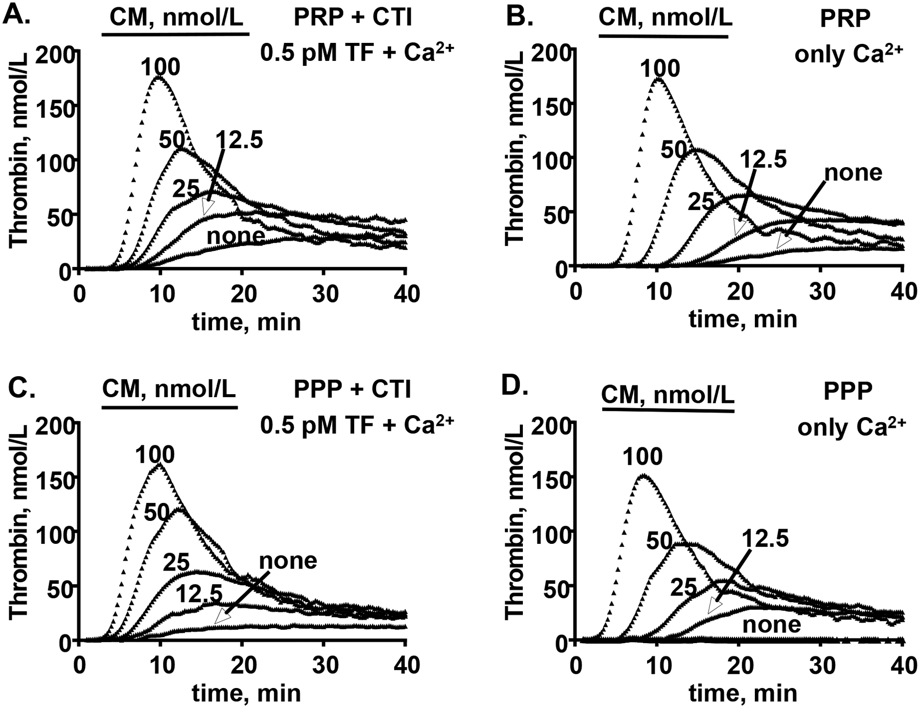
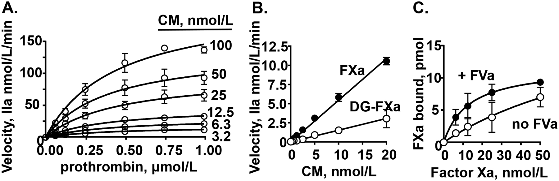
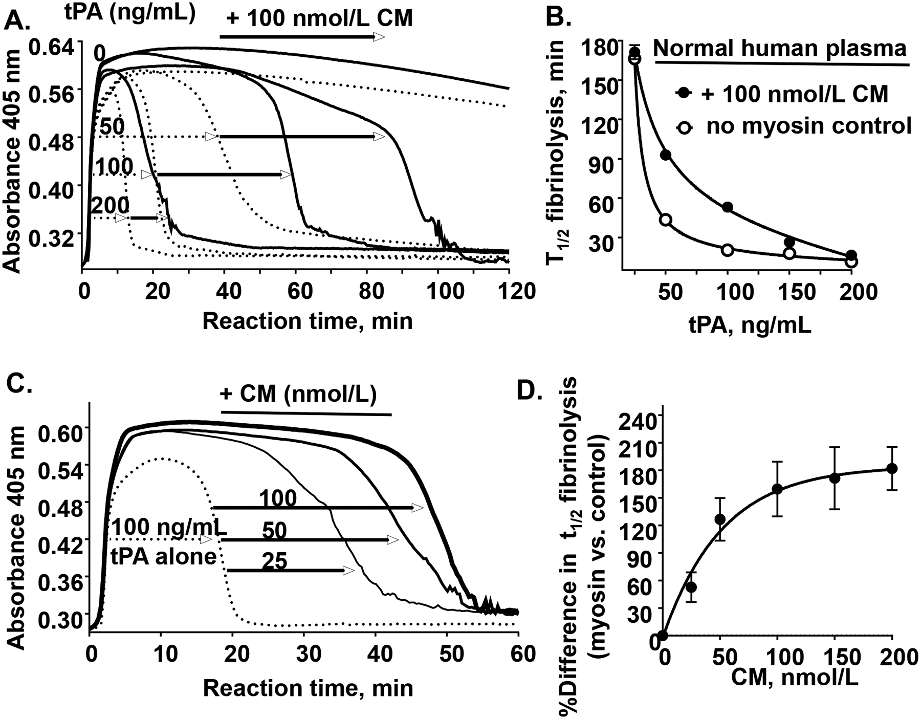
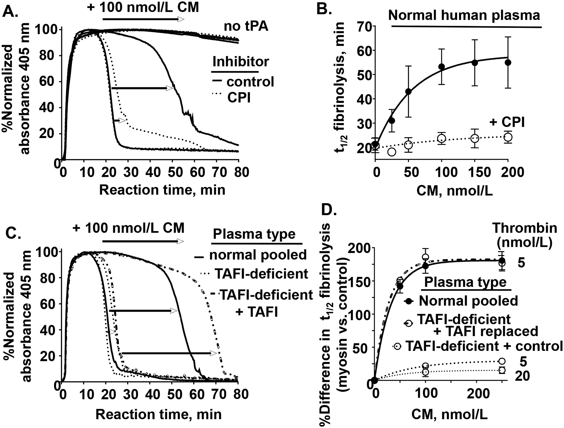
Objective: Cardiac myosin (CM) is structurally similar to skeletal muscle myosin, which has procoagulant activity. Here, we evaluated CM's ex vivo, in vivo, and in vitro activities related to hemostasis and thrombosis. Approach and Results: Perfusion of fresh human blood over CM-coated surfaces caused thrombus formation and fibrin deposition. Addition of CM to blood passing over collagen-coated surfaces enhanced fibrin formation. In a murine ischemia/reperfusion injury model, exogenous CM, when administered intravenously, augmented myocardial infarction and troponin I release. In hemophilia A mice, intravenously administered CM reduced tail-cut-initiated bleeding. These data provide proof of concept for CM's in vivo procoagulant properties. In vitro studies clarified some mechanisms for CM's procoagulant properties. Thrombin generation assays showed that CM, like skeletal muscle myosin, enhanced thrombin generation in human platelet-rich and platelet-poor plasmas and also in mixtures of purified factors Xa, Va, and prothrombin. Binding studies showed that CM, like skeletal muscle myosin, directly binds factor Xa, supporting the concept that the CM surface is a site for prothrombinase assembly. In tPA (tissue-type plasminogen activator)-induced plasma clot lysis assays, CM was antifibrinolytic due to robust CM-dependent thrombin generation that enhanced activation of TAFI (thrombin activatable fibrinolysis inhibitor).

Conclusions: CM in vitro is procoagulant and prothrombotic. CM in vivo can augment myocardial damage and can be prohemostatic in the presence of bleeding. CM's procoagulant and antifibrinolytic activities likely involve, at least in part, its ability to bind factor Xa and enhance thrombin generation. Future work is needed to clarify CM's pathophysiology and its mechanistic influences on hemostasis or thrombosis.

Keywords: cardiac myosins; fibrin; hemostasis; thrombin; thrombosis.


Conflict of interest statement

Disclosures: None. All authors declare no competing financial interest.

Figures

Figure 1.
Figure 1.. CM promotes ex vivo thrombus formation in flowing human blood.
The procoagulant activity of CM was studied in flowing whole blood under initial wall shear of 300 s−1. (A) Whole blood was perfused over CM-coated surfaces or BSA-coated control surfaces and quantification of platelet adhesion and fibrin formation was made. Reperesentative images for CM-coated and BSA-coated surfaces are shown. (B) Amounts of platelet adhesion and fibrin formation were quantified from confocal microscopy images produced by an overlay of maximum fluorescence intensities at four separate positions within a channel. In separate experiments, (C) Whole blood to which either CM (50 nmol/L final) or control buffer was added was perfused past surfaces coated with collagen type I, followed by quantifying platelet adhesion and fibrin formation. P values were obtained by Two-way ANOVA with Tukey’s multiple comparison test, and Tukey method is used to create the whiskers with error bars ranging minimum and maximum.
Figure 2.
Figure 2.. CM exacerbates myocardial injury in vivo in a murine ischemia / reperfusion injury model.
C57BL/6J wildtype mice (n=6 per group) were subjected to myocardial ischemia reperfusion injury and received either vehicle or CM (5.4 mg/kg) via intraarterial infusion 15 min after the initiation of reperfusion. Infarct size (A) and cardiac troponin I (B) were determined as noted in the Method section. Bars indicated median with interquartile range, and p values were calculated by Mann-Whitney test.
Figure 3.
Figure 3.. CM reduces blood loss following tail-cut-induced bleeding in hemophilia A mouse model.
Wild type C57BL/6J mice were injected with 0.25 mg/kg of anti-FVIII (FVIII) antibody (acquired hemophilia A model) (36 female/28 male) or vehicle (saline) (20 female/14 male) at 2 hours prior to tail cut. Then, mice were injected with vehicle or 5.4 mg/kg CM. The distal portion of the tail was surgically removed via a scalpel blade at 1.5-mm tail diameter to induce a moderate bleeding effect. Tails were immersed in 50 mL of saline, pH 7.4 at 37°C. Total blood loss during 10 min was measured as the blood volume collected per mouse weight (μL/g). Bars indicated median with interquartile range, and p values were calculated by Mann-Whitney test.
Figure 4.
Figure 4.. CM promotes thrombin generation in platelet rich and platelet poor plasma.
Whole human blood was collected by venipuncture from normal donors (1:10 sodium citrate) and serially centrifugated to prepare fresh platelet rich plasma (PRP) and platelet poor plasma (PPP). (A, C) After incubation with various indicated concentrations of myosin and CTI for 10 min at 37°C, thrombin generation was initiated by recalcification and addition of TF (0.5 pM final) to either (A) PRP or (C) PPP. (B, D) In parallel experiments, thrombin generation after incubation with various indicated concentrations of myosin alone was initiated with addition of calcium to (B) PRP or (D) PPP. Reactions using blood from a single representative male donor are shown here and are representative of data for three other different donors (one male and two female) studied.
Figure 5.
Figure 5.. CM promotes thrombin generation in purified clotting factor reaction mixtures.
(A) The effect of varying concentrations of myosin and prothrombin on the initial rate of prothrombin activation by factor Xa/factor Va. (B) The effect of varying concentrations of myosin on thrombin generation from prothrombin (0.75 μM final) by factor Xa (FXa) or Gla domain-less factor Xa (DG-FXa) (0.2 nmol/L final) and factor Va (5 nmol/L final). (C) Binding studies of factor Xa to immobilized-myosin in the presence or absence of factor Va (FVa) (10 nmol/L final) where bound factor Xa was determined using factor Xa chromogenic activity assays. Each value represents the mean [SD] of at least triplicate determinations.
Figure 6.
Figure 6.. CM attenutes tPA-induced plasma clot lysis.
(A) As described in Materials and Methods, a 100 μL aliquot containing thrombin, Ca2+, phospholipid vesicles (60% PC: 20% PS: 20% PE), 0.1% BSA and different levels of tPA (as indicated, 0, 50, 100, or 200 ng/mL) was added to a 100 μL aliquot containing citrated normal human pooled plasma which had been pre-incubated for 30 min at 37 °C with CM (100 nmol/L) (solid lines) or control buffer (dotted lines). Following rapid mixing, the reaction mixture was monitored for turbidity at 405 nm at 37°C for 180 min during which a clot was rapidly formed (turbidity increase) and then that clot was subsequently lysed (turbidity decrease). (B) The plasma clot lysis time (t1/2, min for 50 % clot lysis) was quantified in the presence of CM (●) at different tPA levels or control buffer (○). Each value represents the mean [SD] of triplicate determinations. (C) Plasma was pre-incubated with different amounts of CM (as indicated, 0, 25, 50, or 100 nmol/L) followed by thrombin-initiated clot formation (as above), and the clot was lysed in the presence of 100 ng/mL tPA. (D) The percent difference in plasma clot lysis (t1/2) between values for the presence of CM at the indicated concentrations are shown where the plasma clot lysis time at no CM was defined as 100%. Each value represents the mean [SD] of at least triplicate determinations.
Figure 7.
Figure 7.. TAFI activity is required for CM’s inhibition of tPA-induced plasma clot lysis.
(A) Normal human pooled plasma was pre-incubated with either carboxypeptidase inhibitor (CPI) to inhibit TAFI (dotted lines) or control buffer (solid line) and clot lysis studies were done, as in Figure 6, with with 100 nmol/L CM and with 100 ng/mL tPA or without tPA (as indicated by “no tPA”). (B) Plasma clot lysis times (t1/2), determined as done in (A), were quantified in the presence of different levels of CM, 5 nmol/L thrombin, and 100 ng/mL tPA in the absence (●, solid line) or presence of the TAFIa inhibitor, CPI (○, dashed line). (C) TAFI-deficient plasma reconstituted with only control buffer or with reconsititued with normal levels of TAFI and normal plasma was used for plasma clot lysis studies (as above, using 5 nmol/L thrombin, and 100 ng/mL tPA) with either 0 or 100 nmol/L CM. (D) The percent difference in plasma clot lysis t1/2 between values for the presence of CM at the indicated concentrations (50, 100, 250 nmol/L) for the different indicated plasma types (normal (solid line), TAFI-deficient + control buffer (dotted lines for 5 nmol/L or 20 nmol/L thrombin), and TAFI-deficient + TAFI replacement (dash-dotted line)) are shown. Each value represents the mean [SD] of at least triplicate determinations.

References

    1. Fuster V, Badimon L, Cohen M, Ambrose JA, Badimon JJ and Chesebro J. Insights into the pathogenesis of acute ischemic syndromes. Circulation. 1988;77:1213–1220. - PubMed
    1. Herrick JB. Landmark article (JAMA 1912). Clinical features of sudden obstruction of the coronary arteries. By James B. Herrick. Jama. 1983;250:1757–1765. - PubMed
    1. Gorlin R, Fuster V and Ambrose JA. Anatomic-physiologic links between acute coronary syndromes. Circulation. 1986;74:6–9. - PubMed
    1. Baumgartner HR. New aspects in thrombogenesis. VASA Zeitschrift fur Gefasskrankheiten. 1974;3:60–64. - PubMed
    1. Khaw BA, Gold HK, Fallon JT and Haber E. Detection of serum cardiac myosin light chains in acute experimental myocardial infarction: radioimmunoassay of cardiac myosin light chains. Circulation. 1978;58:1130–1136. - PubMed

Publication types