. 2020 Apr 16;94(9):e01998-19.
doi: 10.1128/JVI.01998-19. Print 2020 Apr 16.

Route of a Multipartite Nanovirus across the Body of Its Aphid Vector

Affiliations

Route of a Multipartite Nanovirus across the Body of Its Aphid Vector

Jérémy Di Mattia et al. J Virol. .

Abstract

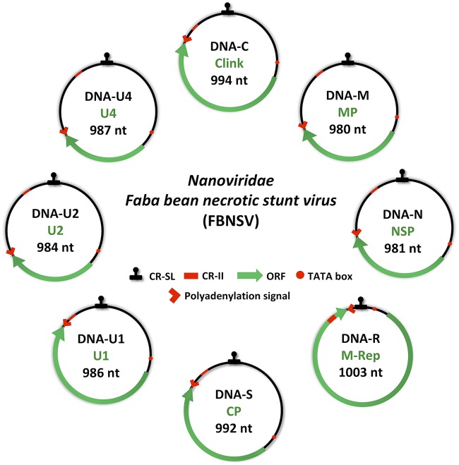
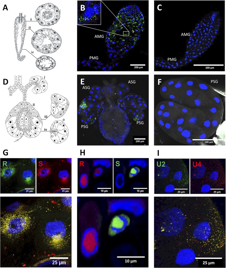
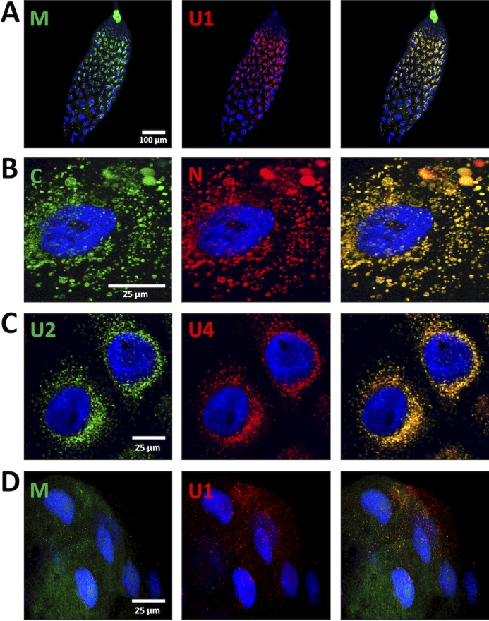
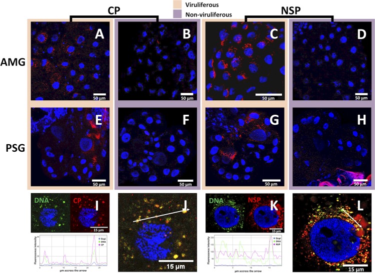
Vector transmission plays a primary role in the life cycle of viruses, and insects are the most common vectors. An important mode of vector transmission, reported only for plant viruses, is circulative nonpropagative transmission whereby the virus cycles within the body of its insect vector, from gut to salivary glands and saliva, without replicating. This mode of transmission has been extensively studied in the viral families Luteoviridae and Geminiviridae and is also reported for Nanoviridae The biology of viruses within these three families is different, and whether the viruses have evolved similar molecular/cellular virus-vector interactions is unclear. In particular, nanoviruses have a multipartite genome organization, and how the distinct genome segments encapsidated individually transit through the insect body is unknown. Here, using a combination of fluorescent in situ hybridization and immunofluorescence, we monitor distinct proteins and genome segments of the nanovirus Faba bean necrotic stunt virus (FBNSV) during transcytosis through the gut and salivary gland cells of its aphid vector Acyrthosiphon pisum FBNSV specifically transits through cells of the anterior midgut and principal salivary gland cells, a route similar to that of geminiviruses but distinct from that of luteoviruses. Our results further demonstrate that a large number of virus particles enter every single susceptible cell so that distinct genome segments always remain together. Finally, we confirm that the success of nanovirus-vector interaction depends on a nonstructural helper component, the viral protein nuclear shuttle protein (NSP), which is shown to be mandatory for viral accumulation within gut cells.IMPORTANCE An intriguing mode of vector transmission described only for plant viruses is circulative nonpropagative transmission, whereby the virus passes through the gut and salivary glands of the insect vector without replicating. Three plant virus families are transmitted this way, but details of the molecular/cellular mechanisms of the virus-vector interaction are missing. This is striking for nanoviruses that are believed to interact with aphid vectors in ways similar to those of luteoviruses or geminiviruses but for which empirical evidence is scarce. We here confirm that nanoviruses follow a within-vector route similar to that of geminiviruses but distinct from that of luteoviruses. We show that they produce a nonstructural protein mandatory for viral entry into gut cells, a unique phenomenon for this mode of transmission. Finally, noting that nanoviruses are multipartite viruses, we demonstrate that a large number of viral particles penetrate susceptible cells of the vector, allowing distinct genome segments to remain together.

Keywords: aphid; circulative; insect vector; multipartite virus; nanovirus; nonpropagative; plant; vector transmission.


Figures

FIG 1
FIG 1
Genome organization of Faba bean necrotic stunt virus (FBNSV). The eight circles represent the different genomic segments. The name and size of each genome segment and the name of the encoded protein are indicated inside circles in black and green, respectively. Clink, cell cycle-linked protein; MP, movement protein; NSP, nuclear shuttle protein; M-Rep, master replication protein; CP, coat protein; U1, U2, or U4, unknown protein 1, 2, or 4. CR-SL, common stem-loop region; CR-II, common region; ORF, open reading frame; nt, nucleotides.
FIG 2
FIG 2
Localization of the DNA segments of Faba bean necrotic stunt virus (FBNSV) in aphid versus that in plant cells. Schematic drawing of the anatomy of the AMG (A) shows longitudinal (i) and transverse sections (ii to iv). Schematic drawing of the anatomy of the salivary glands (D) shows longitudinal sections of the accessory (i) and principal glands (ii) as well as transverse sections of the principal glands (iii and iv). Both panels A and D are adapted from Ponsen (25) describing the anatomy of Myzus persicae. Ponsen’s numbering of distinct cell types (1 to 8) of the salivary glands is indicated in panel D. The accumulation of FBNSV DNA was observed in anterior midgut (AMG) of 64 viruliferous aphids from 8 experiments, and a representative image is shown in panel B. The accumulation of FBNSV DNA in a specific cell type of the principal salivary glands (PSG) was observed in 15 viruliferous aphids from 3 experiments, and a representative image is shown in panel E. In panels B and E, the viral DNA is revealed by FISH (green probe targeting all eight FBNSV segments), and nonviruliferous controls are shown in panels C and F. The respective localizations of R and S segments (probe color as indicated) are compared in AMG cells (G; representative of 24 observed aphids) and in infected faba bean phloem cells (H) (22). The respective localizations of U2 and U4 segments are compared in PSG cells (I; representative of 4 aphids observed). In panels G, H, and I, the merged-color channel image is shown at the bottom, and the corresponding split-color channel images are shown at the top. All images correspond to maximum-intensity projections. Cell nuclei are stained with 4′,6′-diamidino-2-phenylindole (DAPI; blue). PMG, posterior midgut; ASG, accessory salivary glands.
FIG 3
FIG 3
Colocalization of FBNSV segments in AMG and PSG. The colors of the fluorescent probes and the targeted segment pairs are indicated. Three additional pairs of segments were tested in the AMG: 32 aphids from four experiments for the pair M/U1 and 24 aphids from three experiments for the pairs C/N and U2/U4. Illustrative images are, shown, respectively, in panels A, B, and C. In the PSG, the additional segment pair M/U1 was observed in 6 aphids from three experiments, and a representative image is shown in panel D. Split-color channels are shown in the left and middle panels whereas merged images are shown in the right panel. All images correspond to maximum-intensity projections. Cell nuclei are stained with 4′,6′-diamidino-2-phenylindole (DAPI; blue).
FIG 4
FIG 4
Localization of FBNSV DNA, CP, and NSP in AMG and PSG of A. pisum. FBNSV CP is labeled by IF in the AMG (A and B) and PSG (E and F) of viruliferous (A and E) and nonviruliferous (B and F) aphids. NSP is labeled by IF in the AMG (C and D) and PSG (G and H) of viruliferous (C and G) and nonviruliferous (D and H) aphids. When viral DNA and either CP or NSP (FISH and IF) were colabeled in AMG, the DNA probe targeting all eight genome segments appears green whereas the specific CP or NSP antibody appears red (I to L). In each case, 30 aphids from three experiments were observed, and one representative image is shown. Split-color channel images in panels I and K correspond to the merged-color channel images in panels J and L, respectively. The graphics represent the colocalization profiles between either DNA (green curve) and CP (red curve) (I) or DNA (green curve) and NSP (red curve) (K). Fluorescence intensity was measured along the white arrows drawn in panels J and L. Images in panels A to H correspond to maximum-intensity projections, and images in panels I to L correspond to single optical sections. Cell nuclei are stained with 4′,6′-diamidino-2-phenylindole (DAPI; blue).
FIG 5
FIG 5
NSP-dependent accumulation of FBNSV DNA in the AMG of aphid vector. FBNSV DNA is green-labeled in gut cells of aphids fed either on infected plants containing all FBNSV segments (A; representative of 15 aphids from three experiments) or on infected plants lacking segment U4 (B; representative of 5 aphids from one experiment), or on infected plants lacking segment N (C; representative of 15 aphids from three experiments), or on infected plants with all 8 segments but where the N segment has a mutation of the ATG-start codon (D; representative of 5 aphids from one experiment). The presence of the N segment and derivatives His-N and N-His in two replicate infected plants was verified by PCR using primers specific for the coding sequence of the His tag (E, top panel). The expression of NSP, His-NSP, and NSP-His proteins in infected plant tissues was evaluated by Western blotting (E) using antiserum directed against the NSP (middle panel) or His tag (bottom panel). FBNSV NSP (F and G; red) or DNA (H and I; green) are labeled in aphids fed on infected plants expressing wild-type NSP (F and H) or its derivative His-NSP fusion (G and I). Images in panels F and H are representative of 20 aphids observed from two experiments. All confocal images are maximum-intensity projections. Cell nuclei are stained with 4′,6′-diamidino-2-phenylindole (DAPI; blue).

References

    1. Hogenhout SA, Ammar el D, Whitfield AE, Redinbaugh MG. 2008. Insect vector interactions with persistently transmitted viruses. Annu Rev Phytopathol 46:327–359. doi: 10.1146/annurev.phyto.022508.092135. - DOI - PubMed
    1. Ng JC, Perry KL. 2004. Transmission of plant viruses by aphid vectors. Mol Plant Pathol 5:505–511. doi: 10.1111/j.1364-3703.2004.00240.x. - DOI - PubMed
    1. Nault LR. 1997. Arthropod transmission of plant viruses: a new synthesis. Ann Entomol Soc Am 90:521–541. doi: 10.1093/aesa/90.5.521. - DOI
    1. Blanc S, Drucker M, Uzest M. 2014. Localizing viruses in their insect vectors. Annu Rev Phytopathol 52:403–425. doi: 10.1146/annurev-phyto-102313-045920. - DOI - PubMed
    1. Blanc S, Gutierrez S. 2015. The specifics of vector transmission of arboviruses of vertebrates and plants. Curr Opin Virol 15:27–33. doi: 10.1016/j.coviro.2015.07.003. - DOI - PubMed

Publication types