. 2020 May 4;94(10):e01918-19.
doi: 10.1128/JVI.01918-19. Print 2020 May 4.

Identification and Cloning of a New Western Epstein-Barr Virus Strain That Efficiently Replicates in Primary B Cells

Affiliations

Identification and Cloning of a New Western Epstein-Barr Virus Strain That Efficiently Replicates in Primary B Cells

Susanne Delecluse et al. J Virol. .

Abstract

The Epstein-Barr virus (EBV) causes human cancers, and epidemiological studies have shown that lytic replication is a risk factor for some of these tumors. This fits with the observation that EBV M81, which was isolated from a Chinese patient with nasopharyngeal carcinoma, induces potent virus production and increases the risk of genetic instability in infected B cells. To find out whether this property extends to viruses found in other parts of the world, we investigated 22 viruses isolated from Western patients. While one-third of the viruses hardly replicated, the remaining viruses showed variable levels of replication, with three isolates replicating at levels close to that of M81 in B cells. We cloned one strongly replicating virus into a bacterial artificial chromosome (BAC); the resulting recombinant virus (MSHJ) retained the properties of its nonrecombinant counterpart and showed similarities to M81, undergoing lytic replication in vitro and in vivo after 3 weeks of latency. In contrast, B cells infected with the nonreplicating Western B95-8 virus showed early but abortive replication accompanied by cytoplasmic BZLF1 expression. Sequencing confirmed that rMSHJ is a Western virus, being genetically much closer to B95-8 than to M81. Spontaneous replication in rM81- and rMSHJ-infected B cells was dependent on phosphorylated Btk and was inhibited by exposure to ibrutinib, opening the way to clinical intervention in patients with abnormal EBV replication. As rMSHJ contains the complete EBV genome and induces lytic replication in infected B cells, it is ideal to perform genetic analyses of all viral functions in Western strains and their associated diseases.IMPORTANCE The Epstein-Barr virus (EBV) infects the majority of the world population but causes different diseases in different countries. Evidence that lytic replication, the process that leads to new virus progeny, is linked to cancer development is accumulating. Indeed, viruses such as M81 that were isolated from Far Eastern nasopharyngeal carcinomas replicate strongly in B cells. We show here that some viruses isolated from Western patients, including the MSHJ strain, share this property. Moreover, replication of both M81 and of MSHJ was sensitive to ibrutinib, a commonly used drug, thereby opening an opportunity for therapeutic intervention. Sequencing of MSHJ showed that this virus is quite distant from M81 and is much closer to nonreplicating Western viruses. We conclude that Western EBV strains are heterogeneous, with some viruses being able to replicate more strongly and therefore being potentially more pathogenic than others, and that the virus sequence information alone cannot predict this property.

Keywords: Epstein-Barr virus; lytic replication; recombinant virus.


Figures

FIG 1
FIG 1
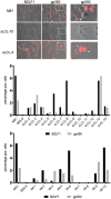
Multiple EBV-positive B-cell lines from Western individuals support lytic replication. Twenty-two sLCLs were immunostained with antibodies specific for BZLF1 and gp350. The pictures show the results of the staining in two of the investigated cases, together with cells infected with rM81 (white arrows, gp350-positive cells; white arrowhead, cells covered with viruses). The graphs give the percentage of BZLF1- and gp350-positive replicating cells and exclude nonreplicating cells coated with viruses.
FIG 2
FIG 2
Multiple EBV-positive B-cell lines from Western individuals produce viruses. (A) A Gardella gel analysis was performed to identify linear viral DNA that is produced during lytic replication. M81 served as a positive control and Raji as a negative control. (B) Electron microscopy pictures showing viruses at the surface of EBV-positive B cells in five cell lines. (C) The panel of cell lines was subjected to a Western blot analysis with a gp350-specific probe. The EBV-negative Elijah cell line served as a negative control.
FIG 3
FIG 3
The ability of virus isolates to replicate is maintained in different B-cell populations and under chemical induction of lytic replication. (A) Thirteen iEBVL and IM sLCLs produced enough virus to infect another unrelated B-cell sample. The replication rate, as assessed by the percentages of BZLF1-positive B cells in the parental and daughter cell lines, is given in a dot plot. The average and standard error is also indicated. (B) We also show an example of immunofluorescence staining and a Western blot for gp350 in parental and daughter cells. (C, right). Fifteen spontaneous LCLs were treated with a combination of tetradecanoyl phorbol acetate (TPA) and butyrate, transforming growth factor β (TGF-β), or ionomycin. The left dot plot shows the fold change in the number of BZLF1-positive B cells after exposure to the first two drugs, relative to that in mock-treated cells, and the right dot plot shows the percentage of BZLF1-positive B cells in the presence of absence of ionomycin. (D) Three independent primary B-cell samples transformed with rMSHJ, rM81, and rB95-8 were treated with antibodies directed against the B-cell receptor, with ionomycin (left), or with cyclosporine (right). The dot plots show the fold change in the number of BZLF1-positive B cells relative to that in mock-treated cells.
FIG 4
FIG 4
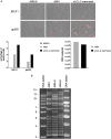
MSHJ replication in marmoset cell lines and its cloning as a BAC. (A) Primary B cells from the peripheral blood of marmosets were infected with sLCL-2. B cells infected by rM81 or by rB95-8 served as positive and negative controls, respectively. The picture shows BZLF1- and gp350-positive cells (red), the first graph gives the percentage of positive cells in the cell lines, and the second graph gives the viral titers in the supernatants of the cell lines, as determined by quantitative PCR (qPCR). (B) The rMSHJ, rM81, and rB95-8 genomes were digested with BamHI, and the resulting fragments separated onto an agarose gel. The size of the fragments is given by the DNA ladders.
FIG 5
FIG 5
IM-3 and rMSHJ are closely related to B95-8. The genomes of IM-3 and MSHJ (each indicated by a dot) were aligned to 130 published EBV genomes, including that of B95-8 (indicated by a square). The genetic tree shows the degree of divergence between the sequences, and the numbers give the branch length percentage and thus the level of divergence.
FIG 6
FIG 6
rMSHJ B-cell tropism and transformation efficiency. (A) Three independent sets of primary B cells were infected with rMSHJ, rM81, and rB95-8 at the same multiplicity of infection (10 genome equivalents per cell). Three days later, cells were stained for the EBNA2 protein. The dot plot shows the percentage of infected B cells. (B) Three independent sets of primary B cells were infected with rMSHJ, rM81, and rB95-8 at the same multiplicity of infection (10 genome equivalents per cell). Three days after infection, cells were stained for EBNA2. Infected cells were seeded in a 96-well cluster plate at a concentration of three EBNA2-positive cells per well. Five weeks later, the percentage of outgrown wells was determined. The results are given in the dot plots. (C) Cells from LCLs generated with rMSHJ, rM81 and rB95-8 (3 × 10⁵) were kept in culture for 4 weeks to generate the growth curves reproduced in the graph.
FIG 7
FIG 7
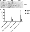
rMSHJ epitheliotropism. Primary epithelial cells were infected with rMSHJ, rM81, and rB95-8 at the same multiplicity of infection (100 genome equivalents per cell), using either direct or transfer infection on primary B cells. Three days later, cells were subjected to in situ hybridization with a probe specific for the highly abundant noncoding RNA EBER. (A) Representative example of transfer infection. (B) The bar graph shows the percentage of infected cells under the different conditions studied after infection of 3 samples. Cells were infected with viruses that express gp110 at high or low levels. We give the mean and standard error from three infection experiments.
FIG 8
FIG 8
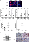
B cells infected with rMSHJ undergo a high level of spontaneous lytic replication. Three sets of independent primary B-cell samples were infected with rMSHJ, rM81, and rB95-8. Expression of BZLF1 and gp350 was monitored once a week for 4 weeks. (A) Example of immunostaining with BZLF1 and gp350. BZLF1 signals were detected in the nucleus, but also in the cytoplasm (white arrowheads), of infected cells. The dot plots show the percentage of cells displaying nuclear (left) or cytoplasmic (right) over time. (B) Six weeks after infection, infected cells were subjected to immunofluorescence staining with antibodies specific for BZLF1 and gp350. The percentage of positive cells is given in the dot plots together with the standard error. (C) BZLF1 and gp350 expression in one B-cell sample infected with the three viruses, as determined by Western blot. Staining for actin expression was used as a loading control. (D) The pictures show expression of BZLF1 and gp350 in EBV-positive lymphoid tumors that developed in immunosuppressed mice after infection with rM81 or rMSHJ.
FIG 9
FIG 9
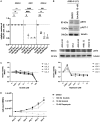
B cells infected with rMSHJ are sensitive to ibrutinib and rapamycin treatment. (A) Three independent primary B-cell samples transformed with rMSHJ, rM81, and rB95-8 were exposed to ibrutinib (10 nM or 100 nM) or rapamycin (10 nM). (B) Western blot showing expression p-Btk and pAKT-1 in rB95.8-transformed B cells with or without 10 nM ibrutinib treatment (upper). Western blot showing p-Btk expression in B cells transformed by viral strains S1 and S2 in blood samples 1 and 2 (lower). (C) Viability of 4 EBV-transformed B-cell samples (LCL 1 to LCL 4) after treatment with increasing concentrations of ibrutinib or rapamycin. (D) Cell growth rate of an EBV-transformed B-cell sample after treatment with different concentrations of ibrutinib or rapamycin.
FIG 10
FIG 10
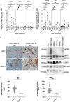
Hypoxia reactivates lytic replication, and lytically replicating cells are located closed to capillaries. (A) Three independent primary B-cell samples, each transformed with rMSHJ, rM81, and rB95-8 were kept under hypoxia for up to 7 days. The percentage of BZLF1-positive cells was determined after 1, 3, 5, and 7 days of hypoxia. The experiment was performed with 30- (left) or 90-day-old (right) transformed B-cell samples. One 90-day-old cell sample set was analyzed after hypoxia treatment by Western blotting using antibodies specific to BZLF1, gp350, LMP1, EBNA2, actin, and PARP. (B) Immunohistochemistry showing CD34-positive small arteries (white arrowhead) and capillaries (arrow), together with BZLF1-positive B cells (arrowhead). We show one sample infected with rM81 and one sample infected with rMSHJ at low (×20) and high (×40) power. (C) We measured the shortest distance between 100 BZLF1-positive B cells and the closest capillary or small artery. The results are given as dot plots for tissues infected with rM81 (left) and rMSHJ (right).

References

    1. Rickinson AB, Kieff E. 2007. Epstein-Barr virus, p 2655–2700. In Knipe DM, Howley PM, Griffin DE, Lamb RA, Martin MA, Roizman B, Strauss SE (ed). Lippincott Williams & Wilkins, Philadelphia, PA.
    1. Dolcetti R, Dal Col J, Martorelli D, Carbone A, Klein E. 2013. Interplay among viral antigens, cellular pathways and tumor microenvironment in the pathogenesis of EBV-driven lymphomas. Semin Cancer Biol 23:441–456. doi:10.1016/j.semcancer.2013.07.005. - DOI - PubMed
    1. Klinke O, Feederle R, Delecluse HJ. 2014. Genetics of Epstein-Barr virus microRNAs. Semin Cancer Biol 26:52–59. doi:10.1016/j.semcancer.2014.02.002. - DOI - PubMed
    1. Tsai M-H, Raykova A, Klinke O, Bernhardt K, Gärtner K, Leung CS, Geletneky K, Sertel S, Münz C, Feederle R, Delecluse H-J. 2013. Spontaneous lytic replication and epitheliotropism define an Epstein-Barr virus strain found in carcinomas. Cell Rep 5:458–470. doi:10.1016/j.celrep.2013.09.012. - DOI - PubMed
    1. Coghill AE, Hildesheim A. 2014. Epstein-Barr virus antibodies and the risk of associated malignancies: review of the literature. Am J Epidemiol 180:687–695. doi:10.1093/aje/kwu176. - DOI - PMC - PubMed

Publication types