. 2020 Apr 1;204(7):1968-1981.
doi: 10.4049/jimmunol.1901168. Epub 2020 Feb 26.

Osteopontin and iCD8α Cells Promote Intestinal Intraepithelial Lymphocyte Homeostasis

Affiliations

Osteopontin and iCD8α Cells Promote Intestinal Intraepithelial Lymphocyte Homeostasis

Ali Nazmi et al. J Immunol. .

Abstract

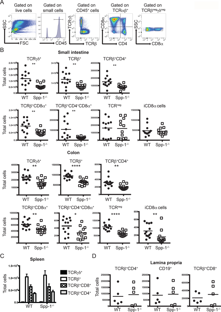
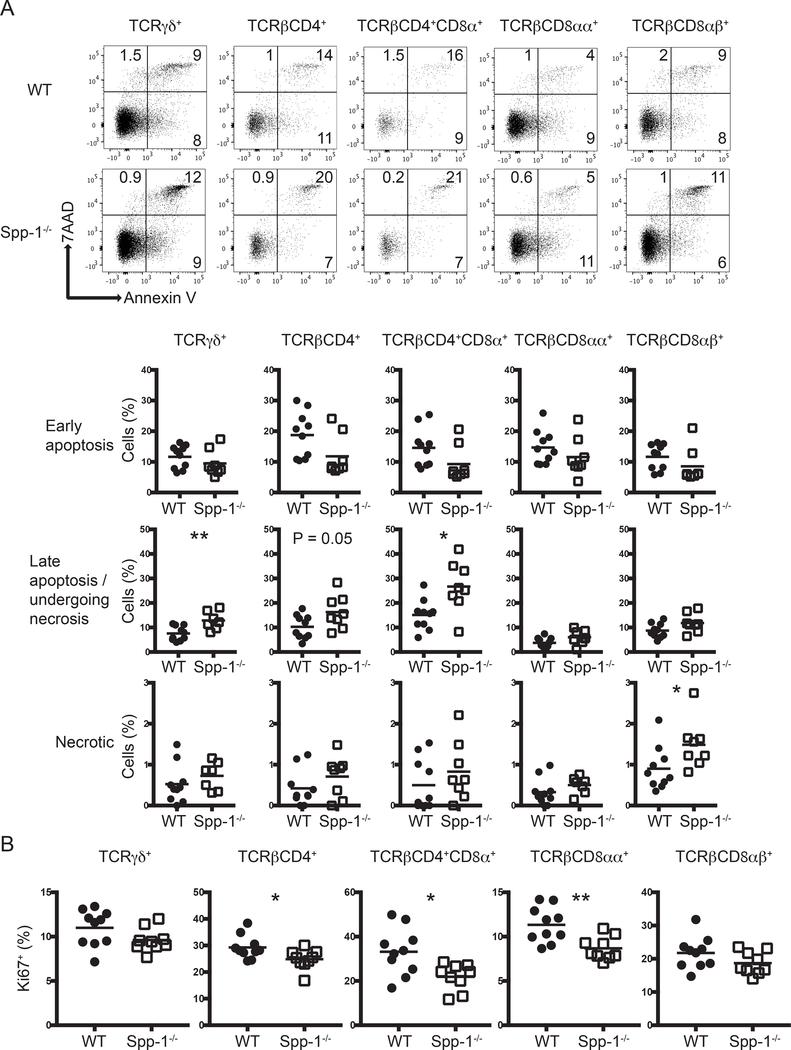
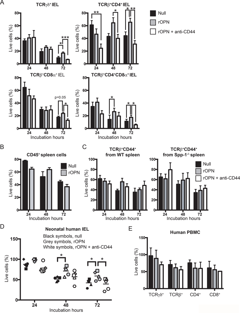
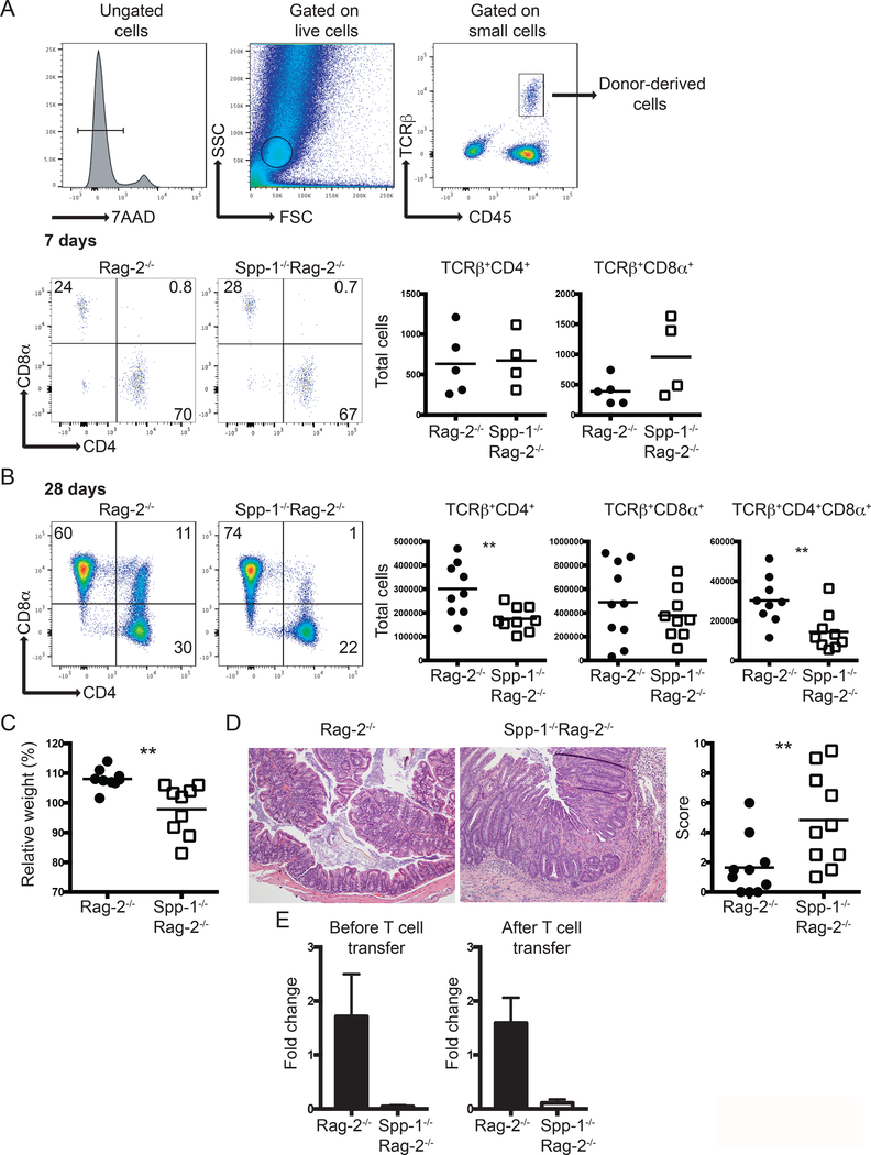
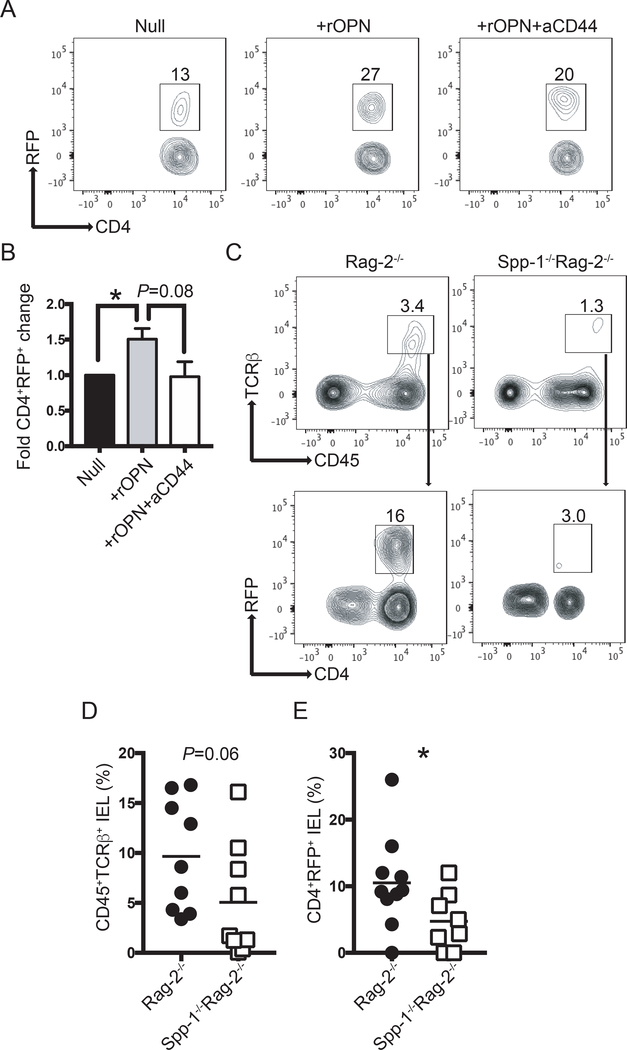
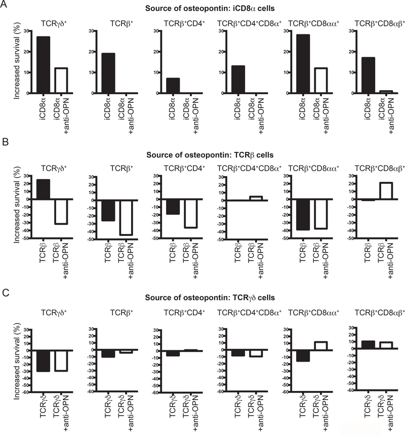
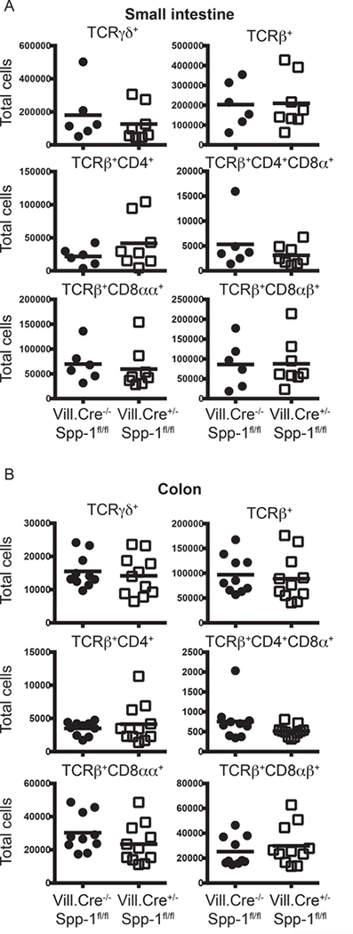
Intestinal intraepithelial lymphocytes (IEL) comprise a diverse population of cells residing in the epithelium at the interface between the intestinal lumen and the sterile environment of the lamina propria. Because of this anatomical location, IEL are considered critical components of intestinal immune responses. Indeed, IEL are involved in many different immunological processes, ranging from pathogen control to tissue stability. However, despite their critical importance in mucosal immune responses, very little is known about the homeostasis of different IEL subpopulations. The phosphoprotein osteopontin is important for critical physiological processes, including cellular immune responses, such as survival of Th17 cells and homeostasis of NK cells among others. Because of its impact in the immune system, we investigated the role of osteopontin in the homeostasis of IEL. In this study, we report that mice deficient in the expression of osteopontin exhibit reduced numbers of the IEL subpopulations TCRγδ+, TCRβ+CD4+, TCRβ+CD4+CD8α+, and TCRβ+CD8αα+ cells in comparison with wild-type mice. For some IEL subpopulations, the decrease in cell numbers could be attributed to apoptosis and reduced cell division. Moreover, we show in vitro that exogenous osteopontin stimulates the survival of murine IEL subpopulations and unfractionated IEL derived from human intestines, an effect mediated by CD44, a known osteopontin receptor. We also show that iCD8α IEL but not TCRγδ+ IEL, TCRβ+ IEL, or intestinal epithelial cells, can promote survival of different IEL populations via osteopontin, indicating an important role for iCD8α cells in the homeostasis of IEL.


Figures

Figure 1.
Figure 1.
Osteopontin-deficient mice have a reduced IEL compartment. (A) Gating strategy utilized in this report. After gating on live cells, IEL were gated as indicated. (B) Total IEL numbers from the small intestine (top) and colon (bottom) of WT and Spp-1−/− mice. Each dot represents an individual mouse (n = 13). (C) Total cell numbers of the indicated populations in the spleens from WT and Spp-1−/− mice (n = 8). Bars indicate SEM. (D) Total cell numbers of the indicated populations in the lamina propria from WT and Spp-1−/− mice. Each dot represents an individual mouse (n = 5). Data from (B) to (D) are representative of two to three independent experiments. * P<0.05; **P<0.01; ****P<0.0001 (Mann-Whitney U test).
Figure 2.
Figure 2.
Differential apoptosis and cell division in IEL from osteopontin-deficient mice. (A) Annexin V staining of different small intestine IEL populations derived from WT and Spp-1−/− mice. After gating cells by size based on forward and side scatter profiles, cells were gated for the expression of CD45 and other surface markers as indicated in Fig. 1A, and then analyzed for annexin V and 7AAD staining. Dot plots show a representative sample. Summary is indicated in the graphs. Data are representative of three independent experiments. Each dot represents an individual sample (n = 8 to 10). (B) Ki67 intracellular staining of different small intestine IEL populations derived from WT and Spp-1−/− mice. Cells were gated as in Figure 1A. Data are representative from two independent experiments. Each dot represents an individual sample (n = 9 to 10). *P<0.05; **P<0.01 (Mann-Whitney U test).
Figure 3.
Figure 3.
Osteopontin promotes in vitro IEL survival. (A) Indicated FACS-enriched IEL populations from WT mice were left untreated or treated with recombinant murine osteopontin (rOPN; 2 μg/ml), with or without anti-mouse CD44 (5 μg/ml). After the indicated time points, cell survival was determined. Data are representative of three independent experiments. Biological replicas consisted of two to three pooled IEL preparations from individual mice; each experiment consisted of 3 biological replicas. (B) Enriched CD45+ splenocytes were treated as in (A). Data are representative of two independent experiments (n = 3). (C) FACS-enriched TCRβ+CD44+ spleen cells from WT and Spp-1−/− mice were treated as in (A). Data are representative of two independent experiments (n = 3). (D) Neonatal human IEL were incubated in the presence or absence of recombinant human osteopontin (2 μg/ml) and anti-human CD44 (5μg/ml). After the indicated time points, cell survival was determined. Each symbol represents an individual human: circle, small intestine from 1-day old patient presenting volvulus and necrosis; square, ileum and colon from 17-day old patient presenting necrotizing enterocolitis; triangle, jejunum from 12-day old patient presenting necrotizing enterocolitis; diamond and hexagon, de-identified individuals. (E) PBMC from adult humans were treated as in (D). Data are representative of two independent experiments (n = 3). *P<0.05, **P<0.01, ***P<0.001 (One-way ANOVA).
Figure 4.
Figure 4.
Environmental osteopontin influences IEL reconstitution and colon inflammation. Enriched total spleen T cells from WT mice were adoptively transferred into Rag-2−/− and Spp-1−/−Rag-2−/− mice. (A) Gating strategy to identify cells derived from donor mice (top). Representative analysis of mice analyzed seven days after transfer (bottom). Dot plots show a representative sample. Results are summarized in the graphs. TCRβ+CD4+CD8α+ cells were not detected above background at this time point in either group. Data are representative from two independent experiments. Each dot represents an individual sample (n = 4 to 5). (B) The same analysis as in (A) performed at 28 days post transfer. Dot plots show a representative sample. Results are summarized in the graphs. Data are representative from three independent experiments. Each dot represents an individual animal (n = 9). (C) Total weight change at 28 days post transfer of mice treated as in (B). (D) Representative H&E stained samples of colon sections from the indicated mice at 28 days (100X magnification). Graph indicates total histological score [immune cell infiltration (3 points), loss of goblet cells (3 points), crypt damage (3 points) and epithelial hyperplasia (3 points)]. For (C) and (D) data are representative from three independent experiments, each dot represents an individual mouse (n = 9). (E) Total mRNA expression from colons of naïve and T cell treated (28 days post-transfer) Rag-2−/− and Spp-1−/−Rag-2−/− mice (n=3–5). **P<0.01 (Mann-Whitney T test).
Figure 5.
Figure 5.
Osteopontin sustains Foxp3 expression. (A) Intestinal T cells from RFP-Foxp3 reporter mice were isolated and cultured in the presence or absence of recombinant osteopontin (2 μg/ml) and anti-CD44 (5 μg/ml). Seventy-two hours later, the percentage of RFP+ cells was determined by flow cytometry. After excluding dead cells, dot plots were gated as CD45+TCRβ+ cells. (B) Bar graph indicates the fold change in CD4+RFP+ cells in relation to the null group. Data are representative of three independent experiments. (C) Enriched splenic RFP+ cells from RFP-Foxp3 reporter mice were adoptively transferred into Rag-2−/− and Spp-1−/−Rag-2−/− recipient mice. Eight weeks after transfer, IEL from small intestine were isolated and the percentage of RFP+ determined. After excluding dead cells, plots were gated as indicated by the arrows. (D) Summary of the percentage of donor-derived cells recovered; each dot represents an individual mouse. Data are representative from three independent experiments. (E) Summary of the percentage of RFP+ cells within the donor-derived cells. *P<0.05 (Mann-Whitney T test). rOPN = recombinant osteopontin; aCD44 = anti-CD44 antibodies.
Figure 6.
Figure 6.
iCD8α cell-derived osteopontin promotes IEL survival. Enriched CD45+ IEL from small intestine and colon derived from Spp-1−/− mice were incubated in the presence or absence of enriched iCD8α IEL (A), TCRβ+ IEL (B), or TCRγδ+ IEL (C) from osteopontin-competent donors. Some cells were cultured in the presence of anti-osteopontin antibodies (2 μg/ml). Four hours after incubation, cells were stained for surface markers, including annexin V, and 7AAD. Cells were gated as in Figure 2A. Graphs indicate “increased survival”, which was determined as 100 - [% of annexin V+ read-out cells in co-culture x 100 / % of annexin V+ cells cultured alone].
Figure 7.
Figure 7.
IEC-derived osteopontin does not promote IEL homeostasis. Small intestine (A) and colon (B) IEL were isolated from Villin-Cre−/−Spp-1fl/fl and Villin-Cre+/−Spp-1fl/fl mice, and the cellularity determined as indicated in Fig. 1A and B.

Similar articles

Cited by

References

    1. Cheroutre H, Lambolez F, and Mucida D. 2011. The light and dark sides of intestinal intraepithelial lymphocytes. Nat Rev Immunol 11: 445–456. - PMC - PubMed
    1. Olivares-Villagomez D, and Van Kaer L. 2017. Intestinal Intraepithelial Lymphocytes: Sentinels of the Mucosal Barrier. Trends Immunol. - PMC - PubMed
    1. Van Kaer L, and Olivares-Villagomez D. 2018. Development, Homeostasis, and Functions of Intestinal Intraepithelial Lymphocytes. J Immunol 200: 2235–2244. - PMC - PubMed
    1. Fuchs A, Vermi W, Lee JS, Lonardi S, Gilfillan S, Newberry RD, Cella M, and Colonna M. 2013. Intraepithelial type 1 innate lymphoid cells are a unique subset of IL-12- and IL-15-responsive IFN-gamma-producing cells. Immunity 38: 769–781. - PMC - PubMed
    1. Talayero P, Mancebo E, Calvo-Pulido J, Rodriguez-Munoz S, Bernardo I, Laguna-Goya R, Cano-Romero FL, Garcia-Sesma A, Loinaz C, Jimenez C, Justo I, and Paz-Artal E. 2016. Innate Lymphoid Cells Groups 1 and 3 in the Epithelial Compartment of Functional Human Intestinal Allografts. Am J Transplant 16: 72–82. - PubMed

Publication types

Substances