. 2020 Feb 26;5(1):e00027-20.
doi: 10.1128/mSphere.00027-20.

Trypanosoma brucei RAP1 Has Essential Functional Domains That Are Required for Different Protein Interactions

Affiliations

Trypanosoma brucei RAP1 Has Essential Functional Domains That Are Required for Different Protein Interactions

Marjia Afrin et al. mSphere. .

Abstract

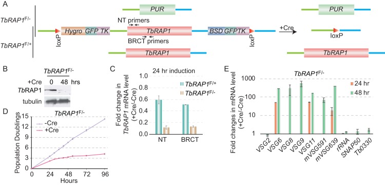
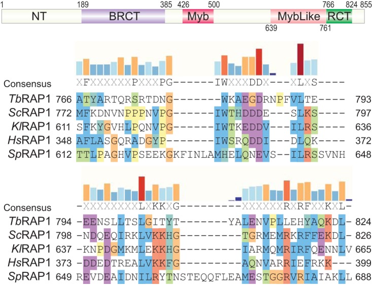
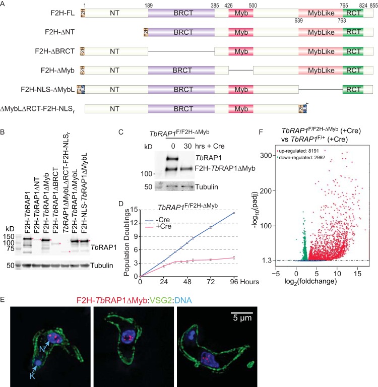
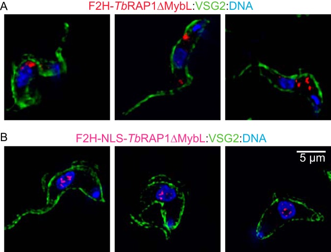
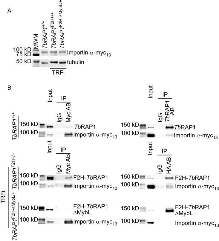
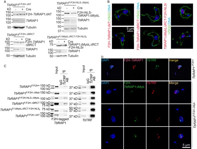
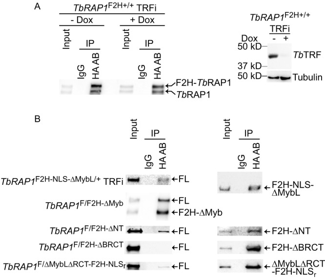
RAP1 is a telomere protein that is well conserved from protozoa to mammals. It plays important roles in chromosome end protection, telomere length control, and gene expression/silencing at both telomeric and nontelomeric loci. Interaction with different partners is an important mechanism by which RAP1 executes its different functions in yeast. The RAP1 ortholog in Trypanosoma brucei is essential for variant surface glycoprotein (VSG) monoallelic expression, an important aspect of antigenic variation, where T. brucei regularly switches its major surface antigen, VSG, to evade the host immune response. Like other RAP1 orthologs, T. brucei RAP1 (TbRAP1) has conserved functional domains, including BRCA1 C terminus (BRCT), Myb, MybLike, and RAP1 C terminus (RCT). To study functions of various TbRAP1 domains, we established a strain in which one endogenous allele of TbRAP1 is flanked by loxP repeats, enabling its conditional deletion by Cre-mediated recombination. We replaced the other TbRAP1 allele with various mutant alleles lacking individual functional domains and examined their nuclear localization and protein interaction abilities. The N terminus, BRCT, and RCT of TbRAP1 are required for normal protein levels, while the Myb and MybLike domains are essential for normal cell growth. Additionally, the Myb domain of TbRAP1 is required for its interaction with T. brucei TTAGGG repeat-binding factor (TbTRF), while the BRCT domain is required for its self-interaction. Furthermore, the TbRAP1 MybLike domain contains a bipartite nuclear localization signal that is required for its interaction with importin α and its nuclear localization. Interestingly, RAP1's self-interaction and the interaction between RAP1 and TRF are conserved from kinetoplastids to mammals. However, details of the interaction interfaces have changed throughout evolution.IMPORTANCETrypanosoma brucei causes human African trypanosomiasis and regularly switches its major surface antigen, VSG, to evade the host immune response. VSGs are expressed from subtelomeres in a monoallelic fashion. TbRAP1, a telomere protein, is essential for cell viability and VSG monoallelic expression and suppresses VSG switching. Although TbRAP1 has conserved functional domains in common with its orthologs from yeasts to mammals, the domain functions are unknown. RAP1 orthologs have pleiotropic functions, and interaction with different partners is an important means by which RAP1 executes its different roles. We have established a Cre-loxP-mediated conditional knockout system for TbRAP1 and examined the roles of various functional domains in protein expression, nuclear localization, and protein-protein interactions. This system enables further studies of TbRAP1 point mutation phenotypes. We have also determined functional domains of TbRAP1 that are required for several different protein interactions, shedding light on the underlying mechanisms of TbRAP1-mediated VSG silencing.

Keywords: RAP1; Trypanosoma brucei; protein-protein interaction; telomere.


Figures

FIG 1
FIG 1
Conditional knockout of T. brucei RAP1 (TbRAP1) led to cell growth arrest and VSG derepression. (A) A diagram showing three different TbRAP1 alleles, including deleted (top), floxed (middle), and WT (bottom) alleles, in TbRAP1F/− (top two) and TbRAP1F/+ (bottom two) cells before (left) and after (right) the Cre induction. NT, N terminus. (B) Western analysis of cell lysates prepared from TbRAP1F/− cells before and after the Cre induction. A rabbit antibody (16) was used to detect TbRAP1 (top). In this and other figures, TAT-1 (107) was used to detect tubulin (as a loading control). (C) Quantitative RT-PCR was performed using TbRAP1 primers that anneal to the N terminus or the BRCT domain of TbRAP1 (marked in panel A) to estimate the change in the TbRAP1 mRNA level in TbRAP1F/+ and TbRAP1F/− strains. (D) Growth curves of TbRAP1F/− cells with and without the doxycycline-induced Cre expression. (E) Quantitative RT-PCR to estimate the changes in mRNA level of a number of ES-linked VSG genes and several control genes. Average values were calculated from three independent inductions. In this and following figures, error bars represent standard deviations.
FIG 2
FIG 2
TbRAP1 has an RCT domain. (Top) TbRAP1 domain structure. The positions of each domain are labeled. NT, N terminus; BRCT, BRCA1 C terminus; RCT, RAP1 C terminus. (Bottom) Sequence alignment of the TbRAP1 RCT domain and the RCT domains in Saccharomyces cerevisiae (Sc) RAP1, Kluyveromyces lactis (Kl) RAP1, Homo sapiens (Hs) RAP1, and Schizosaccharomyces pombe (Sp) RAP1 performed using Clustal X. Alignment is visualized in SnapGene. Amino acid positions are indicated. Amino acids are highlighted based on their properties and level of conservation (Clustal X). Specifically, nonpolar amino acids are highlighted in blue, positively charged amino acids in red, negatively charged amino acids in magenta, G amino acid (lacking of bonding characteristics) in orange, H and Y amino acids (mixture of nonpolar and polar ends to the side chain) in dark turquoise, P amino acid (between polar and nonpolar) in yellow, and S and T amino acids (mostly nonpolar) in green. Levels of conservation at each position (percent identical residues among the five RAP1 orthologs) are shown as bar graphs on top of the aligned sequences.
FIG 3
FIG 3
The Myb domain of TbRAP1 is essential for normal cell growth and VSG silencing. (A) Diagrams of FLAG-HA-HA (F2H)-tagged WT and mutant TbRAP1 used in this study. NLS, simian virus 40 (SV40) large T nuclear localization signal. NLSr, the second half of TbRAP1 nuclear localization signal (aa 737 to 742). (B) Whole-cell lysates from TbRAP1F/F2H-mut cells (after induction of Cre for 30 h) and TbRAP1−/F2H+ cells (as a positive control) were analyzed by Western blotting. HA probe monoclonal antibody (Santa Cruz Biotechnologies) was used to detect the F2H-tagged TbRAP1. Red asterisks indicate the TbRAP1 fragments. F2H-TbRAP1ΔNT ran at a much lower rate than expected. (C) Western blotting of cell lysates prepared from TbRAP1F/F2H-ΔMyb cells before and after induction of Cre. An anti-TbRAP1 rabbit antibody (16) was used to detect TbRAP1 and F2H-TbRAP1ΔMyb. To differentiate WT and mutant TbRAP1, proteins were separated on a 7.5% Tris polyacrylamide gel for 7 h 40 min. (D) Growth curves of TbRAP1F/F2H-ΔMyb cells with and without the Cre induction. (E) F2H-TbRAP1ΔMyb is located in the nucleus. The monoclonal HA antibody 12CA5 (Memorial Sloan Kettering Cancer Center [MSKCC] monoclonal antibody core) was used to detect F2H-TbRAP1ΔMyb. An anti-VSG2 rabbit antibody (16) was used to show the outline of the cell body. DAPI was used to stain DNA. The small DAPI-positive circle represents the kinetoplast (K), and the large DAPI-positive circle represents the nucleus (N). Three different cells are shown in three panels. In this and other figures, IF images are in the same scale and a size bar is shown in one of the panels. (F) Differential gene expression in the TbRAP1ΔMyb mutant was summarized in a volcano plot. TbRAP1F/F2H-ΔMyb and TbRAP1F/+ cells were induced for Cre expression for 30 h and analyzed by RNA-seq. Compared to TbRAP1F/+ cells, more than 8,000 genes were upregulated and nearly 3,000 genes were downregulated in the Cre-induced TbRAP1F/F2H-ΔMyb cells. A log10(adjusted P [padj]) value of 1.3 or higher is considered to be significant.
FIG 4
FIG 4
Immunofluorescent analysis of TbRAP1F/F2H-ΔMybL TRFi and TbRAP1F/F2H-NLS-ΔMybL TRFi cells. F2H-TbRAP1ΔMybL and F2H-NLS-TbRAP1ΔMybL were stained with 12CA5 HA antibody. A rabbit VSG2 antibody was used to show the outline of the cell body. DNA was stained with DAPI. In each strain, three different cells are shown in three panels.
FIG 5
FIG 5
The TbRAP1 MybLike domain is required for interaction with importin α. (A) Expression of importin α-myc13 in TbRAP1+/+, TbRAP1F2H+/+ TRFi, and TbRAP1F2H-ΔMybL/+ TRFi cells. Myc13-tagged proteins were detected by a myc monoclonal antibody, 9E10 (MSKCC monoclonal antibody core). (B) Co-IP of TbRAP1 and importin α. The myc antibody 9E10, an anti-TbRAP1 rabbit antibody (16), and IgG (as a negative control) were used for IP in TbRAP1+/+ cells. Western analysis was performed using the antibodies mentioned above to detect importin α-myc13 and TbRAP1. In TbRAP1F2H+/+ TRFi and TbRAP1F2H-ΔMybL/+ TRFi cells, the 9E10 myc antibody, the 12CA5 HA antibody, and IgG (as a negative control) were used for IP, and Western blotting was performed to detect importin α-myc13 (by 9E10) and F2H-tagged WT and mutant TbRAP1 (by 12CA5 in the left panels and HA probe in the right panels). In this and other figures, input samples represent 1% of the materials used for IP.
FIG 6
FIG 6
The TbRAP1 Myb domain is required for interaction with TbTRF. (A) Western analyses showed the expression levels of the F2H-tagged TbRAP1 mutants (using the HA probe antibody) and WT TbRAP1 (using a rabbit TbRAP1 antibody [16]) before and after the Cre induction in TbRAP1F/F2H-ΔNT, TbRAP1F/F2H-ΔBRCT, TbRAP1F/F2H-NLS-ΔMybL, and TbRAP1F/ΔMybLΔRCT-F2H-NLSr cells. (B) IF analysis showed that F2H-TbRAP1ΔNT, F2H-TbRAP1ΔBRCT, and TbRAP1ΔMybLΔRCT-F2H-NLSr were localized in the nucleus. 12CA5 was used to detect the F2H-tagged TbRAP1 mutants. A rabbit anti-VSG2 antibody was used to show the outline of the cell body. DNA was stained by DAPI. In each strain, two different cells are shown in two panels. (C) Co-IP experiments were performed in a series of TbRAP1F/F2H-mut cells (as labeled on the left). An anti-TbTRF rabbit antibody (89) and IgG (as a negative control) were used for IP. The antibodies used for Western analyses of the IP products were the 12CA5 HA antibody (except for TbRAP1F/F2H-ΔNT, where the HA probe was used) and an anti-TbTRF chicken antibody (16). (D) (Top) F2H-TbRAP1 (detected by 12CA5) partially colocalized with TbTRF (detected by a rabbit TbTRF antibody [89]) in TbRAP1−/F2H+ cells. (Bottom) F2H-TbRAP1ΔMyb (detected by 12CA5) was not colocalized with TbTRF (detected by a chicken TbTRF antibody [16]). DNA was stained by DAPI. Three different TbRAP1F/F2H-∆Myb cells are shown.
FIG 7
FIG 7
TbRAP1 interacts with itself through the BRCT domain. (A) TbRAP1’s self-interaction activity is independent of TbTRF. Left, co-IP experiments were done using the 12CA5 HA antibody and IgG (as a negative control) in TbRAP1F2H+/+ TRFi cells before and after induction of TbTRF RNAi. IP products were analyzed by Western blotting using a rabbit TbRAP1 antibody (16). To differentiate F2H-TbRAP1 and the untagged TbRAP1, proteins were separated on a 7.5% Tris polyacrylamide gel for 7 h. Right, Western analysis showed that TbTRF was efficiently depleted by RNAi after addition of doxycycline (Dox) for 30 h. A rabbit antibody was used to detect TbTRF (89). (B) The BRCT domain of TbRAP1 is critical for its self-interaction. Co-IP experiments were performed in various TbRAP1F/F2H-mut strains (indicated on the left) without Cre induction and in TbRAP1F2H-NLS-ΔMybL/+ TRFi cells. IP experiments were done using the 12CA5 HA antibody and IgG (as a negative control). To analyze the IP products, WT TbRAP1 and F2H-TbRAP1ΔMyb were detected with an anti-TbRAP1 rabbit antibody (16), and other F2H-tagged TbRAP1 mutants were detected by the HA probe antibody in Western blotting. To differentiate TbRAP1 and F2H-TbRAP1ΔMyb, proteins were separated on a 7.5% Tris polyacrylamide gel for 7 h 40 min.

Similar articles

Cited by

References

    1. Maciejowski J, de Lange T. 2017. Telomeres in cancer: tumour suppression and genome instability. Nat Rev Mol Cell Biol 18:175–186. doi:10.1038/nrm.2016.171. - DOI - PMC - PubMed
    1. Podlevsky JD, Bley CJ, Omana RV, Qi X, Chen JJ. 2008. The telomerase database. Nucleic Acids Res 36:D339–D343. doi:10.1093/nar/gkm700. - DOI - PMC - PubMed
    1. Martínez P, Blasco MA. 2015. Replicating through telomeres: a means to an end. Trends Biochem Sci 40:504–515. doi:10.1016/j.tibs.2015.06.003. - DOI - PubMed
    1. Li B, Oestreich S, de Lange T. 2000. Identification of human Rap1: implications for telomere evolution. Cell 101:471–483. doi:10.1016/s0092-8674(00)80858-2. - DOI - PubMed
    1. Scherthan H, Jerratsch M, Li B, Smith S, Hulten M, Lock T, de Lange T. 2000. Mammalian meiotic telomeres: protein composition and redistribution in relation to nuclear pores. Mol Biol Cell 11:4189–4203. doi:10.1091/mbc.11.12.4189. - DOI - PMC - PubMed

Publication types

MeSH terms