. 2020 Feb 26;11(1):1058.
doi: 10.1038/s41467-020-14867-z.

B38-CAP is a bacteria-derived ACE2-like enzyme that suppresses hypertension and cardiac dysfunction

Affiliations

B38-CAP is a bacteria-derived ACE2-like enzyme that suppresses hypertension and cardiac dysfunction

Takafumi Minato et al. Nat Commun. .

Abstract

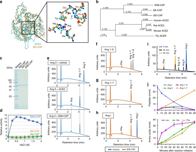
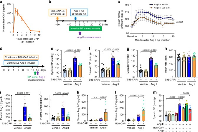
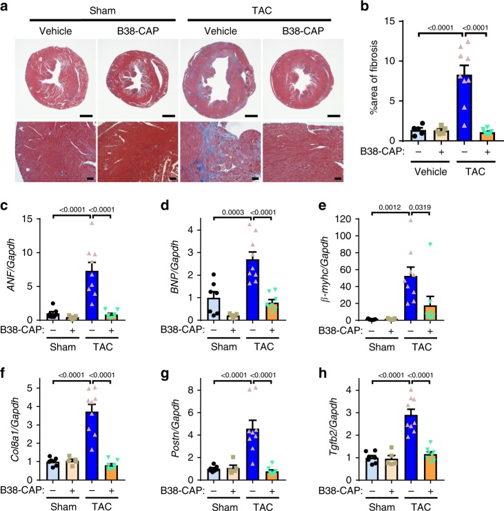
Angiotensin-converting enzyme 2 (ACE2) is critically involved in cardiovascular physiology and pathology, and is currently clinically evaluated to treat acute lung failure. Here we show that the B38-CAP, a carboxypeptidase derived from Paenibacillus sp. B38, is an ACE2-like enzyme to decrease angiotensin II levels in mice. In protein 3D structure analysis, B38-CAP homolog shares structural similarity to mammalian ACE2 with low sequence identity. In vitro, recombinant B38-CAP protein catalyzed the conversion of angiotensin II to angiotensin 1-7, as well as other known ACE2 target peptides. Treatment with B38-CAP suppressed angiotensin II-induced hypertension, cardiac hypertrophy, and fibrosis in mice. Moreover, B38-CAP inhibited pressure overload-induced pathological hypertrophy, myocardial fibrosis, and cardiac dysfunction in mice. Our data identify the bacterial B38-CAP as an ACE2-like carboxypeptidase, indicating that evolution has shaped a bacterial carboxypeptidase to a human ACE2-like enzyme. Bacterial engineering could be utilized to design improved protein drugs for hypertension and heart failure.


Conflict of interest statement

The authors declare no competing interests.

Figures

Fig. 1
Fig. 1. B38-CAP, a bacteria-derived carboxypeptidase, is Angiotensin-converting enzyme 2 (ACE2)-like enzyme.
a Crystal structures of BS-CAP and human ACE2 proteins. Inset: metal-coordinating residues (red) and substrate-binding residues (black) are shown. b Phylogenetic tree of ACE2 and bacterial ACE2-like carboxypeptidases. c SDS-PAGE analysis of recombinant proteins of BS-CAP, BA-CAP, and B38-CAP. d Dependence of ACE2-like proteolytic activity of BS-CAP, BA-CAP, and B38-CAP on anion concentration. ACE2 activity was measured with hydrolysis rate of the fluorogenic ACE2 substrate Nma-His-Pro-Lys(Dnp). eh HPLC analysis of B38-CAP-treated angiotensin peptides. Ang II (e), Ang 1–9 (f), Ang 1–7 (g), or Ang I (h) (5 nmol each) was incubated with vehicle, recombinant B38-CAP protein, or recombinant ACE2 protein (5 μg each) for 90 min, then subjected to HPLC analysis. i, j Kinetic analysis for hydrolysis of Ang I with B38-CAP. HPLC analysis of angiotensin peptides generated after incubating Ang I with B38-CAP (i, j, upper panel). Amino acids in the same samples were quantified with LC-MS system (j, lower panel). Experiments were repeated more than three times and representative chromatography charts are shown. j Values are means ± SEM. n = 3 independent experiments.
Fig. 2
Fig. 2. Effects of B38-CAP on plasma angiotensin II levels and blood pressure in mice.
a Plasma B38-CAP levels in mice after intraperitoneal injection of B38-CAP (2 mg kg−1). B38-CAP activity was measured with the B38-CAP substrate Nma-Leu-Pro-Lys(Dnp) (n = 4, 6, 8, 6, 6, and 6 for 0, 1, 2, 4, 8, and 12 h, respectively). b, c Invasive measurements of arterial blood pressure (BP). Experimental protocol (b); mice pretreated with B38-CAP (2 mg kg−1 i.p.) had liquid-filled catheter inserted into carotid artery for BP measurements and Ang II (0.2 mg kg−1 i.p.) was injected and BP measured. Systolic arterial pressure is shown (c). (n = 6 mice per group). dh Blood pressure measurements with conscious mice. Experimental protocol (d); mice were treated with continuous infusion of vehicle, Ang II (1.5 mg kg−1 per day), B38-CAP (3 mg kg−1 per day), or Ang II (1.5 mg kg−1 per day) plus B38-CAP (3 mg kg−1 per day), and BP was measured by tail-cuff system after 2 weeks. Systolic (e), diastolic (f), and mean (g) BP and heart rate (h) are shown for mice treated vehicle + vehicle (n = 10), vehicle + B38-CAP (n = 11), Ang II + vehicle (n = 11), and Ang II + B38-CAP (n = 11). il Measurements of Ang II and Ang 1–7 in the plasma of mice. The plasma was obtained from the mice treated acutely (i, k) and chronically (j, l) with Ang II, in the cohort of b, c, and dh, respectively. Ang II (i, j) and Ang 1–7 (k, l) levels were measured with ELISA. m BP measurements in conscious mice. The mice pretreated with B38-CAP (2 mg/kg i.p.) at 90 min before acquisition of baseline BP were treated with Ang II (0.2 mg kg−1 i.p.), with or without A779 (0.2 mg kg−1 i.p.). BP was measured every 5 min by tail-cuff system (Supplementary Fig. 9a–d) and the BP at 5 min after the last injection is shown (n = 6 mice per group). All values are means ± SEM. c, em, Two-way ANOVA with Sidak’s multiple-comparisons test. Numbers above square brackets show significant P-values.
Fig. 3
Fig. 3. Effects of B38-CAP on angioteinsin II-induced cardiac hypertrophy and fibrosis.
ac Cardiac hypertrophy in the mice chronically co-treated with Ang II (1.5 mg kg−1 per day) and B38-CAP (3 mg kg−1 per day) in the cohort of Fig. 2d–h. Macroscopic heart images (a), heart weight to body weight ratio (HW/BW) (b), and heart weight to tibia length ratio (HW/TL) (c) in mice treated vehicle + vehicle (n = 10), vehicle + B38-CAP (n = 11), Ang II + vehicle (n = 11), and Ang II + B38-CAP (n = 11). Bars indicate 2 mm. df Echocardiography parameters of left ventricular end-diastolic posterior wall thickness (PWD) (d), end-diastolic interventricular septal wall thickness (IVSD) (e), and %fractional shortening (%FS) (f) in the mouse hearts. Complete echocardiography data are shown in Supplementary Table 2. g, h Histology of hearts. Masson’s trichrome staining (g); bars indicate 2 mm and 100 μm in the upper panels and lower panels, respectively. Quantification of fibrosis in the hearts (h) of mice treated with vehicle + vehicle (n = 5), vehicle + B38-CAP (n = 5), Ang II + vehicle (n = 7), and Ang II + B38-CAP (n = 8). ik qRT-PCR analysis of pro-fibrotic gene expressions in the hearts of mice treated with vehicle + vehicle (n = 5), vehicle + B38-CAP (n = 5), Ang II + vehicle (n = 7), and Ang II + B38-CAP (n = 8); mRNA levels of Collagen 8a (Col8a1) (f), Periostin (Postn) (g), and TGF-β (Tgfb2) (h) normalized with Gapdh. All values are means ± SEM. bf, hk Two-way ANOVA with Sidak’s multiple-comparisons test. Numbers above square brackets show significant P-values.
Fig. 4
Fig. 4. B38-CAP mitigates pressure overload (TAC)-induced cardiac dysfunction and hypertrophy.
a Experimental protocol. The mice were subjected to the surgery of transverse aortic constriction (TAC) surgery and then continuous infusion of B38-CAP (2 mg kg−1 per day) was initiated. bf B38-CAP suppressed cardiac hypertrophy. Representative photograph (b) of the hearts of mice under TAC. Bars indicate 2 mm. HW/BW (c), HW/TL (d), lung weight to body weight ratio (LW/BW) (e), and lung weight to tibia length ratio (LW/TL) (f) in the mice treated with sham + vehicle (n = 7), sham + B38-CAP (n = 5), TAC + vehicle (n = 9), and TAC + B38-CAP (n = 8). gl Echocardiography measurements. Representative M-mode echocardiography images (g), measurements of IVSD (h), PWD (i), LVESD (j), LVEDD (k), and %FS (l) are shown. Complete echocardiography data are shown in Supplementary Table 4. All values are means ± SEM. cf, hl One-way ANOVA with Sidak’s multiple-comparisons test. Numbers above square brackets show significant P-values.
Fig. 5
Fig. 5. B38-CAP suppresses TAC-induced cardiac fibrosis.
a, b Histology. The hearts of B38-CAP or vehicle-treated mice under TAC were stained with Masson’s trichrome. Bars indicate 1 mm (upper) or 100 μm (lower). ch qRT-PCR analysis for the expression of heart failure genes and pro-fibrosis genes; mRNA levels of atrial natriuretic factor (ANF) (c), B-type natriuretic peptide (BNP) (d), β-myosin heavy chain (β-myhc) (e), Collagen 8a (Col8a1) (f), Periostin (Postn) (g), and TGF-β (Tgfb2) (h) in the hearts of mice treated with sham + vehicle (n = 7), sham + B38-CAP (n = 5), TAC + vehicle (n = 9), and TAC + B38-CAP (n = 8). All values are means ± SEM. bh Two-way ANOVA with Sidak’s multiple-comparisons test. Numbers above square brackets show significant P-values.
Fig. 6
Fig. 6. No overt toxic effects of B38-CAP on liver and kidney.
a, b Liver function test with measurements of aspartate aminotransferase (AST) (a) and alanine aminotransferase (ALT) (b) in the blood. c, d Kidney function assessment with measurements of BUN (c) and Creatinine (Cr) (d) in the blood. sham + vehicle (n = 7), sham + B38-CAP (n = 5), TAC + vehicle (n = 9), and TAC + B38-CAP (n = 8). All values are means ± SEM. Two-way ANOVA with Sidak’s multiple-comparisons test. Numbers above square brackets show significant P-values.
Fig. 7
Fig. 7. Therapeutic effects of B38-CAP on established hypertension and cardiac dysfunction.
ae Therapeutic effects of B38-CAP on established hypertension. Experimental protocol (a); Ang II infusion (1 mg/kg/day) was initiated at 7 days before treatment. The mice were injected with B38-CAP (2 mg/kg i.p.) or vehicle twice a day and blood pressure was measured by tail-cuff system at 2 h after injection. Systolic (b), diastolic (c), and mean (d) BP and heart rate (e) in the mice treated Ang II + vehicle (n = 7), Ang II + B38-CAP (n = 7), and vehicle + vehicle (n = 5). fl Therapeutic effects of B38-CAP on established cardiac dysfunction. Experimental protocol (f); the C57BL/6J mice had TAC surgery at 5 weeks before treatment and B38-CAP (2 mg/kg/day) or vehicle was continuously infused with osmotic mini-pumps. Echocardiography parameters of %fractional shortening (%FS) (g) in the mice treated with sham + vehicle (n = 5), TAC + vehicle (n = 6), and TAC + B38-CAP (n = 6). Representative photographs of the hearts of mice under TAC (h). Bars indicate 2 mm. HW/BW (i), HW/TL (j), LW/BW (k), and LW/TL (l) are in the mice treated with sham + vehicle (n = 5), TAC + vehicle (n = 6), and TAC + B38-CAP (n = 6). mq qRT-PCR analysis for the expression of heart failure genes and pro-fibrosis genes in the hearts (n = 5 mice per group). All values are means ± SEM. be Two-way ANOVA with Sidak’s multiple-comparisons test. g One-way ANOVA with Sidak’s multiple-comparisons test for comparison of groups. Two-tailed paired t-test between before and after treatment of the same group. iq One-way ANOVA with Sidak’s multiple-comparisons test. Numbers next to square brackets show significant P-values.

References

    1. Corvol P, Jeunemaitre X, Charru A, Kotelevtsev Y, Soubrier F. Role of the renin-angiotensin system in blood pressure regulation and in human hypertension: new insights from molecular genetics. Recent Prog. Horm. Res. 1995;50:287–308. - PubMed
    1. Skeggs LT, Dorer FE, Levine M, Lentz KE, Kahn JR. The biochemistry of the renin-angiotensin system. Adv. Exp. Med. Biol. 1980;130:1–27. doi: 10.1007/978-1-4615-9173-3_1. - DOI - PubMed
    1. Boehm M, Nabel EG. Angiotensin-converting enzyme 2–a new cardiac regulator. N. Engl. J. Med. 2002;347:1795–1797. doi: 10.1056/NEJMcibr022472. - DOI - PubMed
    1. Donoghue M, et al. A novel angiotensin-converting enzyme-related carboxypeptidase (ACE2) converts angiotensin I to angiotensin 1-9. Circ. Res. 2000;87:E1–E9. doi: 10.1161/01.RES.87.5.e1. - DOI - PubMed
    1. Tipnis SR, et al. A human homolog of angiotensin-converting enzyme. Cloning and functional expression as a captopril-insensitive carboxypeptidase. J. Biol. Chem. 2000;275:33238–33243. doi: 10.1074/jbc.M002615200. - DOI - PubMed

Publication types

MeSH terms

LinkOut - more resources