. 2020 Feb 26;11(1):1073.
doi: 10.1038/s41467-019-14063-8.

Active acetylcholine receptors prevent the atrophy of skeletal muscles and favor reinnervation

Affiliations

Active acetylcholine receptors prevent the atrophy of skeletal muscles and favor reinnervation

Bruno A Cisterna et al. Nat Commun. .

Abstract

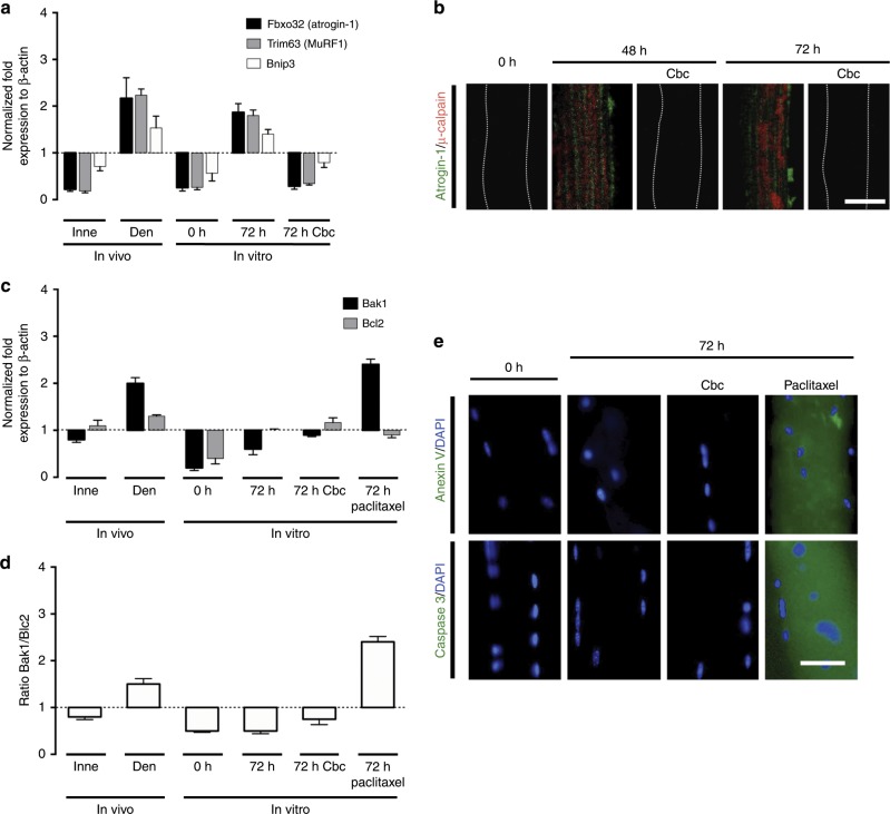
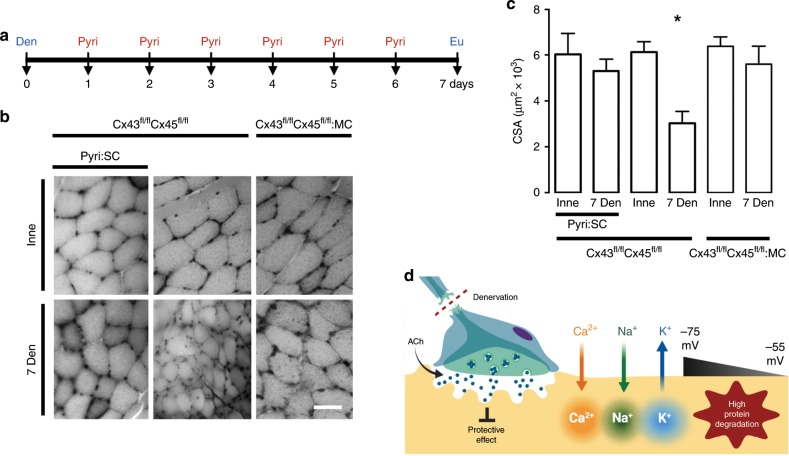
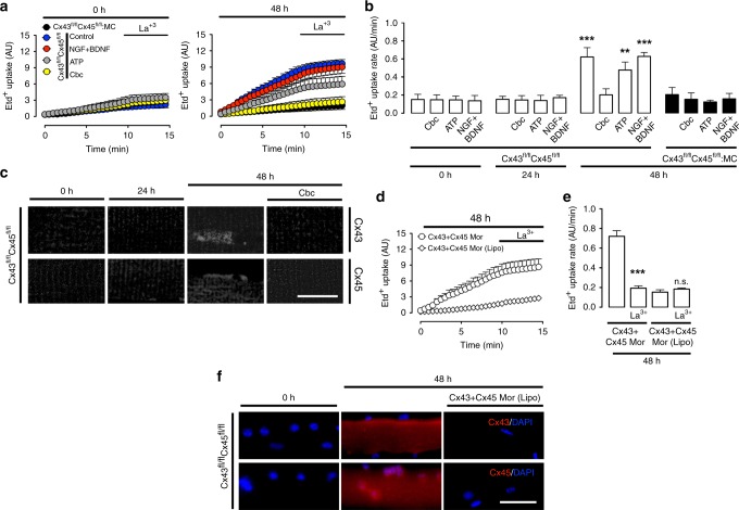
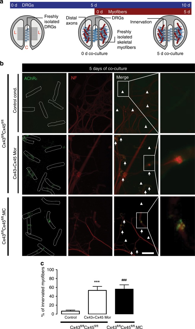
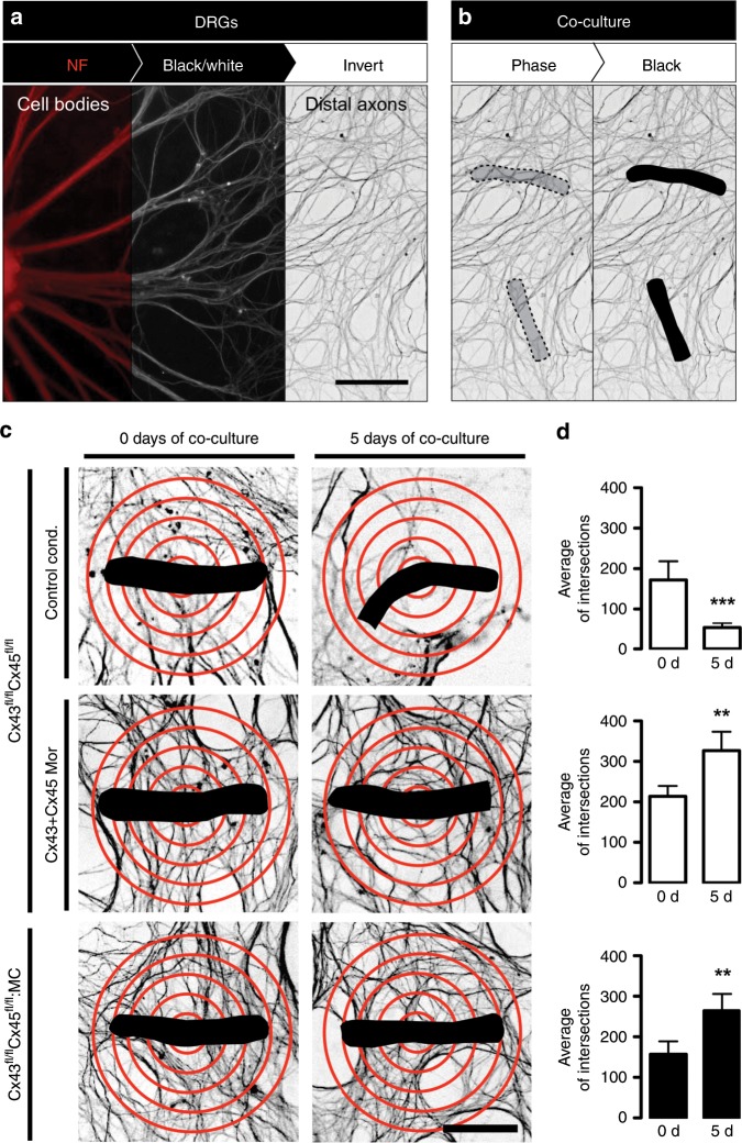
Denervation of skeletal muscles induces severe muscle atrophy, which is preceded by cellular alterations such as increased plasma membrane permeability, reduced resting membrane potential and accelerated protein catabolism. The factors that induce these changes remain unknown. Conversely, functional recovery following denervation depends on successful reinnervation. Here, we show that activation of nicotinic acetylcholine receptors (nAChRs) by quantal release of acetylcholine (ACh) from motoneurons is sufficient to prevent changes induced by denervation. Using in vitro assays, ACh and non-hydrolysable ACh analogs repressed the expression of connexin43 and connexin45 hemichannels, which promote muscle atrophy. In co-culture studies, connexin43/45 hemichannel knockout or knockdown increased innervation of muscle fibers by dorsal root ganglion neurons. Our results show that ACh released by motoneurons exerts a hitherto unknown function independent of myofiber contraction. nAChRs and connexin hemichannels are potential molecular targets for therapeutic intervention in a variety of pathological conditions with reduced synaptic neuromuscular transmission.


Conflict of interest statement

The authors declare no competing interests.

Figures

Fig. 1
Fig. 1. Cultured fibers express genes of atrophy and autophagy, but not of apoptosis.
Unilateral sciatic nerve transections were performed in Cx43fl/flCx45fl/fl mice (control mice); at day 7 post denervation the innervated (Inne) and denervated (Den) flexor digitorum brevis muscles were collected to determine the relative levels of specific mRNAs by qPCR (In vivo). In parallel experiments, myofibers from control mice were cultured for 0, 48, and 72 h under control conditions or treated with 200 nM carbachol (Cbc) or 100 nM Paclitaxel to determine the relative levels of specific mRNAs by qPCR and proteins by immunofluorescence (In vitro). a mRNA relative levels of Fbxo32 (atrogin-1), Trim63 (MuRF1), and Bnip3 normalized to actin β expression. b Relative levels of atrogin-1 (Green) and µ-calpain (Red) evaluated by immunofluorescence using confocal microscopy. Calibration bar: 50 µm. c mRNA relative levels of Bak1 (pro-apoptotic gene) and Bcl2 (anti-apoptotic gene) normalized to actin β expression. d Ratio of Bak1/Bcl2. e Relative levels of anexin V or caspase 3 (Green) evaluated by immunofluorescence (DAPI to stain nuclei in blue). Calibration bar: 50 µm. Bar error is the mean ± SEM. N = 4 independent experiments.
Fig. 2
Fig. 2. Acetylcholine analog prevents cellular alterations in cultured myofibers. Primary cultures of myofibers from flexor digitorum brevis (FDB) muscles were used.
a The permeability of the sarcolemma was measured in time-lapse experiments of ethidium (Etd+) uptake performed after 0, 24, 48, and 72 h of culture. Myofibers were cultured under control conditions (white), or treated with 500 µM ATP (blue) or 50 ng/mL + NGF/50 ng/mL BDNF (NGF + BDNF; yellow), or 200 nM Cbc (red). b Etd+ uptake rate of myofibers. N = 4 independent experiments; six myofibers were recorded in each experiment, each value is the mean ± SEM. ***p < 0.001 compared with myofibers at 0 h of culture; n.s. non-significant difference, by ANOVA with Bonferroni post hoc test. c Resting membrane potential (RMP) was evaluated at 0, 24, 48, and 72 h of culture. Myofibers were cultured under control conditions, or treated with ATP, NGF/BDNF, or Cbc. N = 5 independent experiments with at least twenty myofibers recorded in each independent experiment. *p < 0.05, compared with myofibers at 0 h of culture by ANOVA with Bonferroni post hoc test. d Upper panel, intracellular Ca2+ signal (340/380), lower panel, intracellular Na+ signal was recorded at 0 and 48 h of culture using FURA-2 or SBFI, respectively. The colored scale to the right of the panels depicts the color shifts from blue to green as the dye is bound to Ca2+ or Na+. Parallel cultures were treated at time 0 with 200 nM carbachol (Cbc) and 48 h later the Ca2+ and Na+ signal was evaluated. Parallel cultures were treated at time 0 with 200 nM carbachol (Cbc) and 48 h later the Ca2+ and Na+ signal was evaluated. Scale bar: 50 µm.
Fig. 3
Fig. 3. Acetylcholine analog prevents cellular alterations in vitro, and increasing its half-life prevents the decrease of myofiber size in vivo.
Unilateral denervation of the sciatic nerve in Cx43fl/flCx45fl/fl:Myo-Cre (Cx43fl/flCx45fl/fl:MC) mice were performed. a Experimental design of in vivo acetylcholinesterase blockade. Den: denervation, Pyiri: pyiridostigmine, Eu: euthanasia. b Hematoxylin:eosin-stained cross-section of the FDB at day 7 post denervation. Inne: Innervated. Den: Denervated. c Cross-sectional area (CSA). N = 5; each value is the mean ± SEM. *p < 0.05 for Den compared with Inne muscles by Student’s t test. d Proposed model. Denervation (Den) eliminates the protective effect of ACh and leads to a reduction in acetylcholine (ACh) release resulting in an increase in Ca2+ and Na+ influx via de novo expressed non-selective membrane channels, increasing cytoplasmic concentrations of these ions. Consequently, a reduction in the RMP from −75 to −55 mV occurs due in part by K+ efflux and Ca2+ and Na+ influx; the increase in intracellular Ca2+ signal promotes protein degradation.
Fig. 4
Fig. 4. The activity of nicotinic acetylcholine receptor (nAChR) prevents atrophy of skeletal myofibers.
Primary cultures of myofibers from flexor digitorum brevis (FDB) muscle were used. a Membrane permeability was measured in time-lapse experiments evaluating ethidium (Etd+) uptake performed after 24 h of culture. Myofibers were cultured under control conditions (white), or treated with 15 µM pancuronium (Pcu; green). b N = 4; six myofibers recorded in each independent experiment, each value is the mean ± SEM. ***p< 0.001, for effect of Pcu compared with Control by Student’s t test. c Resting membrane potential (RMP) was evaluated at 24 h of culture. Myofibers were cultured under control conditions or after treatment with 15 µM Pcu. N = 5; at least 20 myofibers recorded in each independent experiment. *p < 0.05, for effect of Pcu compared with Control by Student’s t test. d Upper images, intracellular Ca2+ signals (340/380) and, lower images, Na+ signals were recorded at 24 h of culture with FURA-2 and SBFI, respectively. The colored scale to the right of the panels depicts the color shifts from blue to green as the dye is bound to Ca2+ or Na+. Parallel cultures were treated with 15 µM Pcu and 24 later the Ca2+ and Na+ signals were evaluated. Scale bar: 50 µm. e Proposed model.
Fig. 5
Fig. 5. Acetylcholine analogs repress the expression of connexin hemichannels, which leads to atrophy in denervated skeletal muscles.
Primary cultures of myofibers from flexor digitorum brevis muscles of Cx43fl/flCx45fl/fl and Cx43fl/flCx45fl/fl:Myo-Cre (Cx43fl/flCx45fl/fl:MC) mice were used. a Membrane permeability was evaluated in ethidium (Etd+) uptake experiments at 0 h (left) or 48 h (right) of culture. The first 10 min are baseline, after which myofibers were treated with 200 μM La3+. The sarcolemmal permeability of myofibers from Cx43fl/flCx45fl/fl was measured in culture under control conditions (blue circles), or in the presence of 50 ng/mL NGF + 50 ng/mL BDNF (NGF/BDNF; red circles), 500 µM ATP (gray circles), or 200 nM Cbc (yellow circles). Myofibers of Cx43fl/flCx45fl/fl:MC mice were cultured under control conditions (black circles). b Etd+ uptake rate of myofibers. N = 4; five myofibers recorded in each independent experiment, each value is the mean ± SEM. ***p < 0.001, and **p < 0.005 compared to myofibers at 0 h of culture by ANOVA with Bonferroni post hoc test. Data obtained from Cx43fl/flCx45fl/fl mice are represented by white bars, and from Cx43fl/flCx45fl/fl:MC mice by black bars. c Relative levels of Cx43 and Cx45 evaluated by immunofluorescence using confocal microscopy at 0, 24, and 48 h of culture. Myofibers cultured for 48 h were also treated at time 0 with 200 nM Cbc. Calibration bar: 100 µm. d, Etd+ uptake at 48 h of culture. Myofibers were treated with Cx43 + Cx45 morpholinos (Cx43 + Cx45 Mor) or Cx43 + Cx45 morpholinos with Lipofectamine® 2000 [(Cx43 + Cx45 Mor (Lipo)]. The first 10 min are baseline then myofibers were treated with 200 μM La3+. e Etd+ uptake rate of myofibers. N = 4; five myofibers recorded in each independent experiment, each value is the mean ± SEM. ***p < 0.001, for effect of La3+ on Cx43 + Cx45 Mor compared with Cx43 + Cx45 Mor. n.s. non-significant difference, for La3+ on Cx43 + Cx45 Mor (Lipo) compared Cx43 + Cx45 Mor (Lipo), by ANOVA with Bonferroni post hoc test. f Relative levels of Cx43 and Cx45 (red) evaluated by immunofluorescence in myofibers treated with Cx43 + Cx45 Mor (Lipo) for 48 h. DAPI (blue). Calibration bar: 50 µm.
Fig. 6
Fig. 6. The absence of Cx43 and Cx45 in cultured skeletal myofibers increases the percentage of innervation in co-culture. DRG explants were co-cultured with skeletal myofibers.
a DRG explants were cultured in Campenot chambers in the central compartment (C), at 5 days of DRGs culture; skeletal myofibers are added in the lateral compartments (L) together with the distal axons (0 days of co-culture), up to 10 days of DRGs culture (5 days of co-culture). b Immunofluorescence of myofibers from Cx43fl/flCx45fl/fl mice under control conditions (Control cond.; top row), or treated with morpholinos to Cx43 and Cx45 (Cx43 + Cx45 Mor; middle row), and Cx43fl/flCx45fl/fl:MC mice (down row) at 5 days of co-culture of DRGs and myofibers, using antibodies against neurofilament 200 (NF; red; left column), and nicotinic acetylcholine receptor epsilon subunit (AChRε; green; middle left column). Merged images to identify innervated myofibers (middle right column). Arrows: innervated myofibers, arrowheads: non-innervated myofibers. Scale bar: 100 μm. Dotted rectangle is the magnification of innervated myofibers (right column). c Percentage of innervated myofibers. N = 5; at least 35 myofibers recorded in each independent experiment, each value is the mean ± SEM. The results obtained from Cx43fl/flCx45fl/fl mice are represented by white bars and from Cx43fl/flCx45fl/fl:MC mice by black bars. ***p < 0.001, for Cx43 + Cx45 Mor compared with Control cond. ###p < 0.001, for 43fl/flCx45fl/fl:MC compared with Control cond. by two-way ANOVA with Bonferroni post hoc test.
Fig. 7
Fig. 7. The absence of Cx43 and Cx45 in cultured skeletal myofibers increases the number of axons around the myofibers in co-culture.
a Immunofluorescence on DRGs using antibodies against neurofilament 200 (NF; red; left), converted to monochrome (black and white; middle), and inverted (white and black; right). Scale bar: 100 μm. b Localization of myofibers in co-culture using phase-contrast images (dotted figure; left), converted to black images (dotted figure; right). c Quantification of the number of intersections between distal axons and 5 red concentric circles spaced at 15 μm centered on the myofibers (black). DRGs co-cultured with myofibers from Cx43fl/flCx45fl/fl mice in control conditions (Control cond.; top row), or in presence of Cx43 + Cx45 Mor (middle row), and Cx43fl/flCx45fl/fl:MC mice (bottom row) at 0 days (right column), or 5 days (left column) of co-culture. Scale bar: 50 μm. d Average of intersections. N = 5; at least 55 myofibers recorded in each independent experiment, each value is the mean ± SEM. The results obtained from Cx43fl/flCx45fl/fl mice are represented by white bars and from Cx43fl/flCx45fl/fl:MC mice by black bars. ***p < 0.001, and **p < 0.005, for 5 days of co-culture compared with 0 days of co-culture, by Student’s t test.

References

    1. Burke RE. Sir Charles Sherrington’s the integrative action of the nervous system: a centenary appreciation. Brain. 2007;130:887–894. doi: 10.1093/brain/awm022. - DOI - PubMed
    1. Sandow A. Excitation-contraction coupling in muscular response. Yale J. Biol. Med. 1952;25:176–201. - PMC - PubMed
    1. Dulhunty AF. Excitation-contraction coupling from the 1950s into the new millennium. Clin. Exp. Pharmacol. Physiol. 2006;33:763–772. doi: 10.1111/j.1440-1681.2006.04441.x. - DOI - PubMed
    1. Pellegrino C, Franzini C. An electron microscope study of denervation atrophy in red and white skeletal muscle fibers. J. Cell Biol. 1963;17:327–349. doi: 10.1083/jcb.17.2.327. - DOI - PMC - PubMed
    1. Tomanek RJ, Lund DD. Degeneration of different types of skeletal muscle fibres. I. Denervation. J. Anat. 1973;116:395–407. - PMC - PubMed

Publication types

LinkOut - more resources