. 2020 Apr;39(16):3381-3395.
doi: 10.1038/s41388-020-1225-4. Epub 2020 Feb 26.

MUC1 oncoprotein mitigates ER stress via CDA-mediated reprogramming of pyrimidine metabolism

Affiliations

MUC1 oncoprotein mitigates ER stress via CDA-mediated reprogramming of pyrimidine metabolism

Appolinaire A Olou et al. Oncogene. 2020 Apr.

Abstract

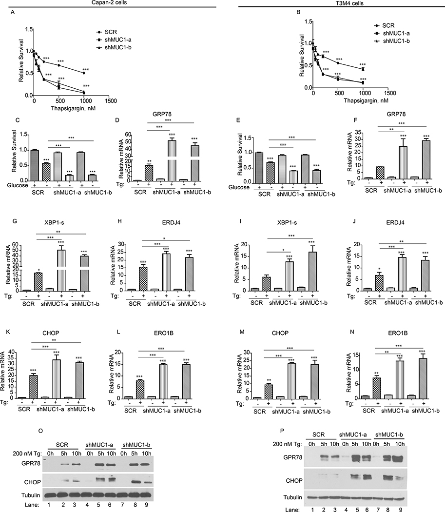
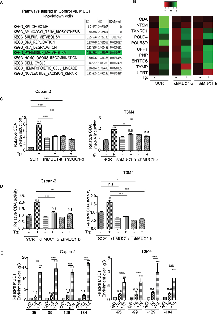
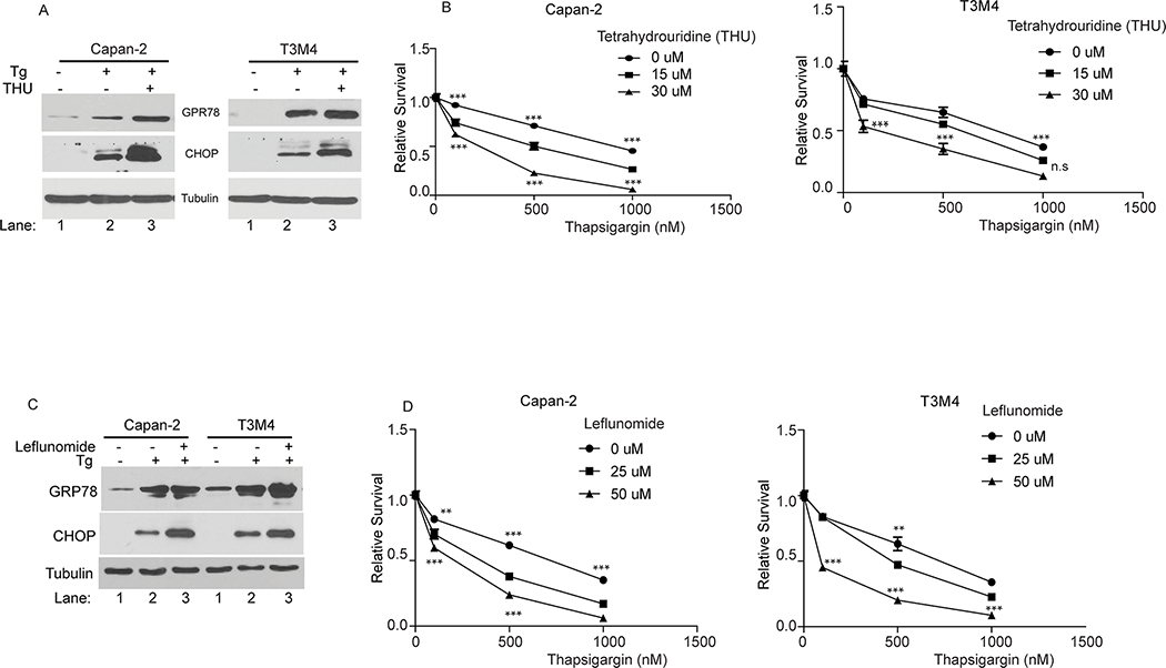
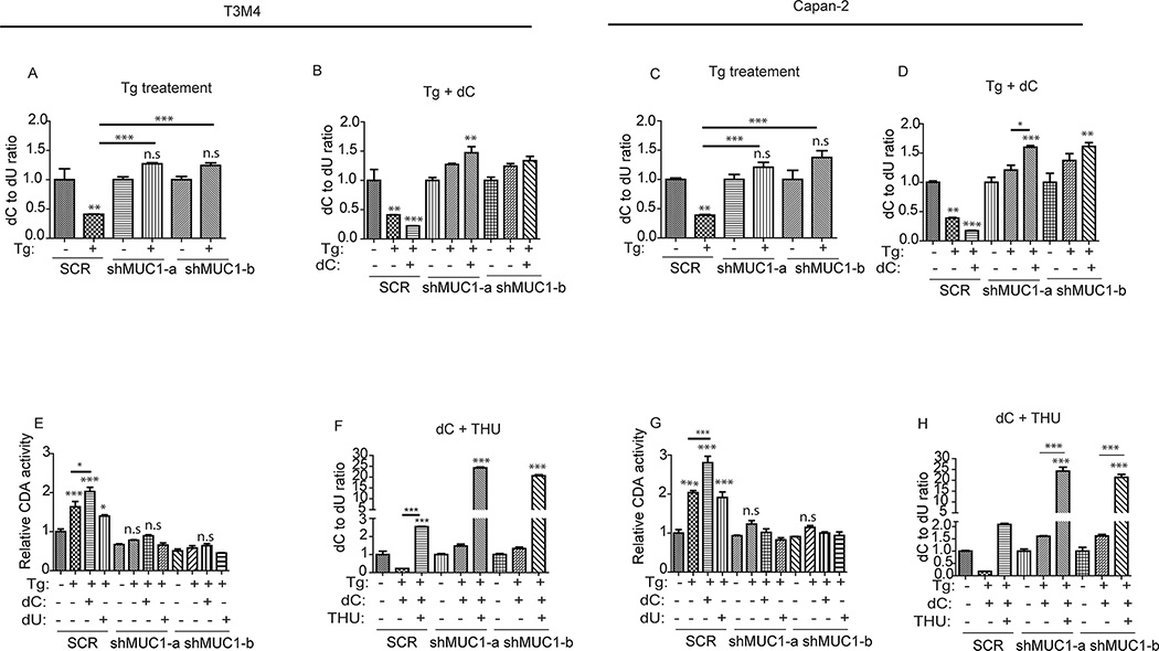
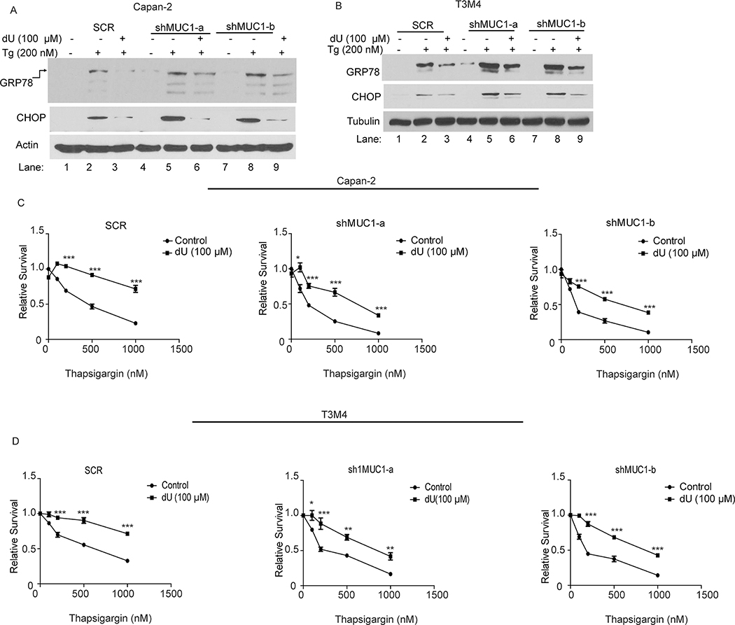
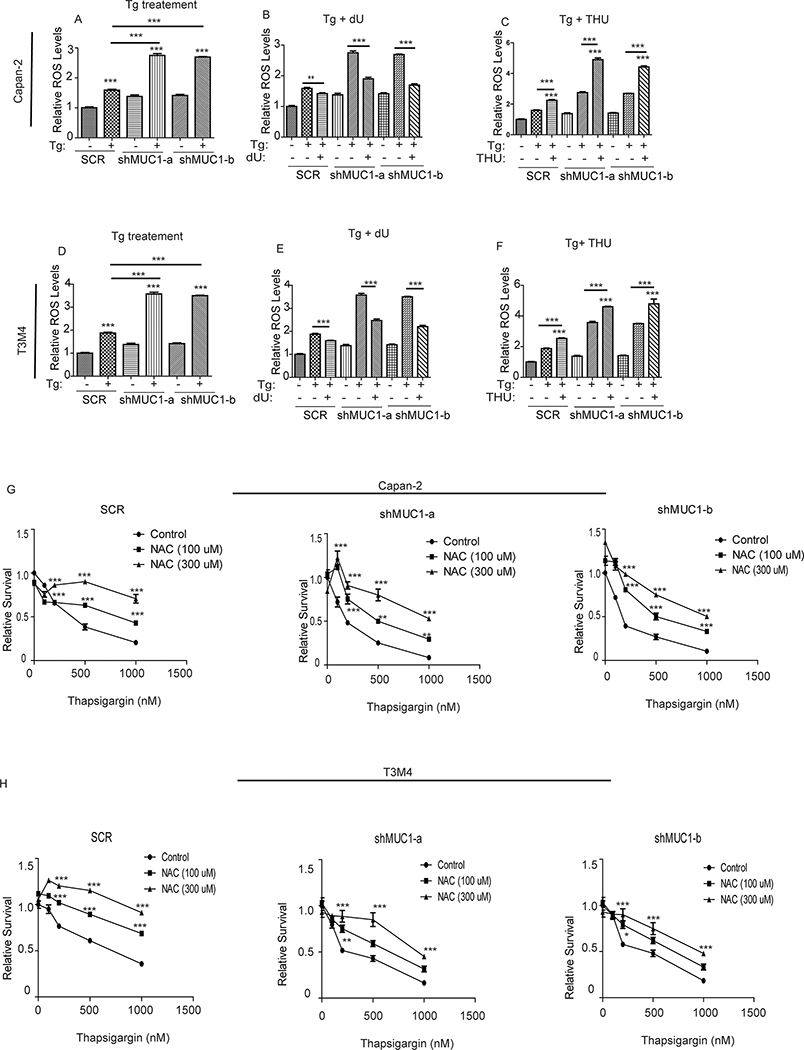
The Mucin 1 (MUC1) protein is overexpressed in various cancers and mediates chemotherapy resistance. However, the mechanism is not fully understood. Given that most chemotherapeutic drugs disrupt ER homeostasis as part of their toxicity, and MUC1 expression is regulated by proteins involved in ER homeostasis, we investigated the link between MUC1 and ER homeostasis. MUC1 knockdown in pancreatic cancer cells enhanced unfolded protein response (UPR) signaling and cell death upon ER stress induction. Transcriptomic analysis revealed alterations in the pyrimidine metabolic pathway and cytidine deaminase (CDA). ChIP and CDA activity assays showed that MUC1 occupied CDA gene promoter upon ER stress induction correlating with increased CDA expression and activity in MUC1-expressing cells as compared with MUC1 knockdown cells. Inhibition of either the CDA or pyrimidine metabolic pathway diminished survival in MUC1-expressing cancer cells upon ER stress induction. Metabolomic analysis demonstrated that MUC1-mediated CDA activity corresponded to deoxycytidine to deoxyuridine metabolic reprogramming upon ER stress induction. The resulting increase in deoxyuridine mitigated ER stress-induced cytotoxicity. In addition, given (1) the established roles of MUC1 in protecting cells against reactive oxygen species (ROS) insults, (2) ER stress-generated ROS further promote ER stress and (3) the emerging anti-oxidant property of deoxyuridine, we further investigated if MUC1 regulated ER stress by a deoxyuridine-mediated modulation of ROS levels. We observed that deoxyuridine could abrogate ROS-induced ER stress to promote cancer cell survival. Taken together, our findings demonstrate a novel MUC1-CDA axis of the adaptive UPR that provides survival advantage upon ER stress induction.


Conflict of interest statement

Conflicts of Interest: The authors declare that they have no conflict of interest.

Figures

Figure 1:
Figure 1:. MUC1 deficiency exacerbates ER stress upon induction
(A-C; E): Cell survival in SCR and MUC1 knockdown cells, in response to the indicated doses of thapsigargin (Tg, A-B) or glucose-starvation (C and E) for 48 hours, by MTT assays. Values were normalized to SCR. (D, F-N): The mRNA levels relative to SCR. Indicated cells were treated with thapsigargin (Tg, 200 nM) for 6 hours followed by total RNA isolation and qPCR with primers for indicated genes. (O-P): Expression levels of UPR marker proteins in cells treated with thapsigargin, by western blotting. Immunoblotting with anti-Tubulin antibody was used as a loading control. P-values are indicated as: p<0.05, *; p<0.01, **; p<0.001, ***.
Figure 2:
Figure 2:. Transcriptomic analysis reveals alterations in the pyrimidine salvage pathway and Cytidine Deaminase (CDA) upon UPR induction.
(A): Top altered pathways in Capan-2 SCR vs. MUC1 knockdown (KD) cells upon ER stress induction with thapsigargin (Tg, 200 nM) for 6 hours. (B): Heatmap of genes altered in the pyrimidine pathway upon Tg treatment. The absolute values in Tg-treated samples were normalized over their base line levels in vehicle-treated cells. (C): CDA mRNA fold change, relative to SCR, upon treatment with thapsigargin (Tg, 200 nM) for 6 hours in indicated cells (SCR and MUC1 knockdown) by qPCR analysis. (D): CDA activity in SCR and MUC1 knockdown cells relative to SCR. Indicated cells were treated with thapsigargin (Tg, 200 nM) for 5 hours and the lysates were then utilized for CDA assay, using equal amounts of protein for each sample. (E): Bar charts representing the occupancy of MUC1 on the indicated regions of CDA promoter by ChIP assays upon Thapsigargin treatment. Cells were treated with thapsigargin (Tg, 200 nM) for 5 hours followed by cross-linking and preparation of protein and chromatin complex. Associated chromatin fragments were then eluted and amplified by qPCR using primers for various CDA promoter regions. IgG antibody pulldowns were used as negative controls. Enrichment is over IgG. P-values are indicated as: p<0.05, *; p<0.01, **; p<0.001, ***.
Figure 3:
Figure 3:. Inhibition of either the CDA enzyme activity or the pyrimidine pathway sensitizes cancer cells to ER stress.
(A and C): UPR marker protein levels in the indicated cells treated with thapsigargin (Tg; 200 nM) alone or in combination with either tetrahydrouridine (THU; 50 μM) or Leflunomide (50 μM) for 10 hours, by western blotting. (B and D): Cell survival in the indicated cells treated with thapsigargin alone or in combination with THU or Leflunomide for 48 hours, by MTT assays relative to untreated cells. P-values are indicated as: p<0.05, *; p<0.01, **; p<0.001, ***.
Figure 4:
Figure 4:. MUC1-mediated CDA activity correlates with deoxycytidine to deoxyuridine reprogramming upon UPR induction.
(A-D, F and H): LC-MS/MS showing change in the levels of intracellular deoxycytidine (dC) and deoxyuridine (dU) assessed by dC to dU ratio relative to respective untreated cells. Indicated cells were treated with thapsigargin, (Tg; 200 nM) alone or in combination with dC (100 μM) or tetrahydrouridine (THU; 10 μM) for 16 hours. Experiments were performed in triplicates. All experiments were run in parallel with same control. (E and G): CDA activity, relative to SCR, is represented by the bar charts. Indicated cells were treated with thapsigargin (Tg, 200 nM) alone for 5 hours or in combination with dC (100 μM) or dU (100 μM). Experiments were done in parallel and used same control as in Fig. 2D. P-values are indicated as: p<0.05, *; p<0.01, **; p<0.001, ***.
Figure 5:
Figure 5:. Deoxyuridine rescues cancer cells from ER stress and provides survival advantage.
(A-B): UPR marker protein levels in the indicated cells treated with deoxyuridine (dU, 100 μM) for 5 hours and subsequently with thapsigargin (Tg; 200 nM) for additional 5 hours, by western blotting. Immunoblotting for Tubulin was used as a loading control. (C-D): Cell survival in the indicated cells treated with thapsigargin alone or in combination with dU for 48 hours, by MTT assays. The values are relative to their respective untreated cells. P-values are indicated as: p<0.05, *; p<0.01, **; p<0.001, ***.
Figure 6:
Figure 6:. Deoxyuridine modulates ER stress-induced Reactive Oxygen Species (ROS) generation and Oxidative stress.
(A-F): ROS levels in the indicated cells treated with thapsigargin (Tg, 200nM) alone (A and D) or in combination with deoxyuridine (dU; B and E) or tetrahydrouridine (THU; C and F) for 12 hours. ROS levels were assessed by utilizing the fluorescent dye 2′,7′–dichlorofluorescin diacetate (DCFDA). All experiments were run in parallel and used same controls. The values were relative to SCR. (G and H): Relative cell survival in cells treated with thapsigargin alone or in combination with N-Acetyl Cysteine (NAC) for 48 hours, as measured by MTT assays. P-values are indicated as: p<0.05, *; p<0.01, **; p<0.001, ***.

Similar articles

Cited by

References

    1. Berridge MJ, Bootman MD, Roderick HL. Calcium signalling: dynamics, homeostasis and remodelling. Nature reviews Molecular cell biology. 2003;4(7):517–29. - PubMed
    1. Fagone P, Jackowski S. Membrane phospholipid synthesis and endoplasmic reticulum function. Journal of lipid research. 2009;50 Suppl:S311–6. - PMC - PubMed
    1. Ikonen E Cellular cholesterol trafficking and compartmentalization. Nature reviews Molecular cell biology. 2008;9(2):125–38. - PubMed
    1. Ron D, Hampton RY. Membrane biogenesis and the unfolded protein response. The Journal of cell biology. 2004;167(1):23–5. - PMC - PubMed
    1. Jun HS, Lee YM, Cheung YY, McDermott DH, Murphy PM, De Ravin SS, et al. Lack of glucose recycling between endoplasmic reticulum and cytoplasm underlies cellular dysfunction in glucose-6-phosphatase-beta-deficient neutrophils in a congenital neutropenia syndrome. Blood. 2010;116(15):2783–92. - PMC - PubMed

Publication types

MeSH terms