. 2020 Jun;10(3):690-705.
doi: 10.1007/s13346-020-00727-2.

Evaluation of the clinical impact of repeat application of hydrogel-forming microneedle array patches

Affiliations

Evaluation of the clinical impact of repeat application of hydrogel-forming microneedle array patches

Rehan Al-Kasasbeh et al. Drug Deliv Transl Res. 2020 Jun.

Abstract

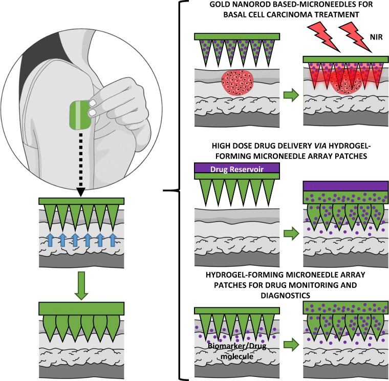
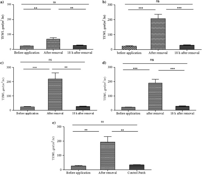
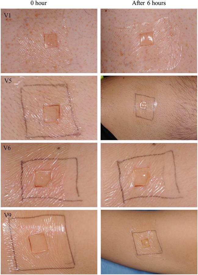
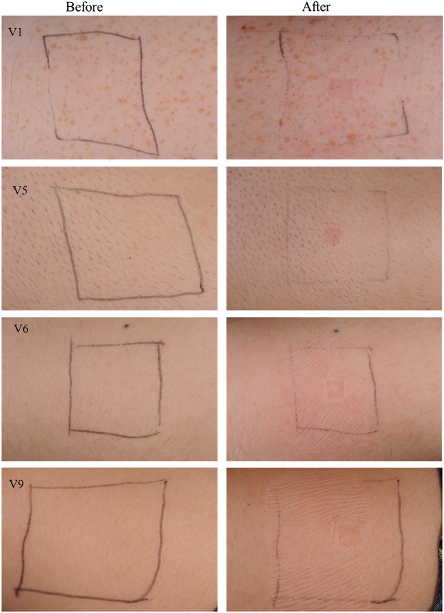
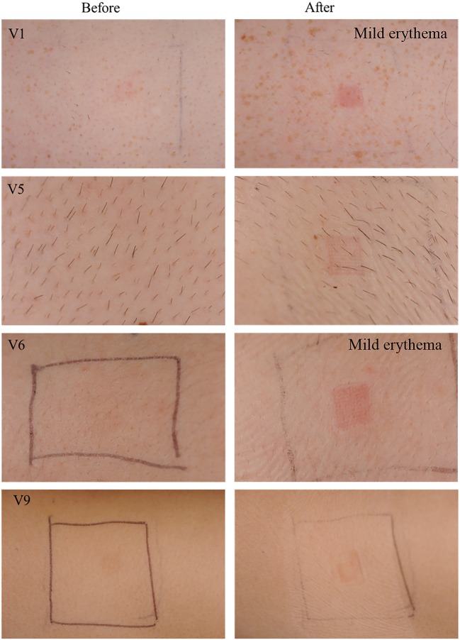
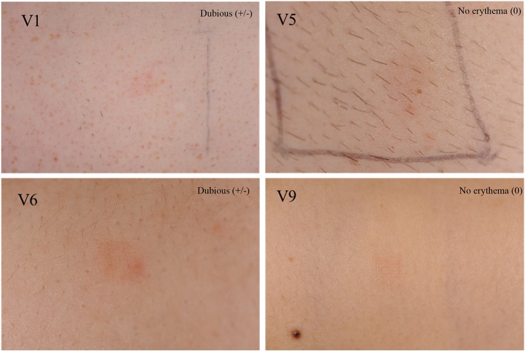
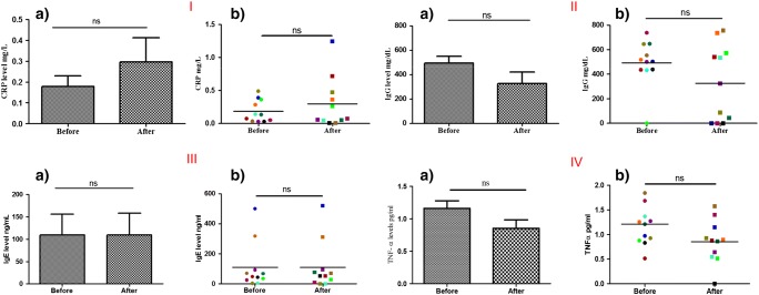
Hydrogel-forming microneedle array patches (MAPs) have been proposed as viable clinical tools for patient monitoring purposes, providing an alternative to traditional methods of sample acquisition, such as venepuncture and intradermal sampling. They are also undergoing investigation in the management of non-melanoma skin cancers. In contrast to drug or vaccine delivery, when only a small number of MAP applications would be required, hydrogel MAPs utilised for sampling purposes or for tumour eradication would necessitate regular, repeat applications. Therefore, the current study was designed to address one of the key translational aspects of MAP development, namely patient safety. We demonstrate, for the first time in human volunteers, that repeat MAP application and wear does not lead to prolonged skin reactions or prolonged disruption of skin barrier function. Importantly, concentrations of specific systemic biomarkers of inflammation (C-reactive protein (CRP); tumour necrosis factor-α (TNF-α)); infection (interleukin-1β (IL-1β); allergy (immunoglobulin E (IgE)) and immunity (immunoglobulin G (IgG)) were all recorded over the course of this fixed study period. No biomarker concentrations above the normal, documented adult ranges were recorded over the course of the study, indicating that no systemic reactions had been initiated in volunteers. Building upon the results of this study, which serve to highlight the safety of our hydrogel MAP, we are actively working towards CE marking of our MAP technology as a medical device.

Keywords: Biomarkers; Clinical translation; Hydrogels; Microneedle array patches; Microneedles; Safety; Skin barrier.


Conflict of interest statement

Ryan Donnelly is an inventor of patents that have been licenced to companies developing microneedle-based products and is a paid advisor to companies developing microneedle-based products. The resulting potential conflict of interest has been disclosed and is managed by Queen’s University Belfast. The companies had no role in the design of the study, in the collection, analyses or interpretation of data, in the writing of the manuscript or in the decision to publish the results.

Figures

Fig. 1
Fig. 1
Schematic representations of hydrogel-forming MAP applications
Fig. 2
Fig. 2
Hydrogel-forming microneedle array patch (MAP) design. Single microneedle array (A); microneedle array attached to the adhesive part of the TegadermTM dressing to form the MAP (B)
Fig. 3
Fig. 3
Schematic representation of the study design
Fig. 4
Fig. 4
Transepidermal water loss values measured before and after applying MAPs. First day (A), second day (B), third day (C), fourth day (D), fifth day (E). On all days, values increased significantly after MAP removal, but returned to baseline levels 18 h post-patch removal (means + SE, n = 11)
Fig. 5
Fig. 5
Optical coherence tomography image of MAP immediately after insertion to the forearm of the researcher. It is clear that the MAP was able to create pores and penetrate into the skin upon manual application
Fig. 6
Fig. 6
Digital images of MAPs after removal from volunteers’ upper arms. Microneedles and baseplates were flexible and rubbery (A). The array centres showed a higher degree of swelling compared with the baseplates (B). Microscope images of swollen MAPs after removal from a volunteer’s arm. MAPs became elastic as a result of imbibing interstitial fluid from the volunteers (C, D). All of the microneedles of every MAP were removed intact, without any deformation inside the skin (E)
Fig. 7
Fig. 7
Microscope images of MAPs showing the dimensions of microneedle array (A) before and (B) after application to human volunteers. The area of the array was calculated by measuring the distance between the first and the last microneedle of the first line in the array in mm. Thus, the area was calculated in mm2. The application time was 6 h
Fig. 8
Fig. 8
Exemplar images of MAPs immediately upon application and after 6 h of wear. Signs of interstitial fluid absorption were noticed as swelling at the centre of the microneedle arrays in some volunteers. The Tegaderm™ adhesive dressing permitted the microneedles to stay in place over the time of wear
Fig. 9
Fig. 9
Clinical photographs taken of the upper arm skin of randomly selected volunteers by a medical photographer on the first day of study. Photographs in the left hand column were taken before application of MAPs, while photographs in the right hand column were taken immediately after patch removal (6-h wear time). The area of patch application was marked with a pen
Fig. 10
Fig. 10
Clinical photographs taken by medical photographer on the fifth day of study. Photographs in the left hand column were taken before application of MAPs, while photographs in the right hand column were taken immediately after patch removal. It is notable that erythema is directly centred to the site of microneedle insertion, rather than in the border area where the adhesive was
Fig. 11
Fig. 11
Clinical photographs taken after 18 h of MAP removal. These images were taken on the fifth day of study (i.e. after four MAP applications; two to this approximate site). Erythema degree as assessed by the dermatologist is assigned to each image
Fig. 12
Fig. 12
I: levels of plasma CRP at the beginning and the end of the study, as determined using ELISA (means ± SE, n = 11) (A) and individual volunteer values (B). II: levels of plasma IgG at the beginning and the end of the study, as determined using ELISA (means ± SE, n = 11) (A) and individual volunteer values (B). III: levels of plasma IgE at the beginning and the end of the study, as determined using ELISA (means ± SE, n = 11) (A) and individual volunteer values (B). IV: levels of plasma TNF-α at the beginning and the end of the study, as determined using ELISA (means ± SE, n = 11) (A) and individual volunteer values (B). ns, non-significant

References

    1. Donnelly RF, Thakur RRS, Garland MJ, Migalska K, Majithiya R, McCrudden CM, Kole PL, Mahmood TMT, McCarthy HO, Woolfson AD. Hydrogel-forming microneedle arrays for enhanced transdermal drug delivery. Adv Funct Mater. 2012;22:4879–4890. - PMC - PubMed
    1. Donnelly RF, McCrudden MTC, Zaid-Alkilani A, Larraňeta E, McAlister E, Thakur RRS, McCarthy HO, Kett VL, Woolfson AD. Hydrogel-forming microneedles prepared from “super swelling” polymers enhance transdermal drug and protein delivery when combined with lyophilised wafers. PLOS One. 2014;9(10):e111547. - PMC - PubMed
    1. Leboulanger B, Guy RH, Delgado-Charro MB. Reverse iontophoresis for non-invasive transdermal monitoring. Physiol Meas. 2004;25:R35–R50. - PubMed
    1. Brunner M, Derendorf H. Clinical microdialysis: current applications and potential use in drug development. Trends Anal Chem. 2006;25(7):674–680.
    1. Caffarel-Salvador E, Brady AJ, Eltayib A, Meng T, Alonso-Vicentre A, Gonzalez-Vazquez P, Torrisi BM, Vicente-Perez EM, Mooney K, Jones DS, Bell SEJ, McCoy CP, McCarthy HO, McElnay JC, Donnelly RF. Hydrogel-forming microneedle arrays allow detection of drugs and glucose in vivo: potential for use in diagnosis and therapeutic drug monitoring. PLoS One. 2005;10(12):e0145644. - PMC - PubMed

Publication types