. 2020 Feb 27;19(1):41.
doi: 10.1186/s12943-020-01165-x.

CCL2-CCR2 axis recruits tumor associated macrophages to induce immune evasion through PD-1 signaling in esophageal carcinogenesis

Affiliations

CCL2-CCR2 axis recruits tumor associated macrophages to induce immune evasion through PD-1 signaling in esophageal carcinogenesis

Hui Yang et al. Mol Cancer. .

Abstract

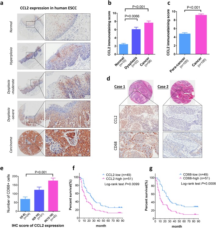
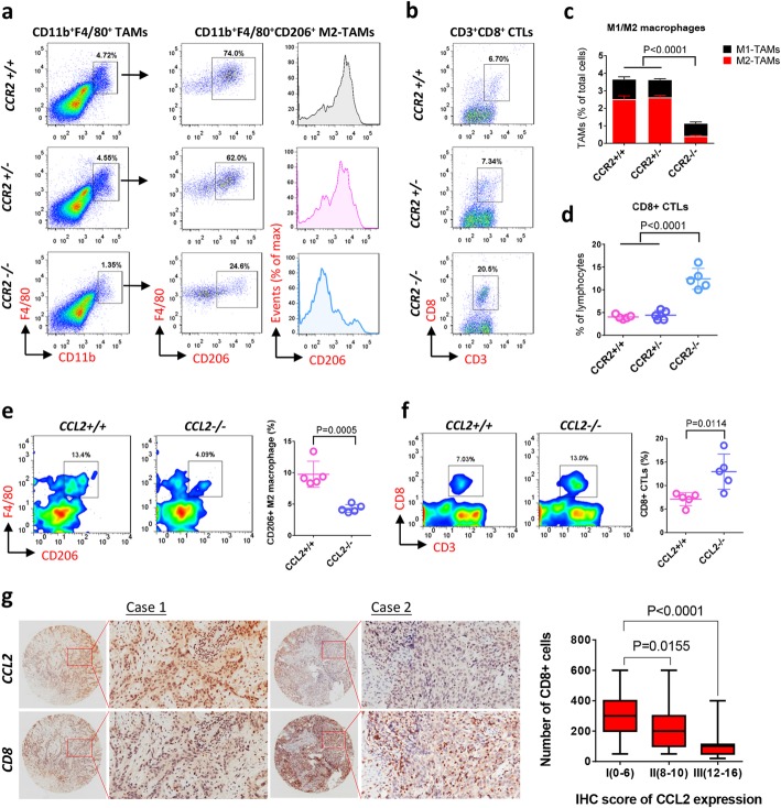
Background: The poor prognosis of esophageal squamous cell carcinoma (ESCC) highlights the need for novel strategies against this disease. Our previous study suggested the involvement of CCL2 and tumor associated macrophages (TAMs) in esophageal carcinogenesis. Despite the recognition of TAMs as a promising target for cancer treatment, mechanisms underlying its infiltration, activation and tumor-promotive function in ESCC remain unknown.

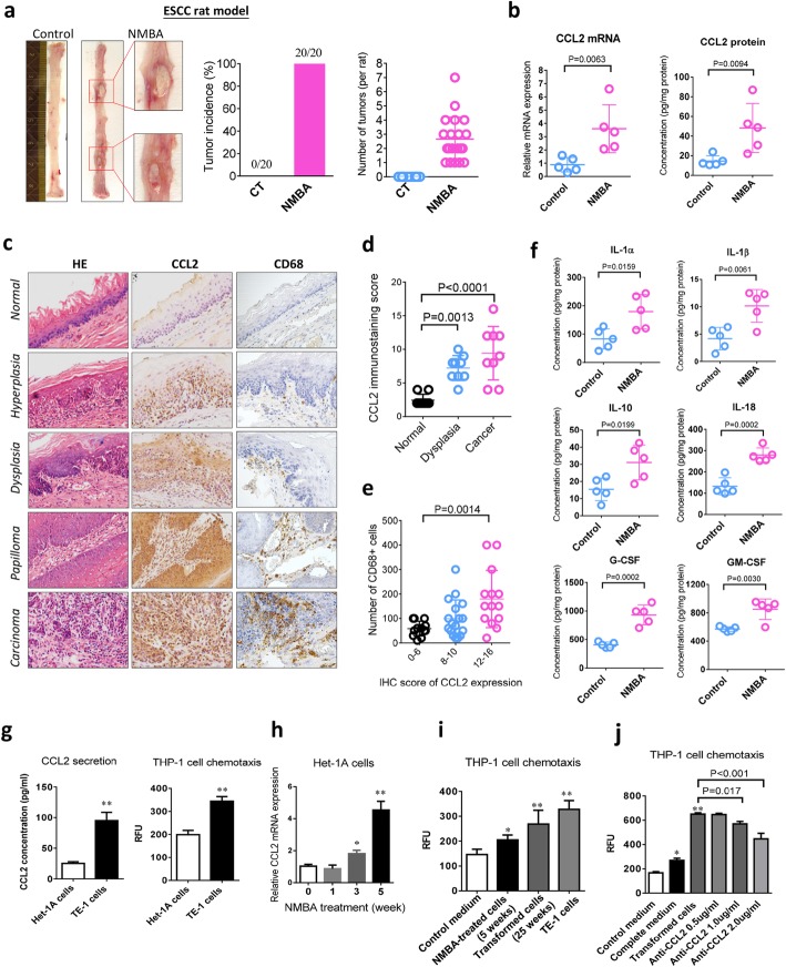
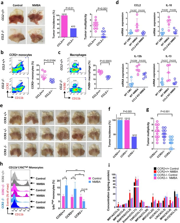
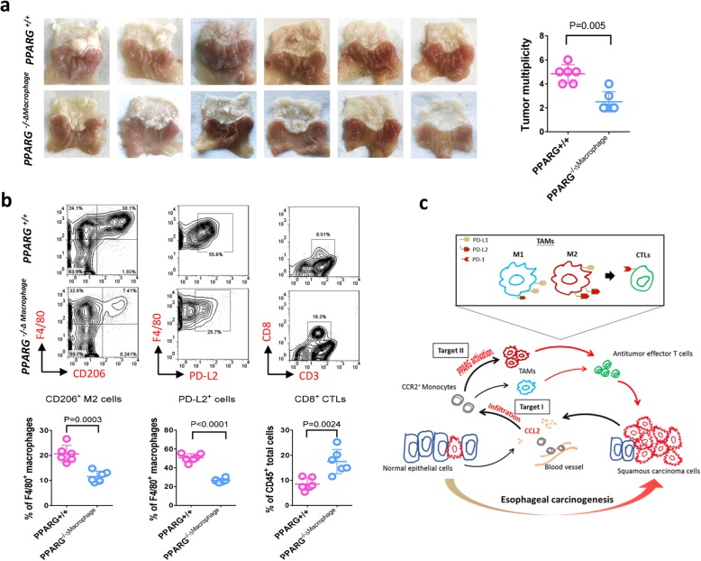
Methods: Human esophageal tissue array and TCGA database were used to evaluate the clinical relevance of CCL2 and TAMs in ESCC. F344 rats and C57BL/6 mice were treated with N-nitrosomethylbenzylamine (NMBA) to establish orthotopic models of esophageal carcinogenesis. CCL2/CCR2 gene knockout mice and macrophage-specific PPARG gene knockout mice were respectively used to investigate the role of infiltration and polarization of TAMs in ESCC. CCL2-mediated monocyte chemotaxis was estimated in malignantly transformed Het-1A cells. THP-1 cells were used to simulate TAMs polarization in vitro. RNA-sequencing was performed to uncover the mechanism.

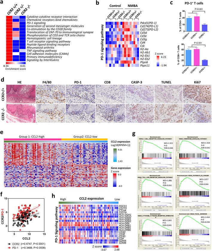
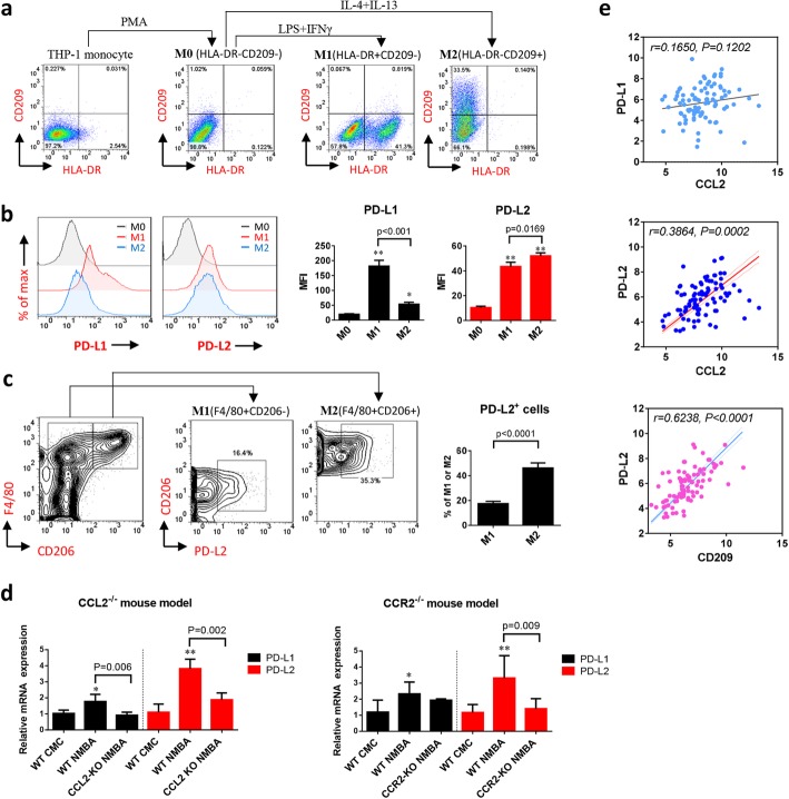
Results: Increasing expression of CCL2 correlated with TAMs accumulation in esophageal carcinogenesis, and they both predicts poor prognosis in ESCC cohort. Animal studies show blockade of CCL2-CCR2 axis strongly reduces tumor incidence by hindering TAMs recruitment and thereby potentiates the antitumor efficacy of CD8+ T cells in the tumor microenvironment. More importantly, M2 polarization increases PD-L2 expression in TAMs, resulting in immune evasion and tumor promotion through PD-1 signaling pathway.

Conclusion: This study highlights the role of CCL2-CCR2 axis in esophageal carcinogenesis. Our findings provide new insight into the mechanism of immune evasion mediated by TAMs in ESCC, suggesting the potential of TAMs-targeted strategies for ESCC prevention and immunotherapy.

Keywords: CCL2; CCR2; Carcinogenesis; Esophageal squamous cell carcinoma (ESCC); Programmed death-1(PD-1); Tumor associated macrophages (TAMs).


Conflict of interest statement

The authors declare that they have no competing interests.

Figures

Fig. 1
Fig. 1
CCL2 expression correlates with TAMs accumulation, cancer progression and poor prognosis in human ESCC. a Representative IHC staining indicates escalating expression of CCL2 with histopathologic progression. b Expression of CCL2 in different pathologic grades of cohort I patients including normal mucosa (10 cases), dysplasia (22 cases), and ESCC (58 cases). c Expression of CCL2 in para-cancer and cancer tissues of cohort II patients (n = 100). d Representative IHC staining indicates correlated expression of CCL2 and CD68 in ESCC. e CCL2 expression is correlated with accumulation of CD68+ TAMs. f and g High-expression of CCL2 (f) and CD68 (g) predicts reduced overall survival of ESCC patients
Fig. 2
Fig. 2
CCL2 correlates with TAMs accumulation and tissue inflammation in nitrosamine-induced esophageal carcinogenesis. a NMBA induces notable tumors in ESCC rat model. b Expression of CCL2 in rat esophageal epithelium is increased at mRNA and protein levels during carcinogenesis (n = 5). c Representative IHC staining indicates the expression of CCL2 and CD68 during rat esophageal carcinogenesis resembling human ESCC. d Expression of CCL2 increases with pathologic progression in rat model (n = 9). e Accumulation of CD68+ TAMs is correlated with CCL2 expression. f Increased expression of inflammatory cytokines during esophageal carcinogenesis (n = 5). g The basal levels of CCL2 between normal human esophageal epithelium cells (Het-1A) and the ESCC cells (TE-1). h NMBA treatment continuously increases CCL2 expression over time. i Chemotaxis of THP-1monocyte is increased by conditioned medium from the transformed cells and TE-1 cells. j Monocyte chemotaxis induced by transformed cells is antagonized by CCL2-neutralizing antibody in a dose-dependent manner. Data is shown by mean ± standard deviation from three independent experiments. When compared to the control group, * indicates P < 0.05, ** indicates P < 0.01
Fig. 3
Fig. 3
Blockade of CCL2-CCR2 axis suppresses monocyte infiltration, TAMs accumulation and tumorigenesis in ESCC mouse model. a Gene knockout of CCL2 in mouse reduces tumor incidence and multiplicity (n = 10). b Deletion of CCL2 in mouse suppresses infiltration of CD11b+CCR2+ monocyte and CD11b+F4/80+ TAMs in forestomach tumors (n = 5). c Deletion of CCL2 in mouse inhibited production of TAMs-associated cytokines (CCL2, IL-10, IL-12b, and IL-13) (n = 5). d, e and f Gene knockout of CCR2 reduces tumor incidence (e) and tumor numbers (f) in mouse forestomach (n = 12). g Infiltration of CD11b+Ly6Chigh inflammatory monocyte elevated in CCR2+/+ wild type mice and CCR2+/− heterozygous mice is blocked in CCR2−/− animals (n = 6). h CCR2 knockout suppresses production of inflammatory cytokines in tumors (n = 6). Data is shown by mean ± standard deviation, * indicates P < 0.05, ** indicates P < 0.01, when compared to the CCR2+/+ control
Fig. 4
Fig. 4
TAMs mediate immune evasion through PD-1 signaling pathway. a Heatmap of enriched pathways (Top 15) from differentially expressed genes from CCR2+/+ wild type mice, CCR2+/− heterozygous mice, and CCR2−/− animals. b Heatmap shows the expression of genes that associated with PD-1 signaling pathway is activated in carcinogenesis but repressed by CCR2 knockout. c Flow cytometry analysis demonstrates suppressed PD-1 expression in CD4+ and CD8+ T cells by CCR2 deletion (n = 6). d Representative IHC staining of F4/80, PD-1, CD8, cleaved CASP-3, Ki67 and TdT-mediated dUTP nick end labeling (TUNEL) in tumor microenvironment of CCR2 knockout ESCC mouse model. e Heatmap of gene expression profiles of human ESCC cases (n = 90) from TCGA database. f Pearson correlation analysis shows tight association of CCL2 with CCR2 and PD-1. g Gene set enrichment analysis (GSEA) between group high (n = 52) and low (n = 38) of CCL2 expression shows enriched pathways associated with PD-1 signaling. h Heatmap shows that expression of PD-1 signaling pathway associated genes is correlated with CCL2 expression in human ESCC
Fig. 5
Fig. 5
M2-polorization of TAMs is associated with depletion of antitumor effector T cells in ESCC carcinogenesis. a and c) Flow cytometry shows inhibition of TAMs (CD11b+F4/80+) accumulation as well as M2 polarization (F4/80+CD206+) by CCR2 knockout. Data is shown by mean ± standard deviation (n = 6). b and d CCR2 knockout increases antitumor effector T cells (CD3+CD8+ CTLs) in tumors (n = 6). e and f CCL2 deletion inhibits TAMs M2-polorization (e) and antitumor effector T cells depletion (f) during carcinogenesis (n = 5). g Representative IHC staining indicates expression of CCL2 inversely correlated with CD8 (effector T cells) in human ESCC cohort II (n = 100)
Fig. 6
Fig. 6
M2 polarization of TAMs facilitates immunosuppression through the elevated expression of PD-L2. a THP-1 monocyte is induced into M1-type by LPS and IFN-γ, and into M2-type by IL-4 and IL-13 following PMA treatment. The polarized M1 and M2 macrophages were distinguished from inactive M0 macrophage with the biomarker HLA-DR and CD209, respectively. b Flow cytometry analysis shows discrepant expression of PD-L1 and PD-L2 between HLA-DR+CD209 M1 macrophages and HLA-DRCD209+ M2 macrophages. Data is shown by mean ± standard deviation from three independent experiments. When compared to the control group, * indicates P < 0.05, ** indicates P < 0.01. c PD-L2 expression is higher in M2 (F4/80+CD206+) than M1 (F4/80+CD206) macrophages in ESCC mouse model (n = 5). d Blockade of CCL2-CCR2 axis in animal models reduces expression of PD-L2 during carcinogenesis (n = 5). e Pearson correlation analysis with TCGA data indicates strong association of PD-L2 with CCL2 expression and the level of M2 marker CD209 in human ESCC
Fig. 7
Fig. 7
Blockade of TAMs M2-polarization via macrophage-specific PPARG deletion reduces PD-L2 expression and inhibits tumorigenesis. a Macrophage-specific gene knockout of PPARG in mouse reduces tumor growth in ESCC mouse model (n = 6). b Flow cytometry analysis shows deficiency of PPARG in macrophage suppresses M2-polarization and PD-L2 expression in TAMs and increases CD8+ CTLs in forestomach tumors (n = 6). c Schematic figure indicates esophageal carcinogenesis can be blocked by targeting TAMs infiltration via CCL2/CCR2 signaling (Target I) and M2 polarization via PPARG activation (Target II)

References

    1. Bray F, Ferlay J, Soerjomataram I, Siegel RL, Torre LA, Jemal A. Global cancer statistics 2018: GLOBOCAN estimates of incidence and mortality worldwide for 36 cancers in 185 countries. CA Cancer J Clin. 2018;68:394–424. doi: 10.3322/caac.21492. - DOI - PubMed
    1. Arnold M, Soerjomataram I, Ferlay J, Forman D. Global incidence of oesophageal cancer by histological subtype in 2012. Gut. 2015;64:381–387. doi: 10.1136/gutjnl-2014-308124. - DOI - PubMed
    1. Abnet CC, Arnold M, Wei WQ. Epidemiology of esophageal squamous cell carcinoma. Gastroenterology. 2018;154:360–373. doi: 10.1053/j.gastro.2017.08.023. - DOI - PMC - PubMed
    1. Pennathur A, Gibson MK, Jobe BA, Luketich JD. Oesophageal carcinoma. Lancet. 2013;381:400–412. doi: 10.1016/S0140-6736(12)60643-6. - DOI - PubMed
    1. Cancer Genome Atlas Research N, Analysis working group. Asan U, Agency BCC. Brigham, Women's H. Broad I, Brown U, Case Western reserve U. Dana-Farber Cancer I. Duke U, et al. Integrated genomic characterization of oesophageal carcinoma. Nature. 2017;541:169–175. doi: 10.1038/nature20805. - DOI - PMC - PubMed

Publication types

MeSH terms